

COVID-19 integrated imaging: our experience and literature review
- PMID: 33758632
- PMCID: PMC7976231
- DOI: 10.5114/pjr.2021.103861
COVID-19 integrated imaging: our experience and literature review
Abstract
Purpose: To investigate the imaging features of emerging COVID-19 pneumonia on chest ultrasound, radiographs and computed tomography examinations performed at admission. In addition, we provide a review of the literature and compare our results with recent evidence regarding the imaging characteristics of this novel disease.
Material and methods: From March 17, 2020 to April 25, 2020, 23 patients with real-time polymerase chain reaction (RT-PCR) assay confirmed COVID-19 were identified. All 23 patients were evaluated and admitted at San Giuseppe Moscati Hospital in Aversa, Italy. Multi-modality imaging findings were evaluated and compared. Literature research was conducted through a methodical search on PubMed.
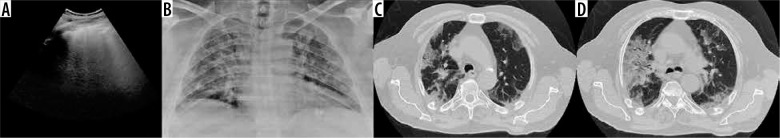
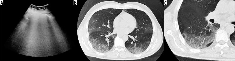
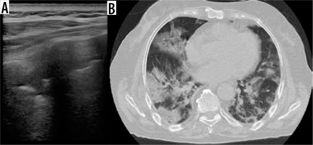
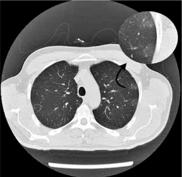
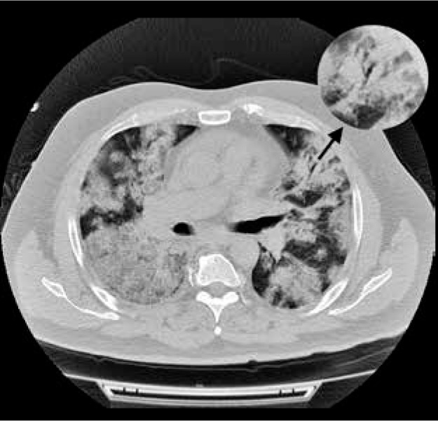
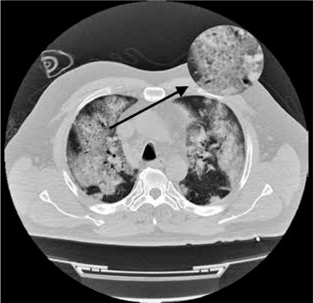
Results: Twenty-three patients were included in the study. Chest transthoracic ultrasound (US), chest X-ray (CXR), and computed tomography (CT) were performed respectively in 11, 16 and 21 patients. Chest US findings were consistent with diffuse B lines (91%), subpleural consolidations (45%), and thickened pleural line (18%). CXR showed prevalent manifestations of consolidations (50%) and hazy increased opacities (37%). Typical CT features are bilateral and multilobar ground-glass opacities (GGO). Indeed GGO were present in 100% of our patients. Consolidations were visible in 76% of our study population. Notably both GGO and consolidations had a peripheral distribution in all our patients. Other CT imaging features included crazy-paving pattern, fibrous stripes, subpleural lines, architectural distortion, air bronchogram sign, vascular thickening and nodules. Our literature review identified thirty original studies supporting our imaging chest findings.
Conclusions: At admission, COVID-19 pneumonia can manifest in chest imaging as B-lines and consolidations on US, hazy opacities and consolidations on CXR, multiple GGO and consolidations on CT scan.
Keywords: COVID-19; SARS-CoV-2; computed tomography (CT); coronavirus disease; pneumonia; radiographic chest examination (CXR).
© Pol J Radiol 2021.
Conflict of interest statement
The authors report no conflict of interest.
Figures
References
-
- WHO. Novel Coronavirus (2019-nCoV). Situation report–7. Available from: https://www.who.int/publications-detail/global-surveillance-for-human-in... (Accessed: 27.02.2020).
LinkOut - more resources
Full Text Sources
Other Literature Sources
Miscellaneous