

Intra-articular long head of the biceps tendon: magnetic resonance-arthrography classification and review of literature
- PMID: 33758634
- PMCID: PMC7976233
- DOI: 10.5114/pjr.2021.104206
Intra-articular long head of the biceps tendon: magnetic resonance-arthrography classification and review of literature
Abstract
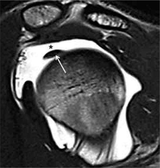
Purpose: Anatomical variants of the long head of the biceps (LHB) and diseases of the rotator interval structures may contribute to shoulder instability. The rotator interval and the LHB tendon are closely associated anatomic structures that confer stability to the shoulder. Anatomical variants around the origins of the long head of the biceps (LHB) are reported to occur with a frequency of 1.9-7.4%. In the past years, many authors have proposed different approaches for the identification and characterization of LHB and rotators interval. Magnetic resonance (MR) arthrography is considered the reference standard in imaging to diagnose superior shoulder diseases. However, few authors have analysed the anatomical variants and the relation between those and shoulder instability. This study aimed to identify the frequency of variants observed during arthroscopic shoulder surgeries, and to classify them based on the Dierickx classification system.
Material and methods: In 326 MR arthrograms we investigated the incidence of LHB anatomical variations and their association with shoulder diseases.
Results: We found 252/326 (77.3%) cases of LHB free, 40/326(12.26%) cases of LHB adherent, 31/326(9.50%) cases of mesotenon, and 3/326(0.9%) cases of split biceps. The prevalence of rotator interval synovitis in the mesotenon group was greater than in the LHB-free group. Moreover, in the LHB-adherent group we observed increased incidence of sublabral recess and SLAP lesions compared with the LHB-free group.
Conclusions: MR-arthrography is useful in the evaluation of superior shoulder structures. A relationship exists between LHB anomalies and superior shoulder instability.
Keywords: MR arthrography; SLAP lesions; anatomical variants; instability shoulder; long head biceps; shoulder.
© Pol J Radiol 2021.
Conflict of interest statement
The authors report no conflict of interest.
Figures


Similar articles
-
MR arthrography: correlation between anatomic intraarticular variants of the long head of the biceps tendon (long head biceps tendon) and superior labral anterior to posterior (SLAP) lesions.J Orthop Traumatol. 2022 Mar 8;23(1):13. doi: 10.1186/s10195-022-00631-0. J Orthop Traumatol. 2022. PMID: 35258708 Free PMC article.
-
Morphological classification of anatomical variants of the intra-articular portion of the long head of the biceps brachii tendon and analysis of the incidence and the relationship with shoulder disease for each subtype.J Orthop Surg (Hong Kong). 2017 Sep-Dec;25(3):2309499017742207. doi: 10.1177/2309499017742207. J Orthop Surg (Hong Kong). 2017. PMID: 29157108
-
MR arthrography of rotator interval, long head of the biceps brachii, and biceps pulley of the shoulder.Radiology. 2005 Apr;235(1):21-30. doi: 10.1148/radiol.2351031455. Epub 2005 Feb 16. Radiology. 2005. PMID: 15716389 Review.
-
[Normal anatomical variants of the superior labrum biceps tendon anchor complex. Anatomical and magnetic resonance findings].Orthopade. 2003 Jul;32(7):586-94. doi: 10.1007/s00132-003-0488-0. Orthopade. 2003. PMID: 12883757 German.
-
Anatomic variations in the long head of biceps: contribution to shoulder dysfunction.Arthroscopy. 2007 Sep;23(9):1012-8. doi: 10.1016/j.arthro.2007.05.007. Arthroscopy. 2007. PMID: 17868842 Review.
Cited by
-
MR arthrography: correlation between anatomic intraarticular variants of the long head of the biceps tendon (long head biceps tendon) and superior labral anterior to posterior (SLAP) lesions.J Orthop Traumatol. 2022 Mar 8;23(1):13. doi: 10.1186/s10195-022-00631-0. J Orthop Traumatol. 2022. PMID: 35258708 Free PMC article.
References
-
- Jakanani GC, Botchu R, Rennie WJ. The MR arthrographic anatomy of the biceps labral insertion and its morphological significance with labral tears in patients with shoulder instability. Eur J Radiol 2012; 81: 3390-3393. - PubMed
-
- Ghalayini SRA, Board TN, Srinivasan MS. Anatomic variations in the long head of biceps: contribution to shoulder dysfunction. Arthroscopy 2007; 23: 1012-1018. - PubMed
-
- Glueck DA, Mair SD, Johnson DL. Shoulder instability with absence of the long head of the biceps tendon. Arthroscopy 2003; 19: 787-789. - PubMed
-
- Harryman DT, Sidles JA, Harris SL, Matsen FA. The role of the rotator interval capsule in passive motion and stability of the shoulder. J Bone Joint Surg Am 1992; 74: 53-66. - PubMed
-
- Bennett WF. Subscapularis, medial, and lateral head coracohumeral ligament insertion anatomy. Arthroscopic appearance and incidence of ‘hidden’ rotator interval lesions. Arthroscopy 2001; 17: 173-180. - PubMed
LinkOut - more resources
Full Text Sources
Other Literature Sources
Miscellaneous