

Radiotherapy of extraosseous nasopharyngeal chordoma: A case report and literature review
- PMID: 33758665
- PMCID: PMC7947947
- DOI: 10.3892/mco.2021.2246
Radiotherapy of extraosseous nasopharyngeal chordoma: A case report and literature review
Abstract
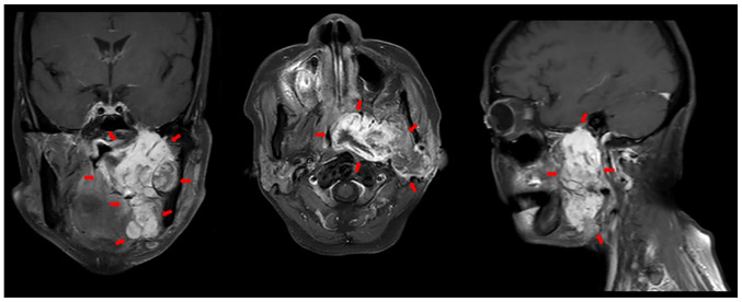
Chordomas are slow-growing aggressive tumors that account for 1-4% of all bone tumors. The anatomical distribution of chordomas includes 50-60% in the sacrococcygeal region, 25-30% in the skull base and 15% in the mobile spine. Virchow was the first to describe and term these tumors as 'ecchordosis physaliphora' in 1857, and Muller established their notochordal origin in 1895. Extraosseous chordomas of the nasopharynx are very rare, and they exhibit similarities with other lesions of the nasopharynx, presenting as a soft tissue mass. Gross total resection combined with postoperative radiotherapy offers the best chance of long-term control. We herein present the case of a 63-year-old female patient with complaints of left temporal headaches, dizziness, left nasal obstruction, left maxillary area numbness, left ear hearing loss and swallowing difficulty. Computed tomography imaging examination revealed an 8.2x3.2x5.7-cm space-occupying lesion with central necrosis in the nasopharynx and oropharynx, partially occluding the pharyngeal lumen; the mass had infiltrated the left parapharyngeal space, the left medial and lateral pterygoid muscle and the left parotid gland, with bone erosion of the left mandible. The patient was diagnosed with extraosseous chordoma of the nasopharynx, conventional type, stage IIB. The patient underwent surgery and high-dose radiotherapy and local control of the chordoma was achieved. However, the patient succumbed to a lung metastasis. The details of the case are discussed, and a review of the current medical literature is presented to provide an updated discussion on the current status of nasopharyngeal chordoma research.
Keywords: chordoma; nasopharynx; radiotherapy; volumetric modulated arc therapy.
Copyright: © Yeh et al.
Conflict of interest statement
The author declares no competing interests.
Figures


References
-
- Bakker SH, Jacobs WCH, Pondaag W, Gelderblom H, Nout RA, Dijkstra PDS, Peul WC, Vleggeert-Lanamo CLA. Chordoma: A systematic review of the epidemiology and clinical prognostic factors predicting progression-free and overall survival. Eur Spine J. 2018;27:3043–3058. doi: 10.1007/s00586-018-5764-0. - DOI - PubMed
-
- Vaz-Guimaraes F, Harsh IV GR. Chapter 5: Demographics, presentation, and diagnosis: Chordomas and chondrosarcomas of the skull base and spine. 2nd edition. Harsh IV GR and Vas-Guimaraes F (eds). Academic press, London, pp45-51, 2018.
LinkOut - more resources
Full Text Sources
Other Literature Sources