[Preprint]. 2021 Mar 16:2021.03.15.435528.
doi: 10.1101/2021.03.15.435528.

Elicitation of broadly protective sarbecovirus immunity by receptor-binding domain nanoparticle vaccines

Affiliations

Elicitation of broadly protective sarbecovirus immunity by receptor-binding domain nanoparticle vaccines

Alexandra C Walls et al. bioRxiv. .

Update in

  • Elicitation of broadly protective sarbecovirus immunity by receptor-binding domain nanoparticle vaccines.
    Walls AC, Miranda MC, Schäfer A, Pham MN, Greaney A, Arunachalam PS, Navarro MJ, Tortorici MA, Rogers K, O'Connor MA, Shirreff L, Ferrell DE, Bowen J, Brunette N, Kepl E, Zepeda SK, Starr T, Hsieh CL, Fiala B, Wrenn S, Pettie D, Sydeman C, Sprouse KR, Johnson M, Blackstone A, Ravichandran R, Ogohara C, Carter L, Tilles SW, Rappuoli R, Leist SR, Martinez DR, Clark M, Tisch R, O'Hagan DT, Van Der Most R, Van Voorhis WC, Corti D, McLellan JS, Kleanthous H, Sheahan TP, Smith KD, Fuller DH, Villinger F, Bloom J, Pulendran B, Baric RS, King NP, Veesler D. Walls AC, et al. Cell. 2021 Oct 14;184(21):5432-5447.e16. doi: 10.1016/j.cell.2021.09.015. Epub 2021 Sep 15. Cell. 2021. PMID: 34619077 Free PMC article.

Abstract

Understanding the ability of SARS-CoV-2 vaccine-elicited antibodies to neutralize and protect against emerging variants of concern and other sarbecoviruses is key for guiding vaccine development decisions and public health policies. We show that a clinical stage multivalent SARS-CoV-2 receptor-binding domain nanoparticle vaccine (SARS-CoV-2 RBD-NP) protects mice from SARS-CoV-2-induced disease after a single shot, indicating that the vaccine could allow dose-sparing. SARS-CoV-2 RBD-NP elicits high antibody titers in two non-human primate (NHP) models against multiple distinct RBD antigenic sites known to be recognized by neutralizing antibodies. We benchmarked NHP serum neutralizing activity elicited by RBD-NP against a lead prefusion-stabilized SARS-CoV-2 spike immunogen using a panel of single-residue spike mutants detected in clinical isolates as well as the B.1.1.7 and B.1.351 variants of concern. Polyclonal antibodies elicited by both vaccines are resilient to most RBD mutations tested, but the E484K substitution has similar negative consequences for neutralization, and exhibit modest but comparable neutralization breadth against distantly related sarbecoviruses. We demonstrate that mosaic and cocktail sarbecovirus RBD-NPs elicit broad sarbecovirus neutralizing activity, including against the SARS-CoV-2 B.1.351 variant, and protect mice against severe SARS-CoV challenge even in the absence of the SARS-CoV RBD in the vaccine. This study provides proof of principle that sarbecovirus RBD-NPs induce heterotypic protection and enables advancement of broadly protective sarbecovirus vaccines to the clinic.


Conflict of interest statement

Declaration of interest A.C.W, N.P.K. and D.V., are named as inventors on patent applications filed by the University of Washington based on the studies presented in this paper. N.P.K. is a co-founder, shareholder, paid consultant, and chair of the scientific advisory board of Icosavax, Inc. and has received an unrelated sponsored research agreement from Pfizer. D.V. is a consultant for and has received an unrelated sponsored research agreement from Vir Biotechnology Inc. R.R., D.T.O., and R.V.D.M are employees of GlaxoSmithKline. C.-L.H. and J.S.M. are inventors on U.S. patent application no. 63/032,502 “Engineered Coronavirus Spike (S) Protein and Methods of Use Thereof. The other authors declare no competing interests.

Figures

Extended Data Fig. 1:
Extended Data Fig. 1:. SARS-CoV-2 S 2P binding titers elicited by RBD-NP vaccination in mice.
Antigen-specific Abs were measured 2 (a) or 5 (b) weeks post-prime; teal squares: 2×0.1 μg group; filled teal circles: 2×1 μg group; open teal circles: 1×1 μg group.
Extended Data Fig. 2:
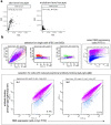
Extended Data Fig. 2:. Effects of mutations on binding of HCP antibodies to RBD and FACS gating strategy.
(a) Correlation plots of site- and mutation-level escape for each of the two independent RBD mutant libraries for the Ab-escape map shown in Fig. 2b. Site-level escape is the sum of the escape fractions for each mutation at a site. (b) Hierarchical FACS gating strategy used for selecting yeast cells expressing antibody-escape RBD variants. First, gates are selected to enrich for single cells (SSC-A vs. FSC-A, and FSC-W vs. FSC-H) that also express RBD (FITC-A vs. FSC-A, cells in pink). Second, cells expressing RBD mutants with reduced polyclonal Ab binding, detected with an anti-IgA+IgG+IgM secondary Ab, were selected with a gate that captured the ~5% of cells with the lowest Ab binding (cells in blue).
Extended Data Fig. 3.
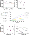
Extended Data Fig. 3.. Evaluation of vaccine-elicited binding and neutralizing Ab titers against SARS-CoV-2 variants and distinct sarbecoviruses.
(a) Wildtype (closed circles) and B.1.351 (open circles) SARS-CoV-2 RBD-specific Ab binding titers of SARS-CoV-2 RBD-NP-elicited sera in pigtail macaques (magenta) and in rhesus macaques (blue), HexaPro-elicited sera (gray), or HCP (orange) analyzed by ELISA. (b) Neutralizing Ab titers against wildtype (D614G) SARS-CoV-2 S, B.1.1.7, and B.1.351 S HIV pseudoviruses from NHPs immunized with 12GS-RBD-NP at day 56 (two immunizations, filled symbols) and 196 (three immunizations, open symbols) (c) Cladogram based on sarbecovirus RBD amino acid sequences. (d) Biolayer interferometry analysis of binding of 1 μM purified polyclonal pigtail macaque IgGs (obtained 70 days post prime) to sarbecovirus RBDs immobilized at the surface of biosensors. (e) SARS-CoV-2 S 2P (circles) or SARS-CoV S 2P (squares) Ab binding titers of SARS-CoV-2 RBD-NP-elicited sera in Pigtail macaques (magenta) or HexaPro-elicited sera in Rhesus macaques (gray) analyzed by ELISA. (f) Competition ELISA between 0.13 nM human ACE2-Fc and RBD-NP-elicited sera in pigtail macaques, rhesus macaques, or HexaPro S-elicited sera in rhesus macaques against SARS-CoV S 2P at various time points following vaccination benchmarked against COVID-19 HCP. Each plot shows the magnitude of inhibition of ACE2 binding to immobilized SARS-CoV S 2P, expressed as reciprocal serum dilution blocking 50% of the maximum binding response.
Extended Data Fig. 4:
Extended Data Fig. 4:. In vitro characterization of sarbecovirus RBD-NP immunogens.
a, Designed models of the various vaccine candidates evaluated. Scale bars, 36 nm. b, SDS-PAGE analysis of purified nanoparticles. DTT, dithiothreitol; F/T, post-freeze/thaw. c, Dynamic light scattering. d, Electron micrographs of negatively stained samples. Scale bars, 50 nm. e, Binding of 100 nM SEC-purified nanoparticle immunogens and the non-assembling cocktail immunogen (which was not purified with SEC) to immobilized hACE2-Fc. f, SEC chromatogram overlay of purified RBD-NP and non-assembling cocktail.
Extended Data Fig. 5:
Extended Data Fig. 5:. Accelerated stability studies of nanoparticle immunogens.
The physical and antigenic stability of mRBD-NP, cRBD-NP, and (SARS-CoV-2) RBD-NP samples incubated at four different temperatures was followed for four weeks. a, The ratio of UV/vis absorbance at 320 nm/280 nm is a measure of turbidity. b, Hydrodynamic diameter of the nanoparticles measured using dynamic light scattering. c, hACE2 binding, measured by comparing the peak amplitude of hACE2 binding for each sample to a reference sample stored at <−70°C using biolayer interferometry. d, Electron micrographs of negatively stained samples after incubation for 28 days at the indicated temperatures. Scale bar, 50 nm.
Extended Data Fig. 6
Extended Data Fig. 6. Confirmation of co-display using sandwich biolayer interferometry.
a, The SARS-CoV-2 S-specific mAb S2H14 immobilized on protein A biosensors was used to capture various nanoparticle immunogens. The captured nanoparticles were subsequently exposed to a Fab derived from the SARS-CoV S-specific mAb S230. b, hACE2-Fc immobilized on protein A biosensors was used to capture various nanoparticle immunogens. The captured nanoparticles were subsequently exposed to a Fab derived from the SARS-CoV S-specific mAb S230.
Extended Data Fig 7:
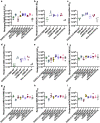
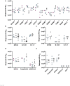
Extended Data Fig 7:. Serum Ab binding titers elicited by mosaic and cocktail RBD-NPs.
(a) ELISA binding to SARS-CoV-2 S at week 5. (b–d) Titers of SARS-CoV-2 S-specific (b) ACE2-Fc (c) CR3022 (d) and S309 competing antibodies in immunized mouse sera. (e) ELISA binding to SARS-CoV S at week 5. (f–i) ELISA binding to RBDs from the (f) SARS-CoV-2 (g) SARS-CoV (h) WIV1 (i) RaTG13 spikes.
Figure 1.
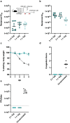
Figure 1.. A single immunization with RBD-NP protects Balb/c cByJ mice from SARS-CoV-2 MA10 challenge.
(a–b) Serum neutralizing Ab titers at 2 (a) or 5 (b) weeks post-prime determined using an MLV pseudotyping system; teal squares: 2×0.1 μg group; filled teal circles: 2×1 μg group; open teal circles: 1×1 μg group. (c) Weight loss following SARS-CoV-2 MA10 challenge up to 4 days post infection (N=6). Black, naïve mice; filled teal circles, 2×1 μg group; open teal circles, 1×1 μg group. (d) Congestion score following SARS-CoV-2 MA10 infection with a score of 0 indicating unchanged lung color and 4 indicating a darkened and diseased lung. (e) Viral titers in the mice lungs (expressed in plaque forming units per lobe) following challenge.
Figure 2:
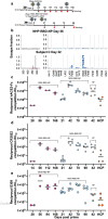
Figure 2:. SARS-CoV-2 RBD-NP vaccination elicits high titers of Abs targeting diverse RBD antigenic sites in NHPs.
(a) Vaccination schedules and RBD antigen doses of the different immunogens. (b) Effects of mutations on RBD binding by polyclonal serum Abs, measured by DMS analysis of serum obtained 8 weeks post-prime from a pigtail macaque vaccinated with 12GS-RBD-NP, compared to a previously reported DMS measurement on plasma Ab binding from a representative SARS-CoV-2 human convalescent individual reproduced here for comparison. The line plots on the left show the summed effect of all mutations at a site in the RBD on sera or plasma binding, with RBD site on the x axis and Ab escape on the y axis. Due to the use of sample-specific FACS gates, the y axes are scaled independently. Sites in the logo plots are colored dark blue if located in the receptor-binding ridge or cyan if located in the RBD 443–450 loop. Larger values indicate more Ab escape. (c–e) Competition ELISA between 0.2 nM human ACE2-Fc (c), 2 nM CR3022 mAb (d), or 0.01 nM S309 mAb (e) and RBD-NP-elicited sera in rhesus/pigtail macaques or HexaPro-elicited sera in rhesus macaques at various time points following vaccination, benchmarked against HCP. Each plot shows the magnitude of inhibition of ACE2/mAb binding to immobilized SARS-CoV-2 S 2P, expressed as reciprocal serum dilution blocking 50% of the maximum binding response.
Figure 3.
Figure 3.. SARS-CoV-2 RBD-NP and HexaPro S elicit Abs with similar neutralization breadth towards SARS-CoV-2 RBD mutants detected in clinical isolates.
(a) Neutralizing Ab titers against wildtype (D614G) SARS-CoV-2 S and RBD point mutants determined using SARS-CoV-2 RBD-NP-elicited sera in pigtail macaques (magenta), in rhesus macaques (blue), and HexaPro-elicited sera in rhesus macaques (gray) with an HIV pseudotyping system. (b) Neutralizing Ab titers against HIV pseudotyped viruses harboring wildtype (D614G) SARS-CoV-2 S, B.1.351 S or B.1.1.7 S determined using SARS-CoV-2 RBD-NP-elicited sera in rhesus macaques (blue), HexaPro-elicited sera in rhesus macaques (gray), or plasma from individuals who received two doses of Pfizer mRNA vaccine (open circles). (c) Neutralizing Ab titers against VSV pseudotyped viruses harboring wildtype (D614G) SARS-CoV-2 S, B.1.351 S or B.1.1.7 S determined using SARS-CoV-2 RBD-NP-elicited sera in rhesus macaques (blue) or HexaPro-elicited sera in rhesus macaques (gray). (d) Neutralizing Ab titers against HIV pseudotyped viruses harboring wildtype (D614G) SARS-CoV-2 S, Pangolin-GD S or SARS-CoV S determined using SARS-CoV-2 RBD-NP-elicited sera in pigtail macaques (magenta), in rhesus macaques (blue), or HexaPro-elicited sera in rhesus macaques (gray). (e) Neutralizing Ab titers against VSV pseudotyped viruses harboring wildtype (D614G) SARS-CoV-2 S, Pangolin-GD S, RaTG13 S, SARS-CoV S or WIV1 S determined using SARS-CoV-2 RBD-NP-elicited sera in rhesus macaques (blue) or HexaPro-elicited sera in rhesus macaques (gray).
Figure 4.
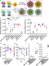
Figure 4.. Mosaic and cocktail RBD-NPs elicit neutralizing Ab against multiple sarbecoviruses and protect against heterotypic challenge in 15 week old Balb/c cByJ mice.
a, Schematic of in vitro assembly of mRBD-NP and cRBD-NP. b, Neutralizing Ab titers against wildtype (D614G) SARS-CoV-2 S MLV pseudovirus at week 5 (2 weeks post-boost) elicited by monovalent, mosaic, and cocktail RBD-NPs. c, Neutralizing Ab titers against SARS-CoV S MLV pseudovirus at week 5 (2 weeks post-boost) elicited by monovalent, mosaic, and cocktail RBD-NPs. d, Neutralizing Ab titers against SARS-CoV-2 B.1.351 S MLV pseudovirus at week 5 (2 weeks post-boost) elicited by monovalent, mosaic, and cocktail RBD-NPs. e, Weight loss following SARS-CoV MA15 challenge up to 4 days post infection (N=6). f, Congestion score following SARS-CoV MA15 infection with a score of 0 indicating unchanged lung color and 4 indicating a darkened and diseased lung. g, Viral titers in the mice lungs (expressed in plaque forming units per lobe) following challenge.

Similar articles

References

    1. Zhou P. et al. A pneumonia outbreak associated with a new coronavirus of probable bat origin. Nature (2020) doi:10.1038/s41586-020-2012-7. - DOI - PMC - PubMed
    1. Zhou H. et al. A Novel Bat Coronavirus Closely Related to SARS-CoV-2 Contains Natural Insertions at the S1/S2 Cleavage Site of the Spike Protein. Curr. Biol. 30, 2196–2203.e3 (2020). - PMC - PubMed
    1. Menachery V. D. et al. SARS-like WIV1-CoV poised for human emergence. Proc. Natl. Acad. Sci. U. S. A. 113, 3048–3053 (2016). - PMC - PubMed
    1. Menachery V. D. et al. A SARS-like cluster of circulating bat coronaviruses shows potential for human emergence. Nat. Med. 21, 1508–1513 (2015). - PMC - PubMed
    1. Walls A. C. et al. Structure, Function, and Antigenicity of the SARS-CoV-2 Spike Glycoprotein. Cell 181, 281–292.e6 (2020). - PMC - PubMed

Publication types