[Preprint]. 2021 Mar 9:2021.03.05.434135.
doi: 10.1101/2021.03.05.434135.

SARS-CoV-2 evolution in animals suggests mechanisms for rapid variant selection

Affiliations

SARS-CoV-2 evolution in animals suggests mechanisms for rapid variant selection

Laura Bashor et al. bioRxiv. .

Update in

Abstract

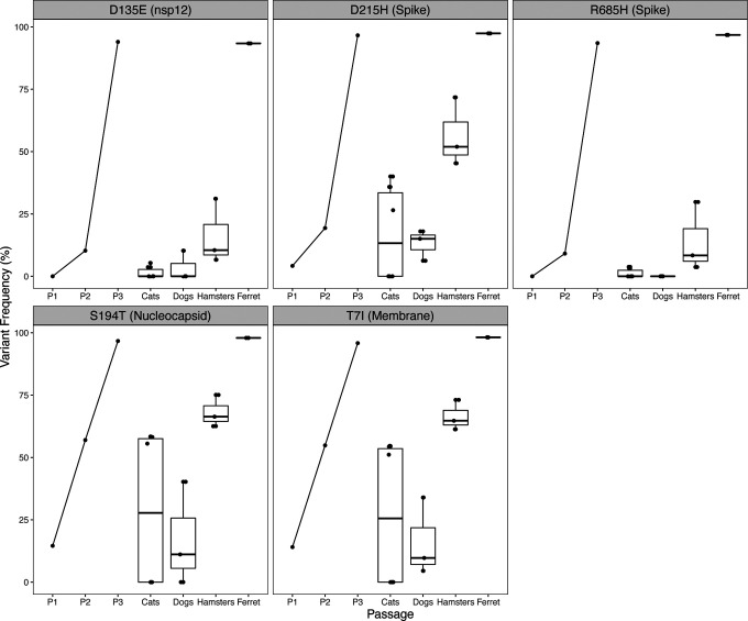
SARS-CoV-2 spillback from humans into domestic and wild animals has been well-documented. We compared variants of cell culture-expanded SARS-CoV-2 inoculum and virus recovered from four species following experimental exposure. Five nonsynonymous changes in nsp12, S, N and M genes were near fixation in the inoculum, but reverted to wild-type sequences in RNA recovered from dogs, cats and hamsters within 1-3 days post-exposure. Fourteen emergent variants were detected in viruses recovered from animals, including substitutions at spike positions H69, N501, and D614, which also vary in human lineages of concern. The rapidity of in vitro and in vivo SARS-CoV-2 selection reveals residues with functional significance during host-switching, illustrating the potential for spillback reservoir hosts to accelerate evolution, and demonstrating plasticity of viral adaptation in animal models.

One-sentence summary: SARS-CoV-2 variants rapidly arise in non-human hosts, revealing viral evolution and potential risk for human reinfection.


Conflict of interest statement

Competing interests: Authors declare that they have no competing interests.

Figures

Fig. 1.
Fig. 1.. Eight-eight unique variants were detected in >3% of sequences of in vitro- and in vivo- derived SARS-CoV-2.
(A) Position, predicted effect and (B) allele frequency of single nucleotide (SNV) and structural (SV) variants detected across the SARS-CoV-2 genome in sequences obtained from 13 experimentally inoculated animals and three passages of the viral inoculum. Each point represents a SNV (circle) or SV (triangle). (A) All variants detected in ≥3% of sequences, demonstrating a majority of SNVs and a slightly increased occurrence of modifications in the spike protein. (B) All variants detected in ≥ 25% of sequences, revealing the presence of higher frequency variants in the spike protein of all datasets excluding the Passage 1 stock virus, and the prevalence of higher frequency variants across the entire genome of SARS-CoV-2 recovered from dogs. Frequency indicated by darkness scale. Schematic of SARS-CoV-2 genome illustrated for orientation.
Fig. 2.
Fig. 2.. SARS-CoV-2 cell culture variants revert rapidly during in vivo experimental infection.
SARS-CoV-2 isolate USA-WA1/2020 was passaged three times in Vero E6 cell line. Five single nucleotide variant substitutions across the genome reached >93% frequency; variant proportion recovered from each supernatant stock indicated by P1, P2, P3. Cats, dogs, hamsters and ferret (n=6, 3, 3, and 1, respectively) were inoculated with 105 – 106 pfu intranasally. Virus was recovered 1–3 days PI, sequenced using a tiled amplicon technique and analyzed with a pipeline for calling single nucleotide and structural variants in viral populations. Cell culture variants decreased in frequency in all animals with the exception of the ferret (n=1). Variants are indicated in reference to the consensus residue in USA-WA1/2020 and their position within the coding sequence of a SARS-CoV-2 protein. Each point represents the mean of two technical replicates, aside from one cat for which a replicate was not sequenced.
Fig. 3.
Fig. 3.. SARS-CoV-2 viral evolution differs across species, gene regions, and individuals.
(A) Each point indicates the number of unique variants detected at ≥ 3% frequency in SARS-CoV-2 genomes recovered from individual animals. There is no significant difference in the number of unique variants detected in different species (ANOVA, p=0.22). (B) Analysis of variant distribution within species reveals that the majority of variants were detected in just one individual within each species. Subplot (a) shows distribution for all variants, while (b) illustrates only variants not occurring in the P3 inoculum at >3%. (C) Variants are distributed across viral genes in relation to each gene’s length as a proportion of the entire genome length (linear model, R2=0.69, p<0.0001). The spike protein contained a notably higher proportion of all variants in comparison to its share of genome length. Grey shading represents the 95% confidence interval for the slope of the regression line. (D) SARS-CoV-2 spike protein variant “residues of concern” are in N terminal domain (NTD), receptor binding domain (RBD) and furin cleavage site. Residues described in text and Table 1 in the SARS-CoV-2 spike trimer are highlighted on structure 6VXX. Blue indicates NTD, Yellow indicates RBD. The furin cleavage site and adjacent residue 686 are in the indicated loop, which was not resolved in this structure.
Fig. 4.
Fig. 4.. Signatures of positive selection are detected in SARS-CoV-2 genome sequences recovered from experimentally inoculated cats, dogs, hamsters and ferret.
(A) Comparison of nonsynonymous (πN) and synonymous (πS) nucleotide diversity reveals that πN is significantly greater than πS, indicating positive selection. Each point represents a measurement πN (red) or πS (blue) calculated for the entire SARS-CoV-2 genome from sequences recovered from an individual animal, relative to the reference sequence of USA-WA1/2020. Analysis of the same measures within each species reveals πN > πS for viral genomes is greater in dogs and hamsters. (B) Orf1ab, S, and M are undergoing positive selection in mammalian hosts. Each point represents πN or πS calculated for a specific SARS-CoV-2 gene or open reading frame from sequences recovered from an individual animal.

References

    1. Sauter D., Kirchhoff F., Key Viral Adaptations Preceding the AIDS Pandemic. Cell Host and Microbe 25, 27–38 (2019). doi: 10.1016/j.chom.2018.12.002 - DOI - PubMed
    1. Andersen K. G., Rambaut A., Lipkin W. I., Holmes E. C., Garry R. F., The proximal origin of SARS-CoV-2. Nature Medicine 26, 450–452 (2020). doi: 10.1038/s41591-020-0820-9 - DOI - PMC - PubMed
    1. Cross P. C., Prosser D. J., Ramey A. M., Hanks E. M., Pepin K. M., Confronting models with data: The challenges of estimating disease spillover. Philosophical Transactions of the Royal Society B: Biological Sciences 374, 1782 (2019). doi: 10.1098/rstb.2018.0435 - DOI - PMC - PubMed
    1. Zhou P., lou Yang X., Wang X. G., Hu B., Zhang L., Zhang W., Si H. R., Zhu Y., Li B., Huang C. L., Chen H. D., Chen J., Luo Y., Guo H., di Jiang R., Liu M. Q., Chen Y., Shen X. R., Wang X., Zheng X. S., Zhao K., Chen Q. J., Deng F., Liu L. L., Yan B., Zhan F. X., Wang Y. Y., Xiao G. F., Shi Z. L., A pneumonia outbreak associated with a new coronavirus of probable bat origin. Nature 579, 270–273 (2020). doi: 10.1038/s41586-020-2012-7 - DOI - PMC - PubMed
    1. Lam T. T. Y., Jia N., Zhang Y. W., Shum M. H. H., Jiang J. F., Zhu H. C., Tong Y. G., Shi Y. X., Ni X. B., Liao Y. S., Li W. J., Jiang B. G., Wei W., Yuan T. T., Zheng K., Cui X. M., Li J., Pei G. Q., Qiang X., Cheung W. Y. M., Li L. F., Sun F. F., Qin S., Huang J. C., Leung G. M., Holmes E. C., Hu Y. L., Guan Y., Cao W. C., Identifying SARS-CoV-2-related coronaviruses in Malayan pangolins. Nature 583, 282–285 (2020). doi: 10.1038/s41586-020-2169-0 - DOI - PubMed

Publication types

LinkOut - more resources