[Preprint]. 2021 Mar 10:2021.03.10.434840.
doi: 10.1101/2021.03.10.434840.

Memory B cell repertoire for recognition of evolving SARS-CoV-2 spike

Affiliations

Memory B cell repertoire for recognition of evolving SARS-CoV-2 spike

Pei Tong et al. bioRxiv. .

Update in

  • Memory B cell repertoire for recognition of evolving SARS-CoV-2 spike.
    Tong P, Gautam A, Windsor IW, Travers M, Chen Y, Garcia N, Whiteman NB, McKay LGA, Storm N, Malsick LE, Honko AN, Lelis FJN, Habibi S, Jenni S, Cai Y, Rennick LJ, Duprex WP, McCarthy KR, Lavine CL, Zuo T, Lin J, Zuiani A, Feldman J, MacDonald EA, Hauser BM, Griffths A, Seaman MS, Schmidt AG, Chen B, Neuberg D, Bajic G, Harrison SC, Wesemann DR. Tong P, et al. Cell. 2021 Sep 16;184(19):4969-4980.e15. doi: 10.1016/j.cell.2021.07.025. Epub 2021 Jul 23. Cell. 2021. PMID: 34332650 Free PMC article.

Abstract

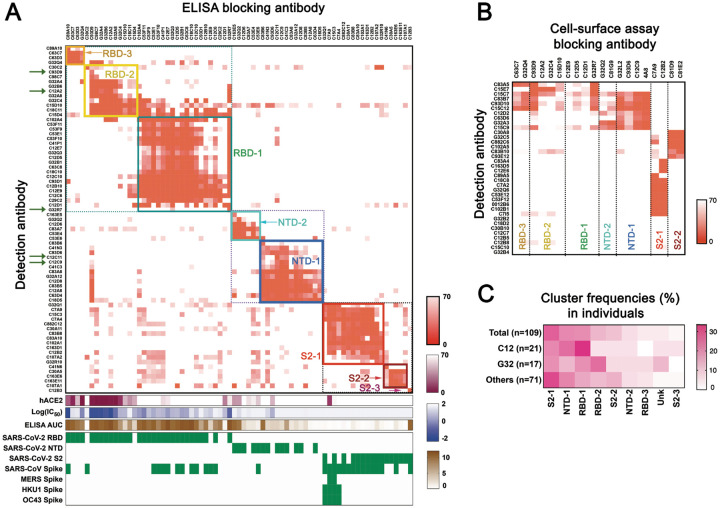
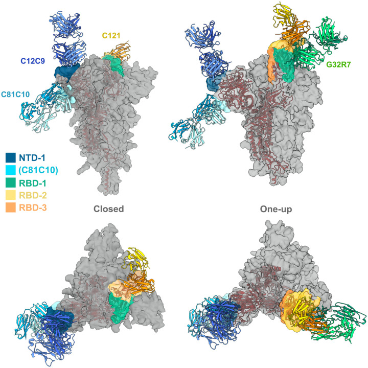
Memory B cell reserves can generate protective antibodies against repeated SARS-CoV-2 infections, but with an unknown reach from original infection to antigenically drifted variants. We charted memory B cell receptor-encoded monoclonal antibodies (mAbs) from 19 COVID-19 convalescent subjects against SARS-CoV-2 spike (S) and found 7 major mAb competition groups against epitopes recurrently targeted across individuals. Inclusion of published and newly determined structures of mAb-S complexes identified corresponding epitopic regions. Group assignment correlated with cross-CoV-reactivity breadth, neutralization potency, and convergent antibody signatures. mAbs that competed for binding the original S isolate bound differentially to S variants, suggesting the protective importance of otherwise-redundant recognition. The results furnish a global atlas of the S-specific memory B cell repertoire and illustrate properties conferring robustness against emerging SARS-CoV-2 variants.


Conflict of interest statement

Competing interests The authors declare no competing interests.

Figures

Fig. 1.
Fig. 1.. SARS-CoV-2 surface glycoprotein (spike) specificities of memory B cells from convalescent subjects.
(A) Cells recovered from two sorting strategies, shown in dot plots as percentages of total CD19+ cells. Left: IgG+CD27+ cells from 18 donors (one dot per donor) and the subset of those that expressed spike-binding BCRs. Right: cells from 3 donors expressing spike-binding BCRs and sorted to recover principally those that did not bind recombinant receptor-binding domain (RBD). Sorting protocols as described in Methods and shown in Fig. S1. (B) Summary of all antibodies (expressed as recombinant IgG1) screened by ELISA (with recombinant spike ectodomain trimer) and cell-surface expression assays (both 293T and yeast cells). Total numbers in the center of each of pie chart; numbers and color codes for the indicated populations shown to next to each chart. To the right of the charts for the two alternative sorting strategies are bar graphs showing frequencies of SARS-CoV-2 RBD and NTD binding antibodies for those subjects from whom at least 10 paired-chain BCR sequences were recovered. (C) Binding to a panel of spike proteins and SARS-CoV-2 subdomains, listed on the left, as determined by both ELISA (with recombinant spike ectodomain) and by association with spike expressed on the surface of 293T cells or with RBD or NTD expressed on the surface of yeast cells, for cells sorted just for spike binding (left) and for those sorted for positive spike binding but no RBD binding (right). The rows with pink highlighting are from the ELISA screen; those with blue highlighting, from the cell-based screens. Each short section of a row represents an antibody. The rows labeled VH mutation and VL mutation are heat maps of counts (excluding CDR3) from alignment by IgBLAST, with the scale indicated. (D) Dot plots of heavy- and light-chain somatic mutation counts in antibodies that bound RBD, NTD, S2, and a “broad CoV group” that included MERS, HKU1, and OC43. The significantly higher numbers of mutations in the last group suggest recalled, affinity matured memory from previous exposures to seasonal coronaviruses. ****P < 0.0001; one-way ANOVA followed by Tukey’s multiple comparison. Horizontal lines show mean ± SEM.
Fig. 2.
Fig. 2.. Competition epitope mapping.
(A) Cross competition matrix for 73 antibodies from the spike+ sort in Fig. 1 with affinity sufficient for detection by ELISA. Blocking antibodies (columns) added at 100 μg/ml; detection antibodies (rows), at 1μg/ml. Intensity of color shows strength of blocking, from 0 signal (complete blocking) to 70% full signal (top gradient at right of panel: orange). Hierarchical clustering of antibodies by cross competition into 7 groups (plus a singleton labeled S2–3), enclosed in square boxes, with designations shown and in colors from dark blue (NTD-1) to dark red (S2–3). Green arrows on the left designate antibodies newly reported here. The lower parts of the panel show: competition of blocking antibody with soluble, human ACE2 (second gradient at right: dark red); log(IC50) in pseudovirus neutralization assay (third gradient at right: violet); area under the curve for ELISA binding (bottom gradient at right: brown); binding (ELISA) to recombinant domains and heterologous spike proteins. (B) Competition in cell-based assay for 36 antibodies with binding in ELISA format too weak for reliable blocking measurement (rows). Blocking antibodies (columns) selected from each of the 7 clusters in the ELISA assay (fig. S2). Strength of blocking shown as intensity of orange color, as in (A). (C) Distribution of antibodies from three individual subjects (expressed as percent of sequence pairs recovered from that subject) into the 7 principal clusters, plus a non-assigned (unknown) category (unk) and S2–3. Data are shown for only those subjects from whom we recovered at least 10 heavy- and light-chain sequence pairs. Heat map scale shown at right of panel. Top row shows total distribution, from panel (A) and (B).
Fig. 3.
Fig. 3.. Ab contact regions.
Surface regions of the SARS-CoV-2 spike protein trimer contacted by antibodies in four of the seven principal clusters, according to the color scheme shown (taken from the color scheme in Fig. 2), with a representative Fab for all except RBD-3. The C81C10 Fab defines an epitope just outside the margin of NTD-1, but it does not compete with any antibodies in RBD-2. The RBD-2 Fv shown is that of C121 (PDB ID: 7K8X: Barnes et al, 2020), which fits most closely, of the many published RBD-2 antibodies, into our low-resolution map for C12A2. Left: views normal to and along threefold axis of the closed, all-RBD-down conformation; right: similar views of the one-RBD-up conformation. C121 (RBD-2) can bind both RBD down and RBD up; G32R7 (RBD-1) binds only the “up” conformation of the RBD. The epitopes of the several published RBD-3 antibodies are partly occluded in both closed and open conformations of the RBD; none are shown here as cartoons. A cartoon of the polypeptide chain of a single subunit (dark red) is shown within the surface contour for a spike trimer (gray).
Fig. 4.
Fig. 4.. Antibody sequence analyses.
(A) Heavy-chain variable-domain genes of the 167 mAbs characterized by binding SARS-CoV-2 spike in either ELISA or cell-surface expression format. The inner ring of each pie chart shows the VH family and the outer ring, the gene. PBMC repertoire is from 350 million reads of deep sequencing (37). S binders include 167 clones in Table S2. *P < 0.05, **P < 0.01, ***P < 0.001, ****P < 0.0001; Bonferroni correction. Red asterisks: comparing to S binders; black asterisks: comparing to a non-selected B cell repertoire from PBMCs. (B) Maps of pairwise distances of CDRH3 (lower left triangle) and CDRL3 (upper right triangle) for the NTD-2 and S2–1 cluster antibodies from (A). Antibodies in both clusters arranged by VH usage. Clones converging on identical VH/VL alleles and closest distance of CDRL3 from the same cluster are shown. Pairwise distances analyzed by Mega X. Intensity of color shows the distance, from 0 (identical) to 1 (no identity). Sequence alignment for the antibodies from the indicated clusters with identical VH and VJ and similar CDR3s. Differences in CDR3s from the reference sequence (bold) are in red; dashes indicate missing amino acids; dots represent identical amino acids. (C) Summary of convergent sequences of anti-SARS-CoV-2 S and RBD antibodies from independent datasets. Ig sequences derived from binding to DIII of Zika virus E protein, and HA of influenza virus H1N1 were used as control datasets. Convergent sequences had identical VH and VL and >50% identity in CDRH3 and CDRL3. (D) Representative convergent clones from different individuals and independent datasets from Fig. 4C.
Fig. 5.
Fig. 5.. Recognition of naturally occurring deletions and mutations in the spike.
Heat map showing binding of 119 mAbs to Nextstrain cluster 20A.EU1 (A222V), Danish mink variant (Δ69–70 and Y453F), UK B.1.1.7 (Δ69–70, Δ144, N501Y, A570D, P681H, T716I, S982A, D1118H) and SA B.1.351 (L18F, D80A, D215G, Δ242–244, K417N, E484K, N501Y, A701V) (top) and NTD deletion variants (bottom). The Wuhan-Hu-1 S sequence and all variants include the D614G mutation. Binding for each mAb was first normalized (“normalized IgG MFI”) by dividing the MFI for that mAb by the MFI for C81E2 (S2–2 cluster). The normalized MFI of for binding the Wuhan-Hu-1 spike was used as a reference (normalized Wuhan IgG MFI). The relative binding intensities of the tested mAbs for each variant, calculated as the ratio of the normalized variant IgG MFI and the normalized Wuhan IgG MFI, are shown in shades of blue.

Similar articles

Cited by

References

    1. Robbiani D. F. et al., Convergent antibody responses to SARS-CoV-2 in convalescent individuals. Nature, (2020). - PMC - PubMed
    1. Schafer A. et al., Antibody potency, effector function, and combinations in protection and therapy for SARS-CoV-2 infection in vivo. J Exp Med 218, (2021). - PMC - PubMed
    1. Victora G. D., Nussenzweig M. C., Germinal centers. Annu Rev Immunol 30, 429–457 (2012). - PubMed
    1. Akkaya M., Kwak K., Pierce S. K., B cell memory: building two walls of protection against pathogens. Nature Reviews Immunology 20, 229–238 (2020). - PMC - PubMed
    1. Turner J. S. et al., Human germinal centres engage memory and naive B cells after influenza vaccination. Nature 586, 127–132 (2020). - PMC - PubMed

Publication types

LinkOut - more resources