[Preprint]. 2021 Mar 14:2021.03.13.435256.
doi: 10.1101/2021.03.13.435256.

Structural basis for backtracking by the SARS-CoV-2 replication-transcription complex

Affiliations

Structural basis for backtracking by the SARS-CoV-2 replication-transcription complex

Brandon Malone et al. bioRxiv. .

Update in

Abstract

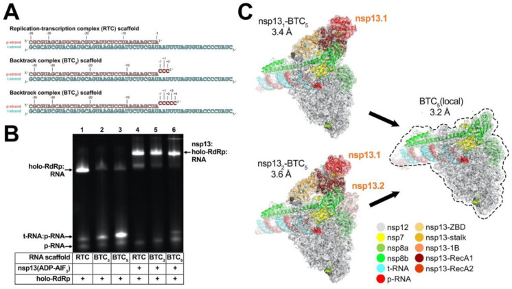
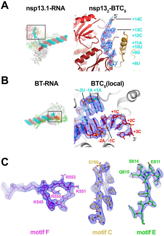
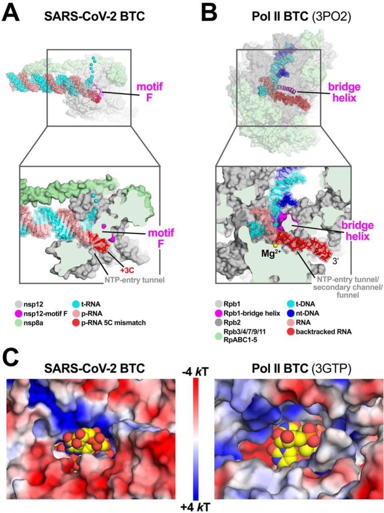
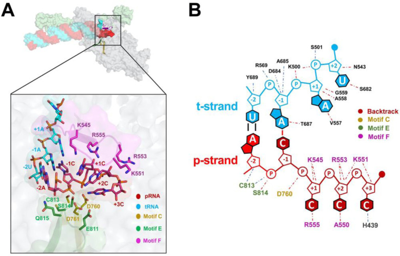
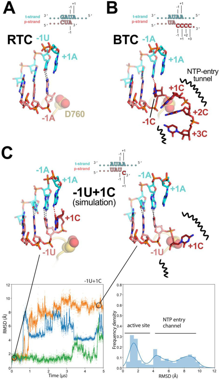
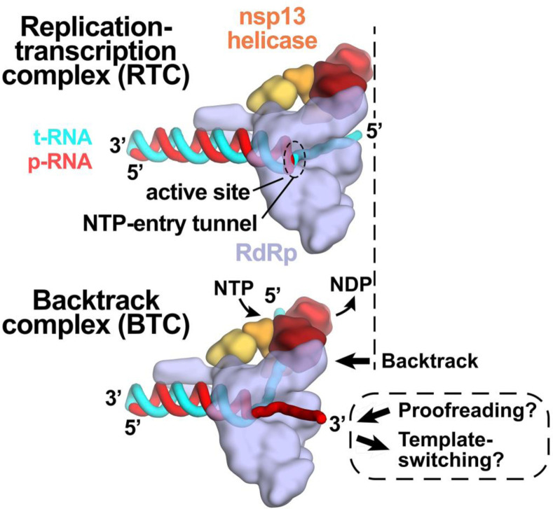
Backtracking, the reverse motion of the transcriptase enzyme on the nucleic acid template, is a universal regulatory feature of transcription in cellular organisms but its role in viruses is not established. Here we present evidence that backtracking extends into the viral realm, where backtracking by the SARS-CoV-2 RNA-dependent RNA polymerase (RdRp) may aid viral transcription and replication. Structures of SARS-CoV-2 RdRp bound to the essential nsp13 helicase and RNA suggested the helicase facilitates backtracking. We use cryo-electron microscopy, RNA-protein crosslinking, and unbiased molecular dynamics simulations to characterize SARS-CoV-2 RdRp backtracking. The results establish that the single-stranded 3'-segment of the product-RNA generated by backtracking extrudes through the RdRp NTP-entry tunnel, that a mismatched nucleotide at the product-RNA 3'-end frays and enters the NTP-entry tunnel to initiate backtracking, and that nsp13 stimulates RdRp backtracking. Backtracking may aid proofreading, a crucial process for SARS-CoV-2 resistance against antivirals.

Keywords: Biological Sciences; Biophysics and Computational Biology; RNA-dependent RNA polymerase; backtracking; coronavirus; cryo-electron microscopy; molecular dynamics.


Conflict of interest statement

Competing Interest Statement: The authors declare no conflict of interest.

Figures

Fig. 1.
Fig. 1.. SARS-CoV-2 backtrack complex.
A. RNA scaffolds: (top) replication-transcription complex (RTC) scaffold (14); (bottom) backtrack complex scaffolds (BTC3 and BTC5). B. A native gel electrophoretic mobility shift assay reveals that holo-RdRp requires nsp13(ADP-AlF3) to bind the BTC scaffolds efficiently. C. Cryo-EM structures of SARS-CoV-2 BTCs. Shown is the transparent cryo-EM density [local-resolution filtered; (47)] with the refined models superimposed (Table S1). The models and density are colored according to the key.
Fig. 2.
Fig. 2.. Cryo-EM density maps.
A. (left) Overall view of nsp131-BTC5. The boxed region is magnified on the right. (right) Magnified view of the t-RNA segment (+14–5’-CCCAUGU-3’-+8) enclosed in the nsp13.1 helicase subunit. The cryo-EM density map (from the nsp132-BTC structure) for the RNA is shown (blue mesh). B. (left) Overall view of the BTC structure. The boxed region is magnified on the right. (right) Magnified view of the region around the RdRp active site, showing the t-RNA (cyan) and p-RNA (red) with the backtracked RNA segment. The cryo-EM density map for the RNA [from BTC5(local)] is shown (blue mesh). C. BTC5(local) cryo-EM density maps around nsp12 conserved motifs F, C, and E. Selected residues are labeled.
Fig. 3.
Fig. 3.. SARS-CoV-2 RdRp and DdRp BTCs.
A, B. SARS-CoV-2 RdRp (A) and DdRp (B) BTCs. (top) Proteins are shown as transparent molecular surfaces, nucleic acids as atomic spheres. The boxed regions are magnified on the bottom. (bottom) Magnified, cross-sectional view. Proteins are shown as molecular surfaces, nucleic acids in stick format with transparent molecular surface. A. The SARS-CoV-2 BTC5(local). Nsp8a and nsp12 are shown (nsp7 and nsp8b are removed for clarity). Nsp12 motif F is shown as a magenta backbone ribbon (top). Backtracked RNA (+1C to +3C of the BTC5-scaffold; Figure 1A) extrude out the NTP-entry tunnel. B. A DdRp (Saccharomyces cerevisiae Pol II) BTC [PDB ID: 3PO2; (29)]. The bridge helix is shown as a magenta backbone ribbon. The backtracked RNA extrudes out the NTP-entry tunnel/secondary channel/funnel. C. Views from the outside into the NTP-entry tunnels of the SARS-CoV-2 (left) and an S. cerevisiae DdRp [PDB ID: 3GTP; (27) BTC. Protein surfaces are colored by the electrostatic surface potential [calculated using APBS; (48)]. Backtracked RNA is shown as atomic spheres with yellow carbon atoms.
Fig. 4.
Fig. 4.. Protein-RNA interactions in the BTC.
A. (top) Overall view of BTC5(local). Proteins are shown as transparent molecular surfaces, nucleic acids as atomic spheres. Nsp8a and nsp12 are shown (nsp7 and nsp8b are removed for clarity). Nsp12 motifs C, E, and F are shown as backbone ribbons (colored according to the key on the bottom. The boxed region is magnified below. (bottom) RNA is shown from −2 to +3. Proteins are shown as transparent molecular surfaces. RdRp motifs C, E, and F are shown as transparent backbone ribbons (colored according to the key) with side chains of residues that approach the backtracked RNA (≤ 4.5 Å) shown. B. Schematic illustrating the same protein-RNA interactions as (A). Drawn using Nucplot (49).
Fig. 5.
Fig. 5.. Comparison of active-site proximal RNA in the RTC and BTC structures, and from simulations of a mismatched nucleotide at the p-RNA 3’-end.
A-B, Comparison of the active-site proximal RNA in the RTC [A; PDB ID: 6XEZ; (14)], BTC5(local) (B), and from selected snapshots of molecular dynamics simulations of a −1U+1C complex (C). The schematics denote the nucleotides shown in the context of the RTC- (A) and BTC5-scaffolds (B; full scaffold sequences shown in Figure 1A) or generated from the BTC5-scaffold for the simulations (C). Carbon atoms of the t-RNA are colored cyan, p-RNA are colored salmon except in the case of mismatched C’s at the 3’-end, which are colored dark red. Watson-Crick base pairing hydrogen-bonds are denoted as dark gray dashed lines, other hydrogen-bonds as red dashed lines. Nsp12 motif C is shown as a yellow-orange backbone ribbon, and the side-chain of D760 is shown as atomic spheres. A. The RTC is in a post-translocated state, with the A-U base pair at the p-RNA 3’-end in the −1 position (14). B. The BTC5(local) RNA is translocated compared to the RTC; the base pair corresponding to A-U at the 3’-end of the RTC RNA in the −1 position is in the −2 position of the BTC RNA. A C-A mismatch occupies the BTC −1 site. The +1, +2 and +3 mismatched C’s trail into the RdRp NTP-entry tunnel (denoted by black squiggly lines). The +4C (present in the BTC5-scaffold; Figure 1A) is exposed to solvent, disordered and not modelled. C. Molecular dynamics simulations of the nsp132-BTC−1U+1C complex. The complex was simulated with 3 replicates. RMSD values plotted as a function of time represent the heavy-atom RMSD of the +1C of the p-RNA compared with the starting configuration (see Methods). The RMSD histograms (plotted on the right) are an aggregate of all three replicates. Two structures taken from one of the simulations are shown, one showing the +1C of the p-RNA in the active site (t = 0 μs) and the other showing the +1C frayed into the NTP-entry tunnel (t = 4.5 μs).
Fig. 6.
Fig. 6.. Role of backtracking in proofreading and template-switching during sub-genomic transcription.
Schematic illustrating the proposed model for backtracking of the SARS-CoV-2 RTC and its potential role in proofreading and template-switching during sub-genomic transcription. The structural models are shown as cartoons (holo-RdRp, light blue; nsp13 helicase, orange shades; RNA strands, colored tubes as indicated). (top) In the RTC, the elongating RdRp moves from left-to-right. The RdRp active site holds the p-RNA 3’-end. The NTP-entry tunnel provides access from solution to the RdRp active site. The downstream (5’) single-stranded t-RNA is not engaged with nsp13. (bottom) In the BTC, nsp13 translocates on the downstream (5’) single-stranded t-RNA, pushing the RdRp backwards (right-to-left) on the RNA. This causes the p-RNA to reverse-thread through the complex, with the resulting single-stranded 3’-fragment extruding out the NTP-entry tunnel. The exposure of the p-RNA 3’-end could facilitate proofreading (9, 10, 12, 50) and also template-switching during sub-genomic transcription (7, 34).

Similar articles

Cited by

References

    1. Wu F., et al., A new coronavirus associated with human respiratory disease in China. Nature 579, 265–269 (2020). - PMC - PubMed
    1. Zhou P., et al., A pneumonia outbreak associated with a new coronavirus of probable bat origin. Nature 579, 270–273 (2020). - PMC - PubMed
    1. Subissi L., et al., One severe acute respiratory syndrome coronavirus protein complex integrates processive RNA polymerase and exonuclease activities. Proceedings of the National Academy of Sciences of the United States of America 111, E3900–9 (2014). - PMC - PubMed
    1. Kirchdoerfer R. N., Ward A. B., Structure of the SARS-CoV nsp12 polymerase bound to nsp7 and nsp8 co-factors. Nature Communications 10, 2342–9 (2019). - PMC - PubMed
    1. U. S. F. and D. Administration, Remdesivir-EUA-Letter-Of-Authorization.pdf. undefined (2020).

Publication types