[Preprint]. 2021 May 1:2021.03.07.21253098.
doi: 10.1101/2021.03.07.21253098.

The plasmablast response to SARS-CoV-2 mRNA vaccination is dominated by non-neutralizing antibodies and targets both the NTD and the RBD

Affiliations

The plasmablast response to SARS-CoV-2 mRNA vaccination is dominated by non-neutralizing antibodies and targets both the NTD and the RBD

Fatima Amanat et al. medRxiv. .

Abstract

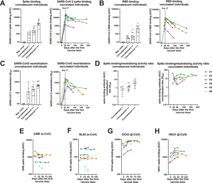
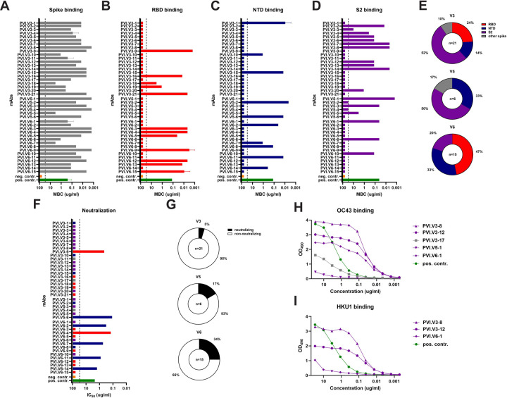
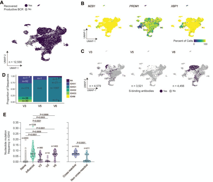
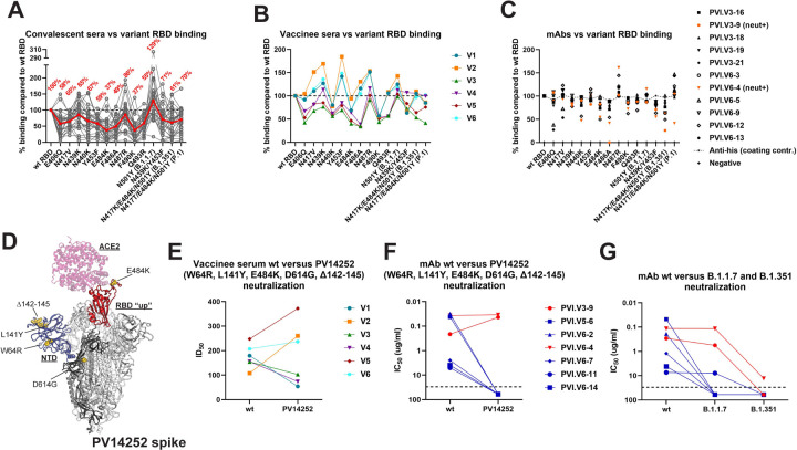
In this study we profiled vaccine-induced polyclonal antibodies as well as plasmablast derived mAbs from individuals who received SARS-CoV-2 spike mRNA vaccine. Polyclonal antibody responses in vaccinees were robust and comparable to or exceeded those seen after natural infection. However, the ratio of binding to neutralizing antibodies after vaccination was greater than that after natural infection and, at the monoclonal level, we found that the majority of vaccine-induced antibodies did not have neutralizing activity. We also found a co-dominance of mAbs targeting the NTD and RBD of SARS-CoV-2 spike and an original antigenic-sin like backboost to seasonal human coronaviruses OC43 and HKU1. Neutralizing activity of NTD mAbs but not RBD mAbs against a clinical viral isolate carrying E484K as well as extensive changes in the NTD was abolished, suggesting that a proportion of vaccine induced RBD binding antibodies may provide substantial protection against viral variants carrying single E484K RBD mutations.


Conflict of interest statement

Conflict of interest statement The Icahn School of Medicine at Mount Sinai has filed patent applications relating to SARS-CoV-2 serological assays and NDV-based SARS-CoV-2 vaccines which list Florian Krammer as co-inventor. Viviana Simon is also listed on the serological assay patent application as co-inventors. Mount Sinai has spun out a company, Kantaro, to market serological tests for SARS-CoV-2. Florian Krammer has consulted for Merck and Pfizer (before 2020), and is currently consulting for Pfizer, Seqirus and Avimex. The Krammer laboratory is also collaborating with Pfizer on animal models of SARS-CoV-2. Ali Ellebedy has consulted for InBios and Fimbrion Therapeutics (before 2021) and is currently a consultant for Mubadala Investment Company. The Ellebedy laboratory received funding under sponsored research agreements that are unrelated to the data presented in the current study from Emergent BioSolutions and from AbbVie.

Figures

Figure 1:
Figure 1:. Antibody responses in individuals vaccinated with mRNA-based SARS-CoV-2 vaccines.
Antibody responses of convalescent individuals and vaccinees to full length spike protein (A) and RBD (B) as measured by ELISA and neutralizing activity of the sera of the same individuals in a microneutralization assay against authentic SARS-CoV-2 (C). Convalescent individuals were grouped based on their initial antibody response (measured in a CLIA laboratory) to spike protein into +, ++, and +++. D shows ratios between binding and neutralizing antibody levels in vaccinees and convalescent individuals. Higher ratios indicate a bias towards non-neutralizing antibodies. E, F, G and H show antibody responses against α-coronavirus 229E and NL63 and β-coronavirus OC43 and HKU1 spike proteins over time.
Figure 2.
Figure 2.. Characterization of mAbs derived from vaccine plasmablasts.
Binding of plasmablasts derived from three vaccinees (V3, V5 and V6) against full length spike (A), RBD (B), NTD (C) and S2 (D). E shows the percentages of the respective antibodies per subject. F shows neutralizing activity of the mABs against authentic SARS-CoV-2 and the proportion of neutralizing antibodies per subject is shown in G. H and I show reactivity of mAbs to spike protein of human β-coronaviruses OC43 and HKU1. MBC = minimal binding concentration.
Figure 3.
Figure 3.. Characterization of bulk sorted plasmablasts via single-cell RNA sequencing.
(A) Uniform manifold approximation and projection (UMAP) of scRNAseq from bulk plasmablast with recovered BCR sequences (purple) or unrecovered (grey). (B) UMAP overlay of percent of cellular population expressing MZB1, PRDM1, and XPB1. Hexbin equals 80 individual cells. (C) UMAP overlay of BCR sequences with confirmed spike binding activity. (D) Proportional composition of heavy chains genes in the spike binding sequences broken down by sample. (E) Comparison of nucleotide-level mutation frequency in immunoglobulin heavy chain variable (IGHV) genes between plasmablasts clonally related to spike binding mAbs from SARS-CoV-2 vaccinees, plasmablasts sorted from PBMCs one week after seasonal influenza vaccination and found in vaccine-responding B cell clones, and naïve B cells found in blood of an influenza vaccinee (left panel); and between plasmablasts from SARS-CoV-2 vaccinees found to be clonally related to spike-binding mAbs that were, respectively, cross-reactive and non-cross-reactive to human β-coronaviruses spike proteins (right panel).
Figure 4.
Figure 4.. Mapping of the amino-acid substitutions and deletions onto the structure of the SARS-CoV-2 spike glycoprotein.
A lists mutations of the three major variants of concern B.1.17, B.1.315 and P.1. B shows these mutations mapped onto the structure of the spike glycoprotein (model generated by superposition of PDB 6M0j and 7C2L (Chi et al., 2020; Lan et al., 2020)). One RBD in the up conformation (red) is bound with ACE2 receptor (pink). The NTD is colored blue and the various amino-acid substitutions are shown as yellow spheres. One spike protomer is shown in bold colors while the other two are colored white. C shows competition between ACE2 and neutralizing RBD targeting mAbs PVI.V3–9 and PVI.V6–4 for binding to RBD. D BLI- measured binding affinities of the RBD mutants to ACE2, as well as the calculated fold change, are shown in the table on the right.
Figure 5.
Figure 5.. Binding and neutralization of SARS-CoV-2 variants.
Binding of serum samples from convalescent individuals, vaccinees and vaccine derived mAbs to a panel of RBD mutants is shown in A, B and C respectively. The red line in A indicates the average reduction. Dotted lines in A and B indicate 100%, the line with smaller dots in C indicated reactivity of the anti-his coating control. For vaccinees late samples (V1=d89, V2=d102, V3=d47, V4=d48, V5=49 and V6=48) were assayed. D shows the spike mutations of virus isolate PVI14252 modelled on a co-crystal structure of the SARS-CoV-2 spike protein with ACE2 (model generated by superposition of PDB 6M0j and 7C2L (Chi et al., 2020; Lan et al., 2020)). E and F show the inhibitory effect of vaccine serum and vaccine derived neutralizing antibodies on both wild type SARS-CoV-2 and PV14252. G shows neutralizing activity of the plasmablast derived neutralizing antibodies aginst wild type, B.1.1.7 and B.1.351 virus isolates. Of note, these comparative assays were always performed side by side but sets are run by different operators and on a different Vero cell clone as the neutralization assays shown in Figure 2.

Similar articles

Cited by

References

    1. Amanat F., Duehr J., Huang C., Paessler S., Tan G.S., and Krammer F. (2020a). Monoclonal Antibodies with Neutralizing Activity and Fc-Effector Functions against the Machupo Virus Glycoprotein. J Virol 94. 10.1128/JVI.01741-19. - DOI - PMC - PubMed
    1. Amanat F., Duehr J., Oestereich L., Hastie K.M., Ollmann Saphire E., and Krammer F. (2018). Antibodies to the Glycoprotein GP2 Subunit Cross-React between Old and New World Arenaviruses. mSphere 3. 10.1128/mSphere.00189-18. - DOI - PMC - PubMed
    1. Amanat F., Stadlbauer D., Strohmeier S., Nguyen T.H.O., Chromikova V., McMahon M., Jiang K., Arunkumar G.A., Jurczyszak D., Polanco J., et al. (2020b). A serological assay to detect SARS-CoV-2 seroconversion in humans. Nat Med 26, 1033–1036. 10.1038/s41591-020-0913-5. - DOI - PMC - PubMed
    1. Amanat F., White K.M., Miorin L., Strohmeier S., McMahon M., Meade P., Liu W.C., Albrecht R.A., Simon V., Martinez-Sobrido L., et al. (2020c). An In Vitro Microneutralization Assay for SARS-CoV-2 Serology and Drug Screening. Curr Protoc Microbiol 58, e108. 10.1002/cpmc.108. - DOI - PMC - PubMed
    1. Annavajhala M.K., Mohri H., Zucker J.E., Sheng Z., Wang P., Gomez-Simmonds A., Ho D.D., and Uhlemann A.C. (2021). A Novel SARS-CoV-2 Variant of Concern, B.1.526, Identified in New York. medRxiv. 10.1101/2021.02.23.21252259. - DOI

Publication types

LinkOut - more resources