. 2021 May 21;49(9):4803-4815.
doi: 10.1093/nar/gkab162.

microRNA-mediated translation repression through GYF-1 and IFE-4 in C. elegans development

Affiliations

microRNA-mediated translation repression through GYF-1 and IFE-4 in C. elegans development

Vinay K Mayya et al. Nucleic Acids Res. .

Abstract

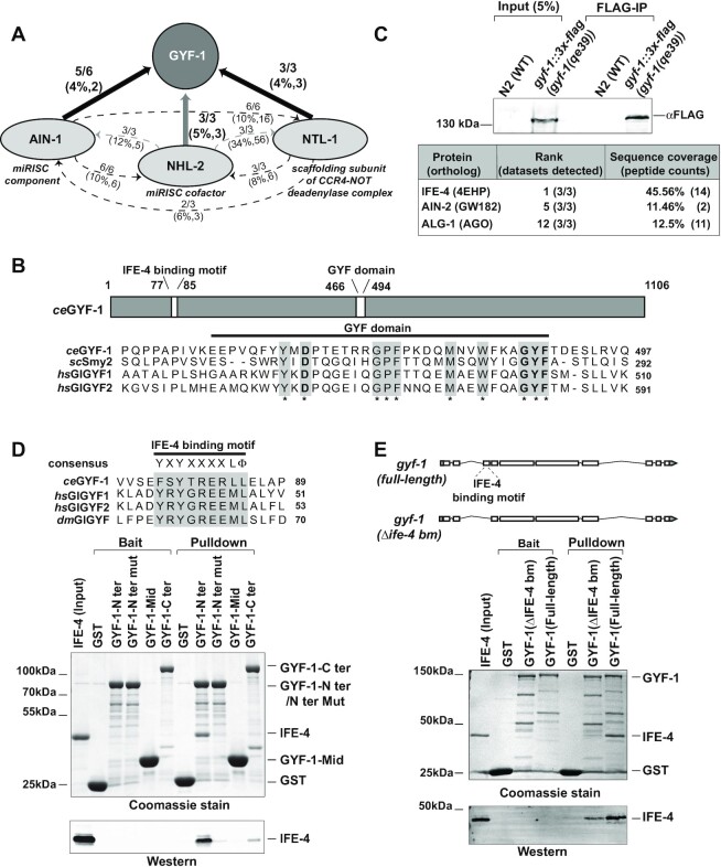
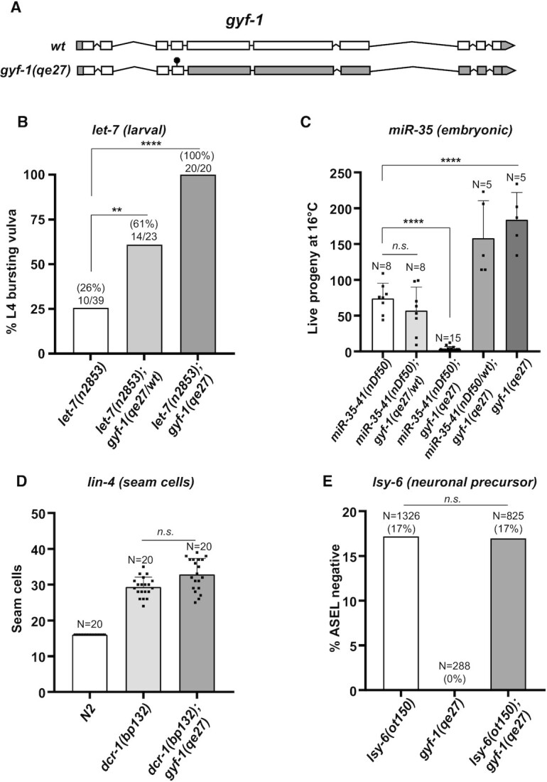
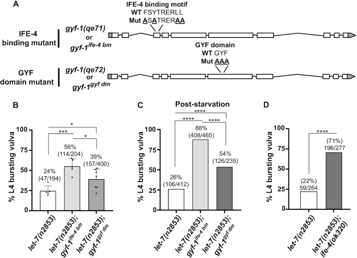
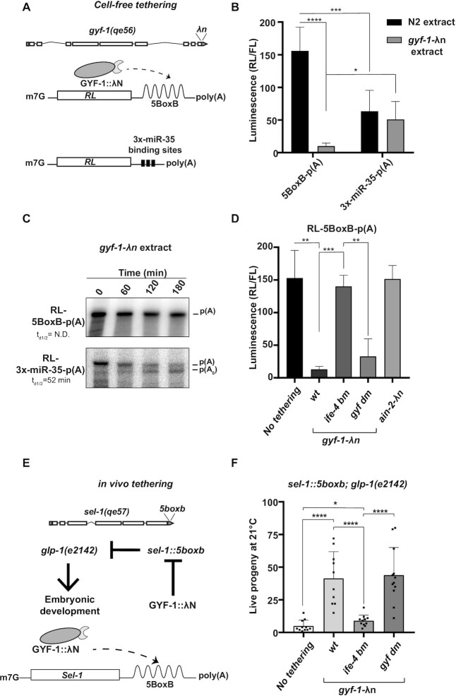
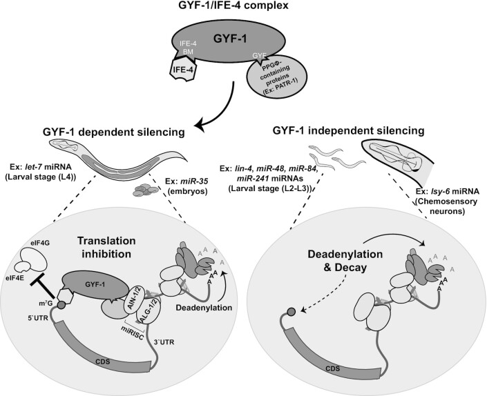
microRNA (miRNA)-mediated gene silencing is enacted through the recruitment of effector proteins that direct translational repression or degradation of mRNA targets, but the relative importance of their activities for animal development remains unknown. Our concerted proteomic surveys identified the uncharacterized GYF-domain encoding protein GYF-1 and its direct interaction with IFE-4, the ortholog of the mammalian translation repressor 4EHP, as key miRNA effector proteins in Caenorhabditis elegans. Recruitment of GYF-1 protein to mRNA reporters in vitro or in vivo leads to potent translation repression without affecting the poly(A) tail or impinging on mRNA stability. Loss of gyf-1 is synthetic lethal with hypomorphic alleles of embryonic miR-35-42 and larval (L4) let-7 miRNAs, which is phenocopied through engineered mutations in gyf-1 that abolish interaction with IFE-4. GYF-1/4EHP function is cascade-specific, as loss of gyf-1 had no noticeable impact on the functions of other miRNAs, including lin-4 and lsy-6. Overall, our findings reveal the first direct effector of miRNA-mediated translational repression in C. elegans and its physiological importance for the function of several, but likely not all miRNAs.


Figures

Figure 1.
Figure 1.
Concerted proteomics identifies GYF-1 association with miRISC and 4EHP. (A) A network of proteins converging on GYF-1, as detected by MuDPIT analyses in C. elegans embryonic extracts. FLAG immunoprecipitations were carried out on endogenously-tagged (genome-edited) AIN-1, NHL-2 and NTL-1. Arrowheads indicate detected interactions. The number of independent IPs in which GYF-1 was detected is indicated along with peptide coverage percentage and counts in brackets. Grey arrowheads indicate RNase A untreated interactions. (B) Schematic representation of the ceGYF-1 protein. The protein contains an N-terminal IFE-4 binding motif and a central GYF domain (top). The protein sequence of the C18H9.3 (ceGYF-1) GYF domain was aligned with other Smy2-type GYF domains (ScSmy2, hsGIGYF1 and hsGIGYF2) (bottom). The conserved amino acids encompassing the GYF domain are highlighted in grey, while the amino acid Aspartate 466 that determines a Smy2-type GYF domain is in bold. (C) Western blot of embryo lysates and FLAG immunoprecipitations (FLAG-IP) from wild-type (N2) and animals expressing FLAG-tagged GYF-1 (top). The table indicates the proteins that were detected in GYF-1 MuDPIT analyses. The proteins were ranked based on NSAF values. (D) Sequence alignment of the IFE-4 binding motif present in ceGYF-1, hsGIGYF1/2, and dmGIGYF proteins. The consensus sequence YXYX4LΦ is highlighted in grey (top). In vitro pull-down assay on GST-tagged WT or mutant fragments of GYF-1 and His-tagged IFE-4 purified recombinants (bottom). (E) Schematic representation of the two gyf-1 isoforms: gyf-1 (full-length) and gyf-1(Δife-4 binding motif) (top). A GST pull-down assay showing the interaction between GST-tagged GYF-1 (full-length or ΔIFE-4 binding motif) with purified His-tagged IFE-4 (bottom). The input, baits, and pull-downs were analyzed by SDS-PAGE and Coomassie staining. Western blotting was performed using an anti-His antibody.
Figure 2.
Figure 2.
gyf-1 is synthetic lethal with let-7 and miR-35 hypomorphs. (A) Schematic representation of the gyf-1 locus, with the white and grey boxes indicating the coding and non-coding regions, respectively. A null allele of gyf-1 was generated by inserting a stop codon (black circle) using the CRISPR/Cas9 gene-editing technique. (B) Percent bursting vulva phenotype was quantified at 16°C for animals with wild-type gyf-1, gyf-1(qe27/wt), gyf-1(qe27) alleles in let-7(n2853) background. The number of bursting animals is indicated over the bars. Statistical significance was assessed using two-tailed chi-square analysis (****P < 0.00005, **P < 0.005). (C) Progeny produced by hermaphrodites of each genotype was counted at 16°C. Each black square within the bars indicates independent replicates. (D) Number of seam cells, quantified by the expression of seam cell-specific reporter scm::gfp in WT, dcr-1(bp132), and dcr-1(bp132);gyf-1(qe27) animals. (E) Loss of ASEL-specific expression of plim-6::gfp reporter was quantified in lsy-6 and gyf-1 single mutants, and lsy-6; gyf-1 double mutants. N = animals scored for each genotype. The error bars represent standard deviation and the P-value (****P < 0.00005) was determined using the two-tailed Student's t-test.
Figure 3.
Figure 3.
Loss of the IFE-4 binding motif or the GYF domain of GYF-1 exacerbate let-7 defects. (A) A schematic representation of the two gyf-1 mutants (gyf-1ife-4 bmand gyf-1gyf dm) generated by the CRISPR/Cas9 gene-editing technique. The residues mutated are shown above the schematics. (B) Homozygous double mutants for both let-7(n2853); gyf-1ife-4 bm/gyf-1gyf dm were monitored for L4-to-adult bursting when maintained at 16°C. The number of bursting animals is indicated over the bars. The error bars represent standard deviation and the P-value (*** P < 0.0005, *P < 0.05) was determined using the two-tailed Student's t-test. (C) Animals grown in a food-deprived condition to induce stress were returned to favorable conditions, and percent L4 bursting vulva was monitored. (D) The percent bursting vulva phenotype was quantified at 16°C for animals with wild-type ife-4 and ife-4(ok320) alleles in let-7(n2853) background. Statistical significance in (C) and (D) was assessed using two-tailed chi-square analysis (****P < 0.0005).
Figure 4.
Figure 4.
GYF-1/4EHP is a potent translational repressor. (A) A schematic representation of the gyf-1 locus encoding a λN-tag at the C-terminus engineered through CRISPR/Cas9 (top). In vitro transcribed reporters were used to monitor translation and deadenylation activity in extracts derived from engineered strains (bottom). (BC) Reporters bearing either 5boxB sites or 3× miR-35 binding sites were incubated in embryonic extracts expressing either wild-type (N2) or λN-tagged GYF-1. RL and FL activities were measured after 3 h using the dual-luciferase reporter assay system (Promega). RL activity was normalized to that of the FL control, n = 6 (B). The RNA was extracted at indicated time points and analyzed by UREA-PAGE (C). p(A) denotes the position of the adenylated reporter mRNA, while p(A0) indicates the position of the deadenylated reporter mRNA. Half-deadenylation rates (td1/2) were quantified using ImageJ. Images are representative of three independent experiments conducted using two different batches of extract preparations. td1/2 = N.D. indicates not detected. (D) Extracts expressing untagged-GYF-1 (no tethering), GYF-1-λN (WT), GYF-1-λN (IFE-4 BM/GYF DM), and AIN-2-λN were incubated with RL-5BoxB-p(A) reporters. RL and FL activities were measured as described in (B). RL activity was normalized to that of the FL control, n = 3. (E) The sel-1 locus was engineered by the CRISPR/Cas9 gene-editing technique to encode 5BoxB sites in its 3′UTR (sel-1(qe57) (top). Sel-1 loss-of-function can suppress the temperature-sensitive embryonic lethality phenotype in the loss-of-function mutation of glp-1(e2142) (middle). Animals expressing untagged-GYF-1 (No tethering) or λN-tagged GYF-1 (WT/IFE-4 BM/ GYF DM) were crossed with sel-1(qe57); glp-1(e2142) alleles. (bottom). (F) Live progeny of each genotype was counted at 21°C. Each black square within the bars indicates independent replicates, n = 10. The error bars represent standard deviation, and the P-value (**** P < 0.00005, *** P < 0.0005, ** P < 0.005, * P < 0.05) was determined using the two-tailed Student's t-test.
Figure 5.
Figure 5.
Model: GYF-1-dependency in miRNA-mediated silencing depends on developmental context. Through interactions with miRISC (larval let-7 or embryonic miR-35), the GYF-1/IFE-4 effector complex inhibits translation by interfering with the recognition of the 5′- cap by the translation initiation complex. For other miRNA/targets such as lin-4, miR-48, miR-84, miR-241 (larval) and lsy-6 (neuronal) miRNAs, GYF-1 is completely dispensable, and other silencing mechanisms such as deadenylation and decay fully compensate for the loss of GYF-1-mediated translation repression.

Similar articles

Cited by

References

    1. Ameres S.L., Zamore P.D.. Diversifying microRNA sequence and function. Nat. Rev. Mol. Cell Biol. 2013; 14:475. - PubMed
    1. Jonas S., Izaurralde E.. Towards a molecular understanding of microRNA-mediated gene silencing. Nat. Rev. Genet. 2015; 16:421. - PubMed
    1. Duchaine T.F., Fabian M.R.. Mechanistic insights into MicroRNA-mediated gene silencing. Cold Spring Harb. Perspect. Biol. 2019; 11:a032771. - PMC - PubMed
    1. Iwakawa H.-o., Tomari Y.. The functions of MicroRNAs: mRNA decay and translational repression. Trends Cell Biol. 2015; 25:651–665. - PubMed
    1. Djuranovic S., Nahvi A., Green R.. miRNA-mediated gene silencing by translational repression followed by mRNA deadenylation and decay. Science. 2012; 336:237–240. - PMC - PubMed

Publication types

MeSH terms

Substances