

3D printing in neurosurgery education: a review
- PMID: 33759067
- PMCID: PMC7989093
- DOI: 10.1186/s41205-021-00099-4
3D printing in neurosurgery education: a review
Abstract
Objectives: The objectives of this manuscript were to review the literature concerning 3D printing of brain and cranial vault pathology and use these data to define the gaps in global utilization of 3D printing technology for neurosurgical education.
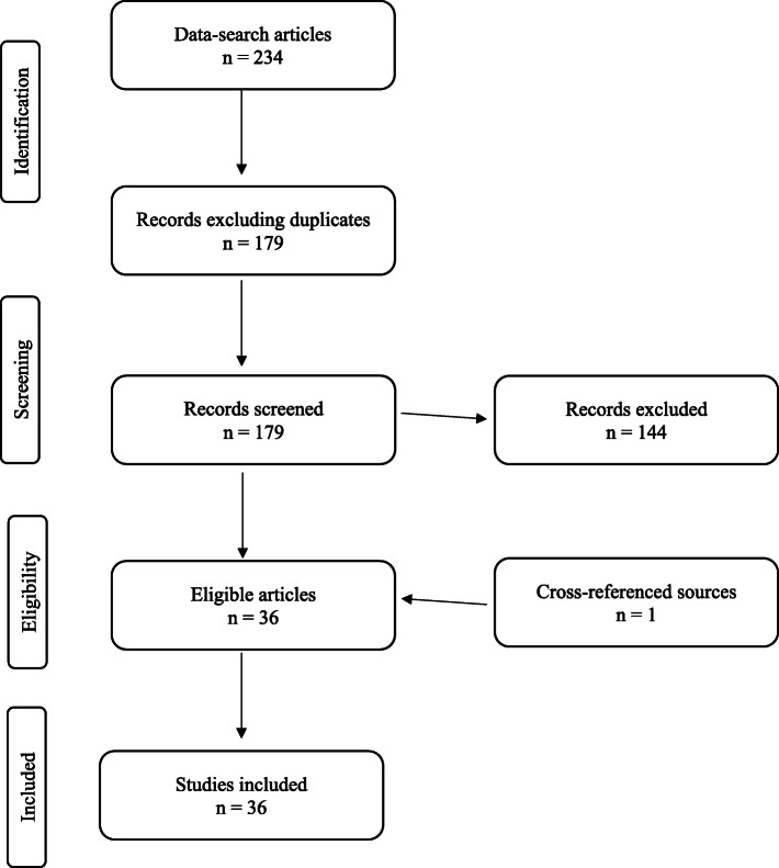
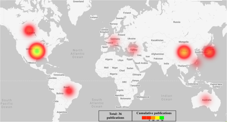
Methods: Using specified criteria, literature searching was conducted to identify publications describing engineered neurosurgical simulators. Included in the study were manuscripts highlighting designs validated for neurosurgical skill transfer. Purely anatomical designs, lacking aspects of surgical simulation, were excluded. Eligible manuscripts were analyzed. Data on the types of simulators, representing the various modelled neurosurgical pathologies, were recorded. Authors' countries of affiliation were also recorded.
Results: A total of thirty-six articles, representing ten countries in five continents were identified. Geographically, Africa as a continent was not represented in any of the publications. The simulation-modelling encompassed a variety of neurosurgical subspecialties including: vascular, skull base, ventriculoscopy / ventriculostomy, craniosynostosis, skull lesions / skull defects, intrinsic brain tumor and other. Finally, the vascular and skull base categories together accounted for over half (52.8 %) of the 3D printed simulated neurosurgical pathology.
Conclusions: Despite the growing body of literature supporting 3D printing in neurosurgical education, its full potential has not been maximized. Unexplored areas of 3D printing for neurosurgical simulation include models simulating the resection of intrinsic brain tumors or of epilepsy surgery lesions, as these require complex models to accurately simulate fine dissection techniques. 3D printed surgical phantoms offer an avenue for the advancement of global-surgery education initiatives.
Keywords: 3D printing; Additive Manufacturing; Neurosurgery Education; Rapid prototyping.
Conflict of interest statement
The authors declare that they have no competing interests.
Figures


References
-
- Horvath J, Horvath JA. Brief History of 3D Printing. Mastering 3D Print. 2014. 10.1007/978-1-4842-0025-4_1.
Publication types
LinkOut - more resources
Full Text Sources
Other Literature Sources