Review
. 2021 May;599(10):2559-2571.
doi: 10.1113/JP281305. Epub 2021 Apr 8.

Chemoreceptor mechanisms regulating CO2 -induced arousal from sleep

Affiliations
Review

Chemoreceptor mechanisms regulating CO2 -induced arousal from sleep

Stephen B G Abbott et al. J Physiol. 2021 May.

Abstract

Arousal from sleep in response to CO2 is a life-preserving reflex that enhances ventilatory drive and facilitates behavioural adaptations to restore eupnoeic breathing. Recurrent activation of the CO2 -arousal reflex is associated with sleep disruption in obstructive sleep apnoea. In this review we examine the role of chemoreceptors in the carotid bodies, the retrotrapezoid nucleus and serotonergic neurons in the dorsal raphe in the CO2 -arousal reflex. We also provide an overview of the supra-medullary structures that mediate CO2 -induced arousal. We propose a framework for the CO2 -arousal reflex in which the activity of the chemoreceptors converges in the parabrachial nucleus to trigger cortical arousal.

Keywords: arousal threshold; interoception; obstructive sleep apnea; optogenetic; respiration; sleep-wake behavior.


Conflict of interest statement

Competing interests

The authors declare they have no competing interests.

Figures

Figure 1.
Figure 1.. RTN neurons contribute to the CO2-arousal reflex: evidence from gain- and loss-of-function experiments
A, RTN and CB ablation blunt the CO2-arousal reflex. Sleep survival curves (mean ± SEM) for rats exposed to CO2 in a plethysmography chamber are shown. Three groups of rats (sham operated n = 10, RTN ablation n = 9; CB ablation n = 6) were subjected to air-to-15% CO2 or air-to-air gas changes. FI,CO2 during CO2 challenges is shown at the bottom of panel A. Ablation of either RTN or CBs reduces the CO2 arousal reflex but the loss of the RTN has a much more pronounced effect. Data from Souza et al. (2019). Significant differences were determined by a Bonferroni test following a two-way repeated measures ANOVA. *P < 0.05, **P < 0.01 for CB ablation vs. sham. *P < 0.05, **P < 0.01***P < 0.001 for RTN ablation vs. sham. Data from Souza et al. 2019. B, optogenetic RTN stimulation causes arousal from sleep. Sleep survival curves (mean ± SEM) during selective stimulation of RTN (n = 8) with increasing frequencies of stimulation are shown. Arousal requires stimulation frequencies of greater than 6–9 Hz, a discharge rate matched by RTN neurons when recording in anaesthetized rats with an end tidal CO2 of 7% (Guyenet et al. 2005). Data from Souza et al. 2020. Significant differences were determined by a Bonferroni test following a two-way repeated measures ANOVA. *P < 0.05 RTN stim vs. no stim.
Figure 2.
Figure 2.. Central organization of CO2-arousal reflex: a hypothesis
The initial trigger to CO2-induced arousal may be the sudden acidification of the CBs followed by that of a small number of lower brainstem structures, especially the RTN. This acidification increases the excitatory drive from the RTN and CBs to respiratory centres in the ventral respiratory column (VRC) and nucleus of the solitary tract (NTS) and pontine respiratory group (not shown). The integrated output of the respiratory centres is communicated to the peripheral respiratory system through premotor and motor neurons located in the medulla and spinal cord. Increased ventilatory motor output also activates sensory afferents from the lungs, chest and airways that provide feedback via cranial and spinal nerves. Serotoninergic neurons in the medullary raphe regulate the activity of the entire central respiratory network, including the RTN. Arousal probably results from an abrupt rise in the activity of the external lateral parabrachial nucleus (PBel), a structure that probably integrates direct or polysynaptic inputs from a variety of sources related to breathing such as the carotid bodies, RTN, VRC, NTS and sensory feedback for the lungs chest and airways. The PBel promotes EEG arousal through a diffuse forebrain network that includes the basal forebrain (BF), lateral hypothalamic area (LHA), and central nucleus of the amygdala (CeA). PBel neurons are regulated by serotonergic inputs from the dorsal raphe. The neurotransmitter phenotype of each neural compartment is indicated; Glu, glutamate; GABA, gamma-aminobutyric acid; Gly, glycine; ACh, acetylcholine; 5HT, serotonin.
Figure 3.
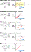
Figure 3.. CO2-arousal reflex: schematic representation of the relative contribution the carotid bodies, central chemoreceptors, serotonergic system and basal forebrain
In the intact brain, CO2 exposure, the CBs and RTN directly or indirectly increase the activity of PBel. PBel activates the basal forebrain which causes EEG desynchronization and arousal when a particular threshold is reached (dashed line). Removal of either the CBs or RTN reduces CO2-related input to the PBel thus delaying the activation of the basal forebrain during CO2 exposure. Based on the findings in Souza et al. (2019), CB ablation causes a rightward shift in the PBel response to CO2 exposure, indicating that the early detection of CO2 is impaired. However, arousal invariably occurs because RTN inputs are intact. RTN ablation, on the other hand, produces a major deficit in the PBel response to CO2 exposure, indicating that the RTN contributes to PBel activity over a broader range of CO2 than the CBs. Hypothetically, removal of both the CBs and RTN may well abolish the CO2 arousal reflex but the extreme hypoventilation resulting from by such a procedure may preclude such an experiment. Removal of the 5HT input to PBel causes a severe deficit in CO2-induced arousal by reducing the excitability of the PBel, and its responsiveness to excitatory inputs from the RTN and CB. Inhibition of the basal forebrain, and other efferent targets of the PBel, raises arousal threshold so that only a very high PBel activity (i.e. a high CO2 stimulus) is sufficient for arousal from sleep.

References

    1. Abbott SB, Stornetta RL, Coates MB & Guyenet PG (2011). Phox2b-expressing neurons of the parafacial region regulate breathing rate, inspiration, and expiration in conscious rats. J Neurosci 31, 16410–16422. - PMC - PubMed
    1. Amiel J, Laudier B, Attie-Bitach T, Trang H, de Pontual L, Gener B, Trochet D, Etchevers H, Ray P, Simonneau M, Vekemans M, Munnich A, Gaultier C & Lyonnet S (2003). Polyalanine expansion and frameshift mutations of the paired-like homeobox gene PHOX2B in congenital central hypoventilation syndrome. Nat Genet 33, 459–461. - PubMed
    1. Banzett RB, Lansing RW, Evans KC & Shea SA (1996). Stimulus-response characteristics of CO2-induced air hunger in normal subjects. Respir Physiol 103, 19–31. - PubMed
    1. Basting TM, Burke PG, Kanbar R, Viar KE, Stornetta DS, Stornetta RL & Guyenet PG (2015). Hypoxia silences retrotrapezoid nucleus respiratory chemoreceptors via alkalosis. J Neurosci 35, 527–543. - PMC - PubMed
    1. Berquin P, Bodineau L, Gros F & Larnicol N (2000). Brainstem and hypothalamic areas involved in respiratory chemoreflexes: a Fos study in adult rats. Brain Res 857, 30–40. - PubMed

Publication types

LinkOut - more resources