. 2021 Apr 6;10(7):e019961.
doi: 10.1161/JAHA.120.019961. Epub 2021 Mar 24.

Recombinant Soluble Corin Improves Cardiac Function in Mouse Models of Heart Failure

Affiliations

Recombinant Soluble Corin Improves Cardiac Function in Mouse Models of Heart Failure

Yayan Niu et al. J Am Heart Assoc. .

Abstract

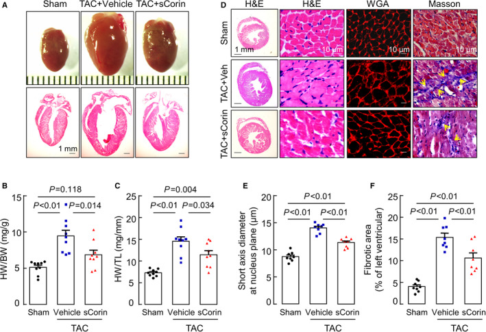
Background Corin is a transmembrane protease that activates ANP and BNP (atrial and B-type natriuretic peptides). Impaired corin expression and function are associated with heart failure. In this study, we characterized a soluble form of corin (sCorin) and examined its effects on cardiac morphology and function in mouse heart failure models. Methods and Results sCorin, consisting of the full-length extracellular fragment of human corin with an engineered activation site, was expressed in Chinese hamster ovary cells, purified from the conditioned medium with affinity chromatography, and characterized in pro-ANP processing assays in vitro and pharmacokinetic studies in mice. Effects of sCorin on mouse models of heart failure induced by left coronary artery ligation and transverse aortic constriction were assessed by ELISA analysis of plasma markers, histologic examination, and echocardiography. We showed that purified and activated sCorin converted pro-ANP to ANP that stimulated cGMP production in cultured cells. In mice, intravenously and intraperitoneally administered sCorin had plasma half-lives of 3.5±0.1 and 8.3±0.3 hour, respectively. In the mouse heart failure models, intraperitoneal injection of sCorin increased plasma ANP, BNP, and cGMP levels; lowered plasma levels of NT-proANP (N-terminal-pro-ANP), angiotensin II, and aldosterone; reduced cardiac hypertrophy and fibrosis; and improved cardiac function. Conclusions We show that sCorin treatment enhanced natriuretic peptide processing and activity, suppressed the renin-angiotensin-aldosterone system, and improved cardiac morphology and function in mice with failing hearts.

Keywords: cardiac function; cardiac hypertrophy; corin; heart failure; mouse models.


Conflict of interest statement

Dr Wu is an inventor on several corin‐related patents that are owned by Bayer Healthcare. Dr Wu does not own Bayer stock and has not and will not receive any royalties from those patents. The remaining authors have no disclosures to report.

Figures

Figure 1
Figure 1. Characterization of soluble corin (sCorin) and pharmacokinetic studies in mice.
A, Corin protein domains are illustrated. The RMNKR sequence at the activation cleavage site (arrowhead) is shown. A disulfide bond (s‐s) linking the propeptide region and the protease domain is indicated. sCorin consists of an Igκ signal peptide and the extracellular fragment of corin, in which the RMNKR sequence is replaced by DDDDK that is cleavable by enterokinase. B, Enterokinase‐activated sCorin (top panel) converted pro‐ANP to ANP (lower panel), as indicated by western blotting under reducing conditions. C, Soluble corin activated ANP‐stimulated cGMP production in cell‐based assays. P values were analyzed by 1‐way ANOVA and Tukey’s post hoc analysis. D and E, Soluble corin was injected intravenously (D) or intraperitoneally (E) in mice (n=7 per group). Plasma samples were collected over time. Soluble corin was analyzed by western blotting under nonreducing conditions (top panels) and ELISA (lower panels) to calculate plasma half‐lives. ANP indicates atrial natriuretic peptide; Fz, frizzled; LDLR, LDL receptor; SR, scavenger receptor; TM, transmembrane; and V, V5 tag.
Figure 2
Figure 2. Left ventricular (LV) function in sham‐operated mice and mice with myocardial infarction (MI) that were treated with vehicle or soluble corin (sCorin).
Echocardiography was done before (0 week) and after (1–4 weeks) the surgery to examine ejection fraction (EF) (A), fractional shortening (FS) (B), LV end‐diastolic dimension (LVEDD) (C) and volume (LVEDV) (D), and LV end‐systolic dimension (LVESD) (E) and volume (LVESV) (F). Data are mean± SEM; n=9‐10 per group. P values among 3 groups at the same time point were analyzed by 1‐way ANOVA and Tukey’s post hoc analysis.
Figure 3
Figure 3. Analysis of heart and lung tissues in sham‐operated mice and mice with MI that were treated with vehicle or soluble corin (sCorin).
A and B, Ratios of heart weight (HW) were normalized to body weight (BW) (A) or tibia length (TL) (B). C and D, Ratios of lung weight (LW) were normalized to BW (C) or TL (D). Data are mean±SEM. P values were analyzed by 1‐way ANOVA and Tukey’s post hoc analysis. E, Serial heart sections from the left coronary artery ligation site toward the apex were stained with Masson’s trichrome. Scale bars: 100 μm. Scar areas (blue) were quantified by Image‐Pro‐Plus software. Quantitative data (mean±SEM) are shown in (F). P values were analyzed by 1‐way ANOVA and Tukey’s post hoc analysis.
Figure 4
Figure 4. Analysis of plasma factors and left ventricular (LV) cGMP levels in sham‐operated mice and mice with myocardial infarction (MI) treated with vehicle or soluble corin (sCorin).
Plasma samples were collected at 4 weeks after surgery. Levels of ANP (A), BNP (B), cGMP (C), NT‐pro‐ANP (D), angiotensin II (E), and aldosterone (F) in plasma samples and cGMP levels in LV tissues (G) were analyzed by ELISA. Data are mean±SEM. P values were analyzed by one‐way ANOVA and Tukey’s post hoc analysis. ANP indicates atrial natriuretic peptide; BNP, B‐type natriuretic peptide; and NT‐pro‐ANP, N‐terminal proatrial natriuretic peptide.
Figure 5
Figure 5. Left ventricular (LV) function in sham‐operated mice and mice with transverse aortic constriction (TAC) that were treated with vehicle or soluble corin (sCorin).
Echocardiography was done before (0 week) and after (2–8 weeks) the surgery to examine ejection fraction (EF) (A), fractional shortening (FS) (B), LV end‐diastolic dimension (LVEDD) (C) and volume (LVEDV) (D), and LV end‐systolic dimension (LVESD) (E) and volume (LVESV) (F). Data are mean±SEM; n=9 per group. P values among 3 groups at the same time point were analyzed by 1‐way ANOVA and Tukey’s post hoc analysis.
Figure 6
Figure 6. Analysis of heart tissues in sham‐operated mice and mice with transverse aortic constriction (TAC) treated with vehicle or soluble corin (sCorin).
A, Representative heart images (top panels) and hematoxylin and eosin (H&E)‐stained sections (lower panels) were from mice at 8 weeks after surgery. Scale bars: 1 mm. B and C, Ratios of heart weight (HW) to body weight (BW) (B) or tibia length (TL) (C) were calculated. Data are mean±SEM. P values were analyzed by one‐way ANOVA and Tukey’s post hoc analysis. D, Representative heart sections stained by hematoxylin and eosin, wheat germ agglutinin (WGA), and Masson’s trichrome. Scale bars are indicated. E, Short‐axis cardiomyocyte diameters at the nucleus plane were measured. Data are mean±SEM from 200 individual cardiomyocytes in >3 randomly selected section from each mouse. n=8 per group. P values were analyzed by 1‐way ANOVA and Tukey’s post hoc analysis. F, Areas of fibrosis (blue in Masson’s trichrome‐stained sections) were quantified by Image‐Pro‐Plus software. Data are mean±SEM. n=8 per group. P values were analyzed by one‐way ANOVA and Tukey’s post hoc analysis.
Figure 7
Figure 7. Analysis of plasma factors and left ventricular (LV) cGMP levels in sham‐operated mice and mice with transverse aortic constriction (TAC) treated with vehicle or soluble corin (sCorin). Plasma samples were collected at 8 weeks after surgery.
Levels of ANP (A), BNP (B), cGMP (C), NT‐pro‐ANP (D), angiotensin II (E), and aldosterone (F) in plasma samples and cGMP levels in LV tissues (G) were analyzed by ELISA. Data are mean±SEM. P values were analyzed by 1‐way ANOVA and Tukey’s post hoc analysis. ANP indicates atrial natriuretic peptide; BNP, B‐type natriuretic peptide; NT‐pro‐ANP, N‐terminal proatrial natriuretic peptide; and Veh, vehicle.

Similar articles

Cited by

References

    1. Goetze JP, Bruneau BG, Ramos HR, Ogawa T, de Bold MK, de Bold AJ. Cardiac natriuretic peptides. Nat Rev Cardiol. 2020;17:698–717. DOI: 10.1038/s41569-020-0381-0. - DOI - PubMed
    1. Kuhn M. Molecular Physiology of membrane guanylyl cyclase receptors. Physiol Rev. 2016;96:751–804. DOI: 10.1152/physrev.00022.2015. - DOI - PubMed
    1. Song W, Wang H, Wu Q. Atrial natriuretic peptide in cardiovascular biology and disease (NPPA). Gene. 2015;569:1–6. DOI: 10.1016/j.gene.2015.06.029. - DOI - PMC - PubMed
    1. Chen W, Spitzl A, Mathes D, Nikolaev VO, Werner F, Weirather J, Špiranec K, Röck K, Fischer JW, Kämmerer U, et al. Endothelial actions of ANP enhance myocardial inflammatory infiltration in the early phase after acute infarction. Circ Res. 2016;119:237–248. DOI: 10.1161/CIRCRESAHA.115.307196. - DOI - PubMed
    1. Holtwick R, van Eickels M, Skryabin BV, Baba HA, Bubikat A, Begrow F, Schneider MD, Garbers DL, Kuhn M. Pressure‐independent cardiac hypertrophy in mice with cardiomyocyte‐restricted inactivation of the atrial natriuretic peptide receptor guanylyl cyclase‐A. J Clin Invest. 2003;111:1399–1407. DOI: 10.1172/JCI17061. - DOI - PMC - PubMed

Publication types