. 2021 Jun 1;320(6):G936-G957.
doi: 10.1152/ajpgi.00053.2021. Epub 2021 Mar 24.

Colon epithelial cell TGFβ signaling modulates the expression of tight junction proteins and barrier function in mice

Affiliations

Colon epithelial cell TGFβ signaling modulates the expression of tight junction proteins and barrier function in mice

Paula Marincola Smith et al. Am J Physiol Gastrointest Liver Physiol. .

Abstract

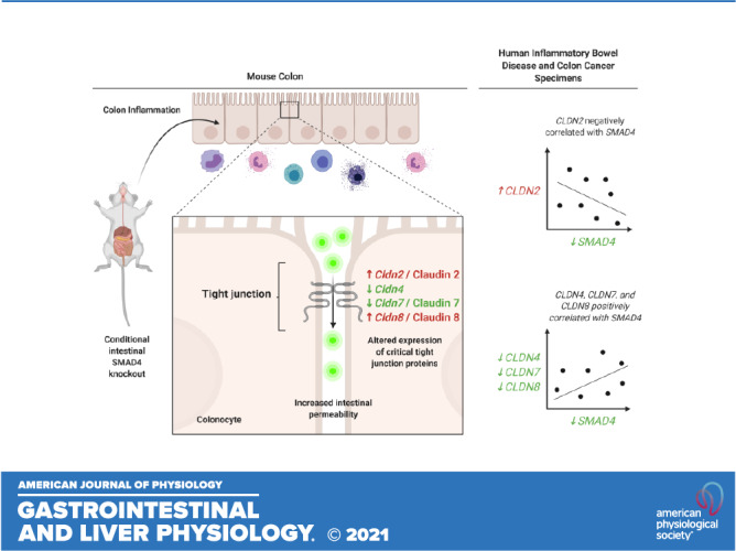
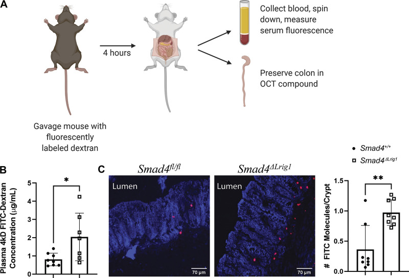
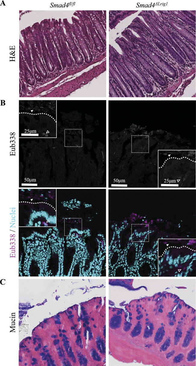
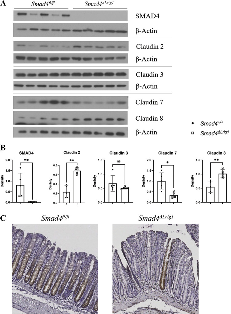
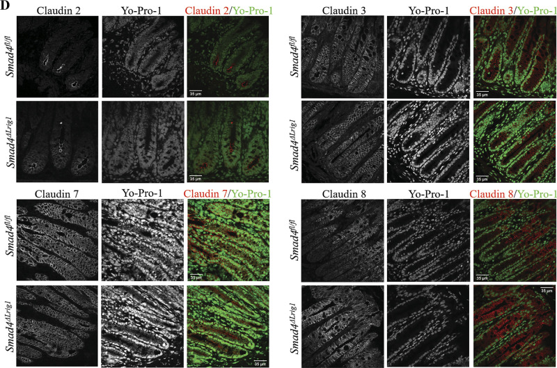
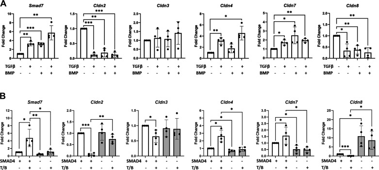
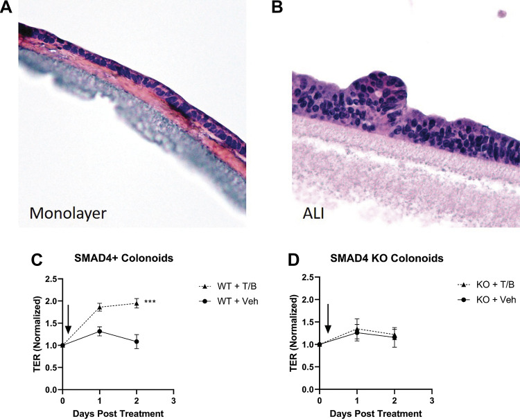
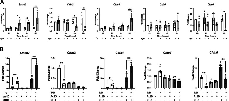
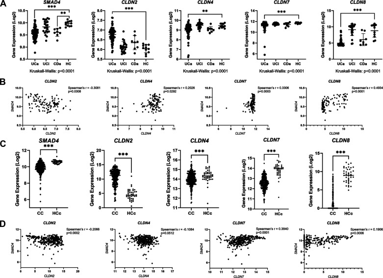
Defective barrier function is a predisposing factor in inflammatory bowel disease (IBD) and colitis-associated cancer (CAC). Although TGFβ signaling defects have been associated with IBD and CAC, few studies have examined the relationship between TGFβ and intestinal barrier function. Here, we examine the role of TGFβ signaling via SMAD4 in modulation of colon barrier function. The Smad4 gene was conditionally deleted in the intestines of adult mice and intestinal permeability assessed using an in vivo 4 kDa FITC-Dextran (FD4) permeability assay. Mouse colon was isolated for gene expression (RNA-sequencing), Western blot, and immunofluorescence analysis. In vitro colon organoid culture was utilized to assess junction-related gene expression by qPCR and transepithelial resistance (TER). In silico analyses of human IBD and colon cancer databases were performed. Mice lacking intestinal expression of Smad4 demonstrate increased colonic permeability to FD4 without gross mucosal damage. mRNA/protein expression analyses demonstrate significant increases in Cldn2/Claudin 2 and Cldn8/Claudin 8, and decreases in Cldn3, Cldn4, and Cldn7/Claudin 7 with intestinal SMAD4 loss in vivo without changes in Claudin protein localization. TGFβ1/BMP2 treatment of polarized SMAD4+ colonoids increases TER. Cldn2, Cldn4, Cldn7, and Cldn8 are regulated by canonical TGFβ signaling, and TGFβ-dependent regulation of these genes is dependent on nascent RNA transcription (Cldn2, Cldn4, Cldn8) but not nascent protein translation (Cldn4, Cldn8). Human IBD/colon cancer specimens demonstrate decreased SMAD4, CLDN4, CLDN7, and CLDN8 and increased CLDN2 compared with healthy controls. Canonical TGFβ signaling modulates the expression of tight junction proteins and barrier function in mouse colon.NEW & NOTEWORTHY We demonstrate that canonical TGFβ family signaling modulates the expression of critical tight junction proteins in colon epithelial cells, and that expression of these tight junction proteins is associated with maintenance of colon epithelial barrier function in mice.

Keywords: claudin; inflammatory bowel disease; tight junction; transforming growth factor β.


Conflict of interest statement

No conflicts of interest, financial or otherwise, are declared by the authors.

Figures

None
Graphical abstract
Figure 1.
Figure 1.
Intestinal Smad4 loss associated with impaired barrier function in mice. A: seven Smad4ΔLrig1 mice and eight control mice were administered 4 kDa fluorescein isothiocyanate-Dextran (FITC-D, FD4) by oral gavage. Blood was collected and colon was fresh frozen 4 h later. B: Smad4ΔLrig1 mice demonstrated a 2.5-fold increase in plasma concentration of FD4 compared with control mice, suggesting an intestinal barrier defect (P = 0.029 by Mann–Whitney test). C: FITC molecules in preserved colon sections from eight Smad4ΔLrig1 mice and eight control mice were detected using an anti-FITC antibody (red), demonstrating more FITC molecules per crypt in Smad4ΔLrig1 mice compared with control (0.187 vs. 0.908, P = 0.007 by Mann–Whitney test). Statistical significance is designated as: *P < 0.05, **P < 0.01. Image created with BioRender.com.
Figure 2.
Figure 2.
Colons from Smad4ΔLrig1 mice show no evidence of gross mucosal damage. A: representative hematoxylin and eosin (H&E) stains from Smad4ΔLrig1 and control mice showing epithelial integrity. B: fluorescence in situ hybridization (FISH) staining with the pan-bacterial probe, Eub338 (red). Nuclei in light blue. Bacterial species indicated by solid arrowhead. Autofluorescent red blood cells indicated by open arrowhead. Border between lumen and epithelium demarcated with dashed line. C: Alcian blue (pH 2.5) stain for mucins (blue).
Figure 3.
Figure 3.
Colonic microbiome composition in five Smad4ΔLrig1 and five control mice. Composition bar plots from stool pellets (A) and colon mucosal scrapings (B) of five Smad4ΔLrig1 and five control mice. Each color represents a unique bacterial species. Heatmaps with unsupervised hierarchical clustering of stool pellets (C) and colon mucosal scrapings (D) from five Smad4ΔLrig1 and five control mice. All mice were littermates. “F” indicates female mice who were housed together, and “M” indicates male mice who were housed together (bedding mixed between the one male and one female cage weekly).
Figure 4.
Figure 4.
Loss of intestinal Smad4 expression is associated with altered levels, but not localization, of several Claudin proteins. A: Western blots (WB) from protein lysates generated from colon epithelium of five Smad4ΔLrig1 and five control mice. B: quantification of relative protein density by WB. Protein density was compared between mouse genotypes by Mann–Whitney test. C: immunohistochemistry for SMAD4 protein (brown) in SMAD4+ control and Smad4ΔLrig1 mice. D: immunofluorescence staining for indicated Claudin proteins. Claudin proteins in red, nuclei in green. Statistical significance is designated as nsP ≥ 0.05, *P < 0.05, **P < 0.01.
Figure 4.
Figure 4.
Loss of intestinal Smad4 expression is associated with altered levels, but not localization, of several Claudin proteins. A: Western blots (WB) from protein lysates generated from colon epithelium of five Smad4ΔLrig1 and five control mice. B: quantification of relative protein density by WB. Protein density was compared between mouse genotypes by Mann–Whitney test. C: immunohistochemistry for SMAD4 protein (brown) in SMAD4+ control and Smad4ΔLrig1 mice. D: immunofluorescence staining for indicated Claudin proteins. Claudin proteins in red, nuclei in green. Statistical significance is designated as nsP ≥ 0.05, *P < 0.05, **P < 0.01.
Figure 5.
Figure 5.
Transforming growth factor-β (TGFβ)/bone morphogenetic protein (BMP) signaling via SMAD4 modulates the expression of Cldn2, Cldn4, Cldn7, and Cldn8 in a cell-autonomous manner. A: wild-type colonoids treated with TGFβ1, BMP2, both, or vehicle for 24 h. B: SMAD4 knockout and control colonoids treated with TGFβ1/BMP2 (T/B) or vehicle for 24 h. Fold change indicates the relative level of mRNA compared with vehicle-treated wild-type controls for indicated genes. Each data point represents a single biological replicate, with biological replicates treated and harvested on different days. Experimental arms were compared using nonparametric Kruskal–Wallis test with post hoc Welch’s t test. Smad7 is used as a positive control to confirm ligand activity. Statistical significance is designated as *P < 0.05, **P < 0.01, ***P < 0.001.
Figure 6.
Figure 6.
Transforming growth factor-β (TGFβ)/bone morphogenetic protein (BMP) signaling via SMAD4 modulates barrier function/transepithelial resistance in a cell-autonomous manner. Representative images of hematoxylin and eosin (H&E) stain demonstrating polarized monolayers of colon epithelial cells grown on collagen-coated transwell membranes before (A) and after (B) initiation of air-liquid interface (ALI). C: normalized transepithelial resistance (TER) for wild-type (SMAD4+) colonoids on collagen-coated transwells in an ALI system. TGFβ/BMP co-treatment in the basolateral chamber was associated with a significant increase in TER compared with vehicle-treated controls at 24- and 48-h (P < 0.001 by repeated-measured ANOVA/mixed effects model). D: normalized TER for SMAD4 knockout colonoids grown on collagen-coated transwells in an ALI system. TGFβ/BMP co-treatment in the basolateral chamber failed to elicit a change in TER compared with vehicle-treated controls at 24- or 48-h. For both C and D, data represent the mean and standard deviation of three biological replicates, with biological replicates being plated, treated, and measured on separate days. Experimental arms were compared using repeat measures ANOVA/Mixed effects model. TER measurements for each well were normalized to the final pretreatment TER for that well (Day 0 on the X-axis).
Figure 7.
Figure 7.
Transforming growth factor-β (TGFβ)/bone morphogenetic protein (BMP) signaling via SMAD4 modulates the expression of Cldn genes by disparate mechanisms. A: wild-type colonoids treated with TGFβ1/BMP2 (gray bars/open boxes) or vehicle (white bars/closed circles) for the indicated periods of time. B: wild-type colonoids treated with TGFβ1/BMP2 (T/B) vs. vehicle (white bars, left), T/B vs. vehicle in the presence of actinomycin D (ActD) (light gray bars, middle), or T/B vs. vehicle in the presence of cyclohexamide (CHX) (dark gray bars, right) for 6.5 h. Fold change indicates the relative level of mRNA for indicated genes compared with vehicle-treated control. Each data point represents a single biological replicate, with biological replicates treated and harvested on different days. Experimental arms were compared using nonparametric Kruskal–Wallis test with post hoc Welch’s t test. Smad7 is used as a positive control to confirm ligand activity. Statistical significance is designated as *P < 0.05, **P < 0.01.
Figure 8.
Figure 8.
SMAD4 and CLDN genes are dysregulated in human inflammatory bowel disease (IBD) and colon cancer. A: in silico analysis of human microarray database (accession number GSE75214). Samples represent human colon biopsy samples from patients with active ulcerative colitis (UCa, n = 74), inactive ulcerative colitis (UCi, n = 23), active Crohn’s disease (CDa, n = 8), and healthy controls (HC, n = 11). Gene expression in human biopsy samples were compared between groups using the nonparametric Kruskal–Wallis test with post hoc Welch’s t test. B: correlation between SMAD4 expression and indicated CLDN gene expression in human colon biopsy specimens from GSE75214. Spearman’s correlation was used to measure correlation between SMAD4 and CLDN gene expression. C: in silico analysis of RNA-sequencing data from The Cancer Genome Atlas (TCGA) database. Samples represent mRNA expression in colon cancer (CC, n = 283) or healthy control colon (HCc, n = 41) specimens. Nonparametric Mann–Whitney test was used to compare gene expression levels between groups. D: correlation between SMAD4 expression and indicated CLDN gene expression in human colon specimens from TCGA. Spearman’s correlation was used to measure correlation between SMAD4 and CLDN gene expression. For AD, gene expression levels are represented on a Log2 scale. Statistical significance is designated as **P < 0.01, ***P < 0.001.

Similar articles

Cited by

References

    1. Allaire JM, Darsigny M, Marcoux SS, Roy SAB, Schmouth J-F, Umans L, Zwijsen A, Boudreau F, Perreault N. Loss of Smad5 leads to the disassembly of the apical junctional complex and increased susceptibility to experimental colitis. Am J Physiol Gastrointest Liver Physiol 300: G586–G597, 2011. doi:10.1152/ajpgi.00041.2010. - DOI - PubMed
    1. Laukoetter MG, Nava P, Lee WY, Severson EA, Capaldo CT, Babbin BA, Williams IR, Koval M, Peatman E, Campbell JA, Dermody TS, Nusrat A, Parkos CA. JAM-A regulates permeability and inflammation in the intestine in vivo. J Exp Med 204: 3067–3076, 2007. doi:10.1084/jem.20071416. - DOI - PMC - PubMed
    1. Martini E, Krug SM, Siegmund B, Neurath MF, Becker C. Mend your fences the epithelial barrier and its relationship with mucosal immunity in inflammatory bowel disease. Cell Mol Gastroenterol Hepatol 4: 33–46, 2017. doi:10.1016/j.jcmgh.2017.03.007. - DOI - PMC - PubMed
    1. McGuckin MA, Eri R, Simms LA, Florin THJ, Radford-Smith G. Intestinal barrier dysfunction in inflammatory bowel diseases. Inflamm Bowel Dis 15: 100–113, 2009. doi:10.1002/ibd.20539. - DOI - PubMed
    1. Sluis M. D, Koning BAED, Bruijn A, Velcich A, Meijerink JPP, Goudoever JBV, Büller HA, Dekker J, Seuningen IV, Renes IB, Einerhand AWC. Muc2-deficient mice spontaneously develop colitis, indicating that MUC2 is critical for colonic protection. Gastroenterology 131: 117–129, 2006. doi:10.1053/j.gastro.2006.04.020. - DOI - PubMed

Publication types