. 2021 May 1;320(5):L958-L968.
doi: 10.1152/ajplung.00121.2020. Epub 2021 Mar 24.

Blockade of PD-1 decreases neutrophilic inflammation and lung damage in experimental COPD

Affiliations

Blockade of PD-1 decreases neutrophilic inflammation and lung damage in experimental COPD

Felix Ritzmann et al. Am J Physiol Lung Cell Mol Physiol. .

Abstract

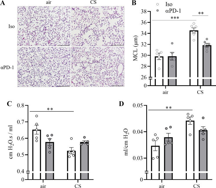
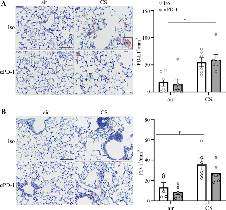
Chronic obstructive lung disease (COPD) and lung cancer are both caused by smoking and often occur as comorbidity. The programmed cell death protein 1/programmed cell death ligand 1 (PD-1/PD-L1) axis is an important canonic immunoregulatory pathway, and antibodies that specifically block PD-1 or PD-L1 have demonstrated efficacy as therapeutic agents for non-small cell lung cancer. The role of the PD-1/PD-L1 axis in the pathogenesis of COPD is unknown. Here, we analyzed the function of the PD-1/PD-L1 axis in preclinical COPD models and evaluated the concentrations of PD-1 and PD-L1 in human serum and bronchoalveolar lavage (BAL) fluids as biomarkers for COPD. Anti-PD-1 treatment decreased lung damage and neutrophilic inflammation in mice chronically exposed to cigarette smoke (CS) or nontypeable Haemophilus influenzae (NTHi). Ex vivo stimulated macrophages obtained from anti-PD-1-treated mice released reduced amounts of inflammatory cytokines. PD-L1 concentrations correlated positively with PD-1 concentrations in human serum and BAL fluids. Lung sections obtained from patients with COPD stained positive for PD-L1. Our data indicate that the PD-1/PD-L1 axis is involved in developing inflammation and tissue destruction in COPD. Inflammation-induced activation of the PD-1 pathway may contribute to disease progression.

Keywords: COPD; PD-1; PD-L1; inflammation; lung damage; macrophages.


Conflict of interest statement

No conflicts of interest, financial or otherwise are declared by the authors.

Figures

Figure 1.
Figure 1.
Anti-PD-1 treatment decreases CS-induced neutrophilic inflammation. C57BL/6 mice were exposed to CS 5 days a week for 5 days and 4 wk and treated with a PD-1-blocking antibody or an isotype antibody two times a week. Numbers of total immune cells (A), neutrophils (B), macrophages (C), and lymphocytes (D), and concentrations of (E) KC and (F) MIP-2 were determined in BAL fluids 24 h after the final exposure to CS; n = 4–8 mice per group. Two independent experiments for each group. Data were compared by two-way ANOVA with Bonferroni posttest and are shown as means ± SE. **P < 0.01 and ***P < 0.001. BAL, bronchoalveolar lavage; CS, cigarette smoke; PD-1, programmed cell death protein 1.
Figure 2.
Figure 2.
Anti-PD-1 treatment decreases CS-induced lung damage. C57BL/6 mice were exposed to CS 5 days a week for 12 wk and treated with a PD-1-blocking antibody or an isotype antibody two times a week. A: lung histology (representative hematoxylin-eosin staining, scale bar = 100 µm), B: mean chord length (MCL), C: respiratory resistance, and D: respiratory system compliance were determined 24 h after the final exposure to CS; n = 5 or 6 mice per group. Two independent experiments for each group. Data were compared by two-way ANOVA with Bonferroni posttest and are shown as means ± SE. **P < 0.01 and ***P < 0.001. CS, cigarette smoke; PD-1, programmed cell death protein 1.
Figure 3.
Figure 3.
Numbers of PD-L1+ and PD-1+ cells are increased in the lungs of CS-exposed mice. C57BL/6 mice were exposed to CS 5 days a week for 12 wk and treated with a PD-1-blocking antibody or an isotype antibody two times a week. Immunohistochemical staining (representative histology, scale bar = 50 µm) for (A) PD-L1 and (B) PD-1 and quantification of the PD-L1+ and PD-1+ cells in lung parenchyma; n = 5 or 6 mice per group. Two independent experiments for each group. Data were compared by two-way ANOVA with Bonferroni posttest and are shown as means ± SE. *P < 0.05. CS, cigarette smoke; PD-1, programmed cell death protein 1; PD-L1, programmed cell death ligand 1.
Figure 4.
Figure 4.
Numbers of PD-1-expressing CD4+ cells and PD-L1-expressing CD68+ cells are increased in the lungs of CS-exposed mice. C57BL/6 mice were exposed to CS 5 days a week for 12 wk and treated with a PD-1-blocking antibody or an isotype antibody two times a week. Double immunohistochemical staining (representative histology, scale bar = 20 µm) for (A) CD4 (red)/PD-1 (green), (B) CD8 (red)/PD-1 (green), and (C) CD68 (red)/PD-L1 (green), and quantification of the cells in lung parenchyma; n = 3 or 4 mice per group. Results are presented for each mouse, were compared by two-way ANOVA with Bonferroni posttest and are shown as means ± SE. *P < 0.05, **P < 0.01, and ***P < 0.001. CS, cigarette smoke; PD-1, programmed cell death protein 1; PD-L1, programmed cell death ligand 1.
Figure 5.
Figure 5.
Anti-PD-1 treatment decreases NTHi-induced lung damage. C57BL/6 mice were exposed to NTHi 3 days a week for 4 wk and treated with a PD-1-blocking antibody or an isotype antibody three times a week. Immunohistochemical staining (representative histology, scale bar = 50 µm) for (A) PD-L1, (B) PD-1, (C) lung histology (representative hematoxylin-eosin staining, scale bar = 100 µm), and (D) MCL were determined 24 h after the final exposure to NTHi; n = 4 or 5 mice per group. Two independent experiments for each group. Data were compared by two-way ANOVA with Bonferroni posttest and are shown as means ± SE. ***P < 0.001. MCL, mean chord length; NTHi, nontypeable Haemophilus influenza; PD-1, programmed cell death protein 1; PD-L1, programmed cell death ligand 1.
Figure 6.
Figure 6.
Anti-PD-1 treatment modulates the inflammatory response of alveolar macrophages. C57BL/6 mice were exposed to CS 5 days a week for 4 wk and treated with a PD-1-blocking antibody or an isotype antibody two times a week. Alveolar macrophages were isolated 2 h after the final exposure to CS and stimulated with inactivated NTHi or control media for 24 h. The concentrations of (A) IL-6, (B) TNF-α, (C) G-CSF, (D) KC, and (E) IL-10 were measured in supernatants. Two independent experiments for each group (n = 6 replicates per group). Data were compared by two-way ANOVA with Bonferroni posttest and are shown as means ± SE. *P < 0.05, **P < 0.01, ***P < 0.001. CS, cigarette smoke; NTHi, nontypeable Haemophilus influenza; PD-1, programmed cell death protein 1.
Figure 7.
Figure 7.
PD-1 and PD-L1 in serum and BAL fluids collected from patients with COPD. A: association of PD-L1 and PD-1 concentrations in serum collected from patients with stable COPD and patients during AECOPD. B: PD-L1 and PD-1 concentrations in serum collected from patients with stable COPD and during AECOPD. C: association of PD-L1 and PD-1 concentrations in BAL fluids collected from stable COPD patients. D: PD-L1 and PD-1 concentrations in BAL fluids collected from stable GOLD I/II and GOLD III/IV patients with COPD. Data were compared by Mann–Whitney and are shown as means ± SE. *P < 0.05. Correlation analysis was performed using the nonparametric Spearman’s correlation test. E: PD-L1 was detected by immunohistochemistry in formalin histological-fixed paraffin-embedded human lung samples from patients with end-stage COPD (representative histology, scale bar = 50 µm). BAL, bronchoalveolar lavage; COPD, chronic obstructive lung disease; GOLD, Global Initiative for Chronic Obstructive Lung Disease; PD-1, programmed cell death protein 1; PD-L1, programmed cell death ligand 1.

Similar articles

Cited by

References

    1. Decramer M, Janssens W. Chronic obstructive pulmonary disease and comorbidities. Lancet Respir Med 1: 73–83, 2013. doi:10.1016/S2213-2600(12)70060-7. - DOI - PubMed
    1. Lopez-Campos JL, Tan W, Soriano JB. Global burden of COPD. Respirology 21: 14–23, 2016. doi:10.1111/resp.12660. - DOI - PubMed
    1. Hoenderdos K, Condliffe A. The neutrophil in chronic obstructive pulmonary disease. Am J Respir Cell Mol Biol 48: 531–539, 2013. doi:10.1165/rcmb.2012-0492TR. - DOI - PubMed
    1. Sethi S. Infection as a comorbidity of COPD. Eur Respir J 35: 1209–1215, 2010. doi:10.1183/09031936.00081409. - DOI - PubMed
    1. Durham AL, Adcock IM. The relationship between COPD and lung cancer. Lung Cancer 90: 121–127, 2015. doi:10.1016/j.lungcan.2015.08.017. - DOI - PMC - PubMed

Publication types

MeSH terms