Review
. 2021 Apr 15;131(8):e142242.
doi: 10.1172/JCI142242.

Type 1 diabetes mellitus: much progress, many opportunities

Review

Type 1 diabetes mellitus: much progress, many opportunities

Alvin C Powers. J Clin Invest. .

Abstract

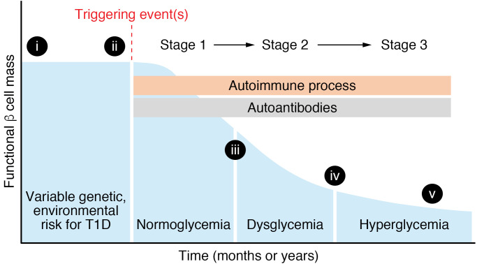
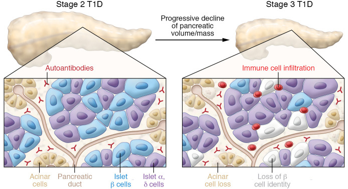
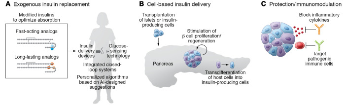
As part of the centennial celebration of insulin's discovery, this review summarizes the current understanding of the genetics, pathogenesis, treatment, and outcomes in type 1 diabetes (T1D). T1D results from an autoimmune response that leads to destruction of the β cells in the pancreatic islet and requires lifelong insulin therapy. While much has been learned about T1D, it is now clear that there is considerable heterogeneity in T1D with regard to genetics, pathology, response to immune-based therapies, clinical course, and susceptibility to diabetes-related complications. This Review highlights knowledge gaps and opportunities to improve the understanding of T1D pathogenesis and outlines emerging therapies to treat or prevent T1D and reduce the burden of T1D.


Conflict of interest statement

Conflict of interest: The author has declared that no conflict of interest exists.

Figures

Figure 1
Figure 1. Model of stages of type 1 diabetes (T1D).
Graph shows functional β cell mass through the stages of T1D. The blue shaded area shows number or insulin secretory capacity of β cells, with time on the x axis reflecting a broad range (could be months or years of T1D development). See text for definition of T1D stages. Roman numerals on the graph refer to questions about T1D pathogenesis shown in Table 1.
Figure 2
Figure 2. Pancreatic changes and immune abnormalities in T1D.
The reduction of pancreas size or volume from normal to stage 2 to stage 3 T1D is shown at the top of the figure. The insets below shows a stylized section of the pancreas with islets and acinar cells in stage 2 and stage 3 T1D. Circulating autoantibodies directed at islet-enriched molecules are present in stage 2 and stage 3 but are not cytotoxic. In stage 3, immune cells are present within islets and exocrine pancreas, and there is a loss of both β cells and acinar cells. Other changes (not shown) in the stage 3 T1D islets include (a) insulin-negative, pseudoatrophic islets with rare islets appearing normal or having β cells; (b) alterations in proinsulin and insulin processing and expression of islet-enriched transcription factors such as PDX-1 and NKX6.1; (c) islet cell hyperexpression of HLA class I and class II molecules; (d) insulitis (immune cell infiltration in some islets) is variable, involving primarily CD8+ T cells, but also B lymphocytes, CD4+ T lymphocytes, and macrophages; CD20+ B lymphocytes are more common in recent-onset T1D in younger individuals; and (e) β cell mass is variable in stage 1, 2, and 3 (see Figure 1). Changes in the exocrine pancreas include (a) reduced pancreatic volume/mass at T1D onset and in autoantibody-positive individuals; progressive decline in pancreas volume in first 5 years of T1D; (b) acinar cell loss, some exocrine fibrosis in stage 3 pancreas; and (c) leukocyte infiltration of exocrine compartment. See text for additional details.
Figure 3
Figure 3. Emerging or future T1D therapies.
(A) Exogenous insulin replacement includes insulin analogs designed to optimize absorption, integrated closed-loop systems combining insulin delivery devices and glucose-sensing technology, and personalized algorithms (AI, artificial intelligence) to tailor insulin replacement. (B) Cell-based insulin delivery options include transplantation of islets or insulin-producing cells (derived from ES or iPS cells), strategies to stimulate β cell proliferation or regeneration, and approaches that encourage transdifferentiation of host cells into insulin-producing cells. (C) Protective strategies include immunomodulatory approaches to block inflammatory cytokines or pathogenic immune cells and prevent damage or loss of β cells. See text for additional information.

Similar articles

Cited by

References

    1. Bliss M. The Discovery of Insulin. University of Toronto Press; 1982.
    1. Cooper T, Ainsberg A. Breakthrough: Elizabeth Hughes, the Discovery of Insulin, and the Making of a Medical Miracle. St. Martin’s Publishing Group; 2010.
    1. Redondo MJ, et al. The clinical consequences of heterogeneity within and between different diabetes types. Diabetologia. 2020;63(10):2040–2048. doi: 10.1007/s00125-020-05211-7. - DOI - PMC - PubMed
    1. Balasubramanyam A. Defining and classifying new subgroups of diabetes. Annu Rev Med. 2021;72:1–74. doi: 10.1146/annurev-med-070119-115459. - DOI - PubMed
    1. Chung WK, et al. Precision medicine in diabetes: a consensus report from the American Diabetes Association (ADA) and the European Association for the Study of Diabetes (EASD) Diabetologia. 2020;63(9):1671–1693. doi: 10.1007/s00125-020-05181-w. - DOI - PMC - PubMed

Publication types

MeSH terms