. 2021 Jul 28;144(6):1661-1669.
doi: 10.1093/brain/awab076.

CSPα reduces aggregates and rescues striatal dopamine release in α-synuclein transgenic mice

Affiliations

CSPα reduces aggregates and rescues striatal dopamine release in α-synuclein transgenic mice

Laura Caló et al. Brain. .

Abstract

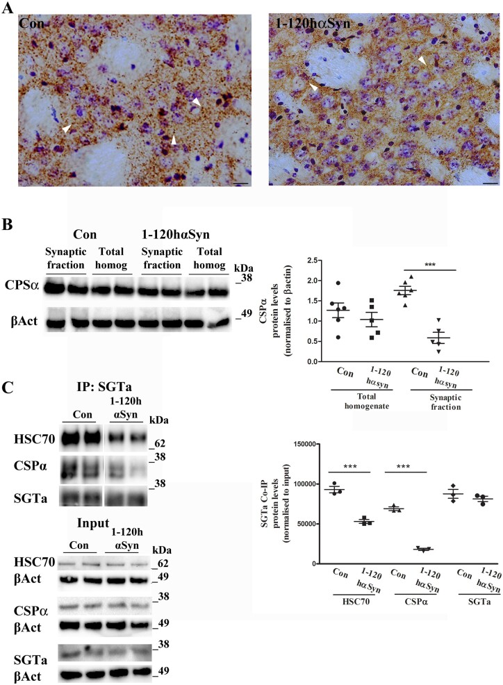
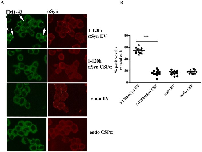
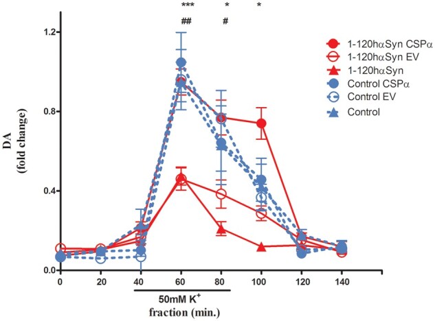
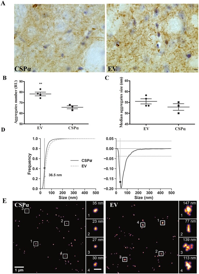
α-Synuclein aggregation at the synapse is an early event in Parkinson's disease and is associated with impaired striatal synaptic function and dopaminergic neuronal death. The cysteine string protein (CSPα) and α-synuclein have partially overlapping roles in maintaining synaptic function and mutations in each cause neurodegenerative diseases. CSPα is a member of the DNAJ/HSP40 family of co-chaperones and like α-synuclein, chaperones the SNARE complex assembly and controls neurotransmitter release. α-Synuclein can rescue neurodegeneration in CSPαKO mice. However, whether α-synuclein aggregation alters CSPα expression and function is unknown. Here we show that α-synuclein aggregation at the synapse is associated with a decrease in synaptic CSPα and a reduction in the complexes that CSPα forms with HSC70 and STGa. We further show that viral delivery of CSPα rescues in vitro the impaired vesicle recycling in PC12 cells with α-synuclein aggregates and in vivo reduces synaptic α-synuclein aggregates increasing monomeric α-synuclein and restoring normal dopamine release in 1-120hαSyn mice. These novel findings reveal a mechanism by which α-synuclein aggregation alters CSPα at the synapse, and show that CSPα rescues α-synuclein aggregation-related phenotype in 1-120hαSyn mice similar to the effect of α-synuclein in CSPαKO mice. These results implicate CSPα as a potential therapeutic target for the treatment of early-stage Parkinson's disease.

Keywords: CSPα; DNAJ chaperone family; Parkinson’s disease; synapse; α-synuclein.


Figures

Figure 1
Figure 1
CSPα expression is altered in the striatum of 12-month-old 1-120hαSyn mice. (A) CSPα staining in striatal sections from controls and 1-120hαSyn mice. Note the loss of CSPα puncta intensity in 1-120hαSyn mice compared to controls (arrowheads indicate typical CSPα puncta). Scale bar = 20 µm. (B) Left: Immunoblot for anti-CSPα and β-actin (βAct) in total homogenates and synaptic fractions from striata of control and 1-120hαSyn mice. Right: RI of CSPα levels normalized to β-actin in total homogenates and synaptic fractions. Data are presented as mean ± standard error of the mean (SEM) of n = 5–6 mice, ***P < 0.0001 (Student’s t-test). (C) Left: immunoblots of HSC70, CSPα, SGTa after co-immunoprecipitation with anti-SGTa antibody with non-hydrolysable ADP. CSPα and HSC70 complexes with SGTa are reduced in 1-120hαSyn mice compared to controls whereas input levels do not change (bottom, CSPα, HSC70 and SGTa and correspondent β-actin). Right: RI of HSC70, CSPα, SGTa relative to their input levels (total homogenates prior to co-immunoprecipitation). Values in graph represent n = 4–5 mice in three independent experiments; ***P < 0.001 (one-way ANOVA with Bonferroni’s multiple comparison test).
Figure 2
Figure 2
CSPα rescues the vesicle cycle impairment in PC12 cells expressing 1-120hαSyn. (A) FM1-43 dye fluorescence (left, green) and α-syn staining (right, red) in cells treated with empty vector (EV) or CSPα. Note the reduction in FM1-43 dye retention in cells stably expressing 1-120hαSyn treated with CSPα compared to their EV-treated counterpart (arrows). Treatment with CSPα or EV had no effect in non-transfected PC12 cells (endo). (B) Quantification of the number of cells that retained FM1-43 dye above basal levels in every treatment group. Values are mean ± SEM of n = 12 independent experiments. ***P < 0.001, two-way ANOVA with Bonferroni’s multiple comparison test. Scale bar = 10 µm.
Figure 3
Figure 3
CSPα restores dopamine release impairment in 1-120hαSyn mice. Striatal DA release after infusion of 50mM KCl for 60 min during in vivo microdialysis. Twelve-month-old 1-120hαSyn mice showed a significant reduction in DA release following KCl stimulation compared with controls (***P < 0.001, *P < 0.05). Values are expressed as fold change normalized to peak control mice value. Increase of CSPα following AAVCSPα injection restored DA release to control levels in 1-120hαSyn mice whereas treatment with AAVEV was ineffective (fractions: 60 min ##P < 0.01, 80 min #P < 0.05). No significant difference was observed in the 60 and 80 min fractions between CSPα and EV-treated or untreated control mice. Values are mean ± SEM of n = 5–7 1-120hαSyn and n = 5–6 control mice per treatment group, two-way ANOVA, Bonferroni’s multiple comparison test.
Figure 4
Figure 4
CSPα reduces α-syn aggregates and increases α-syn monomers in the striatum of 1-120hαSyn mice. (A) α-Syn immunostaining in the striata of CSPα and EV treated 1-120hαSyn mice. Scale bar = 10 µm. (B) Aggregate number (RU) versus monomeric α-syn in AAVEV (EV) and AAVCSPα (CSPα)-treated mice based on dSTORM analysis using anti-α-syn Syn1 antibody. A significant reduction in the number of aggregates versus monomeric α-syn is present after AAVCSPα injection. Two-tailed Student’s t-test **P⩽ ≤ 0.01; n = 4 mice per group. (C) Median size of α-syn aggregates showing no significant difference between EV-and CSPα-injected mice. Two-tailed Student’s t-test P > 0.01, n = 4 mice. (D) Cumulative histograms for α-syn species distribution in EV and CSPα-treated mice (left) and difference between the two distributions (right). Frequency values of α-syn species at the 36.5 nm intercept; <36.5 nm, EV 0.245, CSPα 0.411; >36.5 nm EV 0.754, CSPα 0.588. Kolmogorov-Smirnov test **P < 0.01, number of measured species EV: 4583, CSPα: 2574. (E) Representative dSTORM images of α-syn staining in CSPα or EV injected 1-120hαSyn mouse striatum. Note the difference in size of α-syn aggregates (single α-syn aggregates enlarged in boxed areas; scale bar insets = 200 nm).

References

    1. Lunati A, Lesage S, Brice A.. The genetic landscape of Parkinson's disease. Rev Neurol. 2018;174(9):628–643. - PubMed
    1. Spillantini MG, Crowther RA, Jakes R, Hasegawa M, Goedert M.. Alpha-Synuclein in filamentous inclusions of Lewy bodies from Parkinson's disease and dementia with Lewy bodies. Proc Natl Acad Sci U S A. 1998;95:6469–6473. - PMC - PubMed
    1. Spillantini MG, Schmidt ML, Lee VM, Trojanowski JQ, Jakes R, Goedert M.. Alpha-synuclein in Lewy bodies. Nature. 1997;388:839–840. - PubMed
    1. Baba M, Nakajo S, Tu PH, et al.Aggregation of alpha-synuclein in Lewy bodies of sporadic Parkinson's disease and dementia with Lewy bodies. Am J Pathol. 1998;152:879–884. - PMC - PubMed
    1. Crowther RA, Jakes R, Spillantini MG, Goedert M.. Synthetic filaments assembled from C-terminally truncated alpha-synuclein. FEBS Lett. 1998;436:309–312. - PubMed

Publication types