. 2021 May;47(5):81.
doi: 10.3892/ijmm.2021.4914. Epub 2021 Mar 24.

Endoplasmic reticulum stress/XBP1 promotes airway mucin secretion under the influence of neutrophil elastase

Affiliations

Endoplasmic reticulum stress/XBP1 promotes airway mucin secretion under the influence of neutrophil elastase

Xiaoyan Xu et al. Int J Mol Med. 2021 May.

Abstract

Endoplasmic reticulum (ER) stress is an important reaction of airway epithelial cells in response to various stimuli, and may also be involved in the mucin secretion process. In the present study, the effect of ER stress on neutrophil elastase (NE)‑induced mucin (MUC)5AC production in human airway epithelial cells was explored. 16HBE14o‑airway epithelial cells were cultured and pre‑treated with the reactive oxygen species (ROS) inhibitor, N‑acetylcysteine (NAC), or the ER stress chemical inhibitor, 4‑phenylbutyric acid (4‑PBA), or the cells were transfected with inositol‑requiring kinase 1α (IRE1α) small interfering RNA (siRNA) or X‑box‑binding protein 1 (XBP1) siRNA, respectively, and subsequently incubated with NE. The results obtained revealed that NE increased ROS production in the 16HBE14o‑cells, with marked increases in the levels of ER stress‑associated proteins, such as glucose‑regulated protein 78 (GRP78), activating transcription factor 6 (ATF6), phosphorylated protein kinase R‑like endoplasmic reticulum kinase (pPERK) and phosphorylated (p)IRE1α. The protein and mRNA levels of spliced XBP1 were also increased, and the level of MUC5AC protein was notably increased. The ROS scavenger NAC and ER stress inhibitor 4‑PBA were found to reduce ER stress‑associated protein expression and MUC5AC production and secretion. Further analyses revealed that MUC5AC secretion was also attenuated by IRE1α and XBP1 siRNAs, accompanied by a decreased mRNA expression of spliced XBP1. Taken together, these results demonstrate that NE induces ER stress by promoting ROS production in 16HBE14o‑airway epithelial cells, leading to increases in MUC5AC protein production and secretion via the IRE1α and XBP1 signaling pathways.

Keywords: endoplasmic reticulum stress; mucin 5AC; reactive oxygen species; inositol‑requiring kinase 1α; X‑box‑binding protein 1; respiratory inflammation.


Conflict of interest statement

The authors declare that they have no competing interests.

Figures

Figure 1
Figure 1
Proliferation of cells detected by MTT assay. Cells were exposed to various concentrations of NE (treatment groups were as follows: Negative control, and 25, 50, 100 and 200 ng/ml NE) for different periods of time (8, 12, 24, 36 and 48 h). Data are presented as the means ± SD (n=8 per group). *P<0.05 vs. 100 ng/ml NE group for 36 h;#P<0.01 vs. 100 ng/ml NE group for 48 h. NE, neutrophil elastase.
Figure 2
Figure 2
Effect of NE on ROS generation in 16HBE14o-cells. Cells were treated with various concentrations of NE for 24 h, and the generation of ROS was detected using a ROS detection kit. Data are presented as the means ± SD (n=8 per group). *P<0.05 vs. control (0 ng/ml); #P<0.01 vs. control (0 ng/ml). NE, neutrophil elastase; ROS, reactive oxygen species.
Figure 3
Figure 3
Expression of endoplasmic reticulum stress-related proteins in 16HBE14o-cells. Cells were exposed to 100 ng/ml NE or pre-treated with NAC and 4-PBA prior to NE exposure. GRP78, PERK, p-PERK, IRE1α, p-IRE1α, and ATF6 proteins were assayed by western blot analysis. (A) Relative GRP78 protein expression levels are presented as the ratio of GRP78 to β-actin. (B) Relative p-PERK protein expression levels are presented as the ratio of p-PERK to PERK, β-actin blots are for the loading control. (C) Relative p-IRE1α protein expression levels are presented as the ratio of pIRE1α to IRE1α, β-actin blots are for the loading control. (D) Relative ATF6 protein expression levels are presented as the ratio of ATF6 to β-actin. Data are presented as the means ± SD (n=4 samples per group). *P<0.01 vs. negative control; #P<0.01 vs. the NE group. NE, neutrophil elastase; NAC, N-acetylcysteine; ATF6, activating transcription factor 6; 4-PBA, 4-phenylbutyric acid; GRP78, glucose-regulated protein 78; IRE1α, inositol-requiring kinase 1α; p, phosphorylated; PERK, protein kinase R-like endoplasmic reticulum kinase.
Figure 4
Figure 4
Protein expression of IRE1α and XBP1 in 16HBE14o-cells. (A) Cells were transfected with IRE1α siRNA or negative control siRNA, IRE1α protein was assayed by western blot analysis. Relative IRE1α protein expression levels are presented as the ratio of IRE1α to β-actin. Data are presented as the means ± SD (n=4 samples per group). *P<0.01 vs. negative control and control siRNA (B) Cells were transfected with XBP1 siRNA or negative control siRNA, XBP1 protein was assayed by western blot analysis. Relative XBP1(s) protein expression levels are presented as the ratio of XBP1(s) to β-actin. Relative XBP1(u) protein expression levels are presented as the ratio of XBP1(u) to β-actin. Data are presented as the means ± SD (n=4 samples per group). *P<0.01 vs. negative control and control siRNA. (C) Cells were exposed to 100 ng/ml NE or pre-treated with NAC and 4-PBA prior to NE exposure. XBP1 protein was assayed by western blot analysis. Data are presented as the means ± SD (n=4 samples per group). *P<0.01 vs. negative control; #P<0.01 vs. the NE group. (D) Cells were transfected with IRE1α siRNA or XBP1 siRNA, and then incubated with 100 ng/ml NE. Relative XBP1(s) protein expression levels are presented as the ratio of XBP1(s) to β-actin. Relative XBP1(u) protein expression levels are presented as the ratio of XBP1(u) to β-actin. Data are presented as the means ± SD (n=4 samples per group). #P<0.01 vs. the NE + control siRNA group. IRE1α, inositol-requiring kinase 1α; NE, neutrophil elastase; NAC, N-acetylcysteine; 4-PBA, 4-phenylbutyric acid; XBP1, X-box-binding protein 1; XBP1(s), spliced XBP1; XBP1(u), unspliced XBP1.
Figure 5
Figure 5
Expression of spliced XBP1 mRNA in 16HBE14o-cells. (A) Cells were exposed to 100 ng/ml NE or pre-treated with NAC and 4-PBA prior to NE exposure. XBP1(s) was evaluated by reverse transcription-quantitative PCR. (B) Cells were transfected with IRE1α siRNA or XBP1 siRNA prior to exposure to NE. Data are presented as the means ± SD (n=4 samples per group). *P<0.01 vs. control; #P<0.01 vs. NE group. NE, neutrophil elastase; NAC, N-acetylcysteine; IRE1α, inositol-requiring kinase 1α; 4-PBA, 4-phenylbutyric acid; XBP1, X-box-binding protein 1; XBP1(s), spliced XBP1.
Figure 6
Figure 6
MUC5AC protein production in 16HBE14o-cells. (A) Cells were exposed to 100 ng/ml NE or pre-treated with NAC and 4-PBA prior to NE exposure. (B) Cells were transfected with IRE1α siRNA or XBP1 siRNA prior to exposure to NE. MUC5AC protein in the supernatant and in the cytoplasm were detected by ELISA. Data are presented as the means ± SD (n=4 samples per group). *P<0.01 vs. control; #P<0.01 vs. NE group. NE, neutrophil elastase; NAC, N-acetylcysteine; IRE1α, inositol-requiring kinase 1α; 4-PBA, 4-phenylbutyric acid; XBP1, X-box-binding protein 1; MUC, mucin.
Figure 7
Figure 7
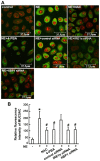
Intracellular MUC5AC expression in 16HBE14o-cells. (A) MUC5AC was detected by immunofluorescence and confocal microscopy. Cell nuclei were stained with propidium iodide (red fluorescence). (B) The fluorescence intensities are expressed as the IOD/area. IOD/area values were calculated using Image pro-plus-6.0 software. *P<0.01 vs. control; #P<0.01 vs. NE group. NE, neutrophil elastase; NAC, N-acetylcysteine; 4-PBA, 4-phenylbutyric acid; MUC, mucin.

Similar articles

Cited by

References

    1. Ma J, Rubin BK, Voynow JA. Mucins, mucus, and goblet cells. Chest. 2018;154:169–176. doi: 10.1016/j.chest.2017.11.008. - DOI - PubMed
    1. Bonser LR, Erle DJ. Airway mucus and asthma: The role of MUC5AC and MUC5B. J Clin Med. 2017;6:112. doi: 10.3390/jcm6120112. - DOI - PMC - PubMed
    1. Thornton DJ, Rousseau K, McGuckin MA. Structure and function of the polymeric mucins in airways mucus. Annu Rev Physiol. 2008;70:459–486. doi: 10.1146/annurev.physiol.70.113006.100702. - DOI - PubMed
    1. Bae CH, Na HG, Choi YS, Song SY, Kim YD. Clusterin induces MUC5AC expression via activation of NF-κB in human airway epithelial cells. Clin Exp Otorhinolaryngol. 2018;11:124–132. doi: 10.21053/ceo.2017.00493. - DOI - PMC - PubMed
    1. Song KS, Yoon JH, Kim KS, Ahn DW. c-Ets1 inhibits the interaction of NF-κB and CREB, and downregulates IL-1β-induced MUC5A coverproduction during airway inflammation. Mucosal Immunol. 2012;5:207–215. doi: 10.1038/mi.2011.67. - DOI - PMC - PubMed

MeSH terms