

Galectin‑3 facilitates the proliferation and migration of nasopharyngeal carcinoma cells via activation of the ERK1/2 and Akt signaling pathways, and is positively correlated with the inflammatory state of nasopharyngeal carcinoma
- PMID: 33760180
- PMCID: PMC7986014
- DOI: 10.3892/mmr.2021.12009
Galectin‑3 facilitates the proliferation and migration of nasopharyngeal carcinoma cells via activation of the ERK1/2 and Akt signaling pathways, and is positively correlated with the inflammatory state of nasopharyngeal carcinoma
Abstract
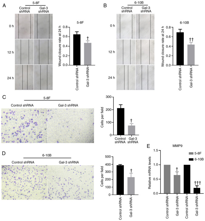
Nasopharyngeal carcinoma (NPC) is an epithelial carcinoma originating from the nasopharyngeal mucosal tissue and is highly prevalent in southeast Asia. Galectin‑3 (gal‑3) serves crucial roles in many cancers but its role in NPC remains to be elucidated. The aim of the present study was to investigate the role of gal‑3 in NPC. Immunohistochemistry and ELISA were used to determine the expression level of gal‑3 in patients with NPC or chronic rhinitis (CR). Gal‑3 short hairpin (sh)RNA was established to knockdown gal‑3 in 5‑8F and 6‑10B cells, allowing for the evaluation of the roles of gal‑3 in proliferation, migration and apoptosis in NPC cell lines. Immunohistochemistry staining of IL‑6 and IL‑8 was applied to access the inflammatory state of tumor tissues, and the correlation between the inflammatory state and gal‑3 was analyzed. The results demonstrated that gal‑3 was upregulated in patients with NPC compared with patients with CR. Knockdown of gal‑3 inhibited proliferation and migration in 5‑8F and 6‑10B cells, as well as promoted apoptosis in these cells. The expression levels of MMP‑9 and IL‑8 were also decreased in 5‑8F and 6‑10B cells after transfection with gal‑3 shRNA. A positive correlation was identified between the expression level of gal‑3 and the inflammatory state of NPC. The phosphorylation levels of ERK1/2 and Akt were downregulated after knockdown of gal‑3 in 5‑8F and 6‑10B cells. In conclusion, the expression level of gal‑3 was upregulated in patients with NPC and was positively correlated with the inflammatory state of NPC. The results suggested that gal‑3 promoted the proliferation and migration of 5‑8F and 6‑10B cells, while inhibiting the apoptosis of these cells. Moreover, activation of ERK1/2 and Akt may be the underlying mechanism of the effects of gal‑3 on NPC.
Keywords: galectin‑3; nasopharyngeal carcinoma; ERK1/2; Akt; inflammation.
Conflict of interest statement
The authors declare that they have no competing interests.
Figures


Similar articles
-
Expression of the Annexin A1 gene is associated with suppression of growth, invasion and metastasis of nasopharyngeal carcinoma.Mol Med Rep. 2014 Dec;10(6):3059-67. doi: 10.3892/mmr.2014.2656. Epub 2014 Oct 15. Mol Med Rep. 2014. PMID: 25322804
-
microRNA-613 exerts anti-angiogenic effect on nasopharyngeal carcinoma cells through inactivating the AKT signaling pathway by down-regulating FN1.Biosci Rep. 2019 Jul 10;39(7):BSR20182196. doi: 10.1042/BSR20182196. Print 2019 Jul 31. Biosci Rep. 2019. Retraction in: Biosci Rep. 2021 Sep 30;41(9):BSR-20182196_RET. doi: 10.1042/BSR-20182196_RET. PMID: 31189740 Free PMC article. Retracted.
-
MiR-214 Mediates Cell Proliferation and Apoptosis of Nasopharyngeal Carcinoma Through Targeting Both WWOX and PTEN.Cancer Biother Radiopharm. 2020 Oct;35(8):615-625. doi: 10.1089/cbr.2019.2978. Epub 2020 Feb 26. Cancer Biother Radiopharm. 2020. PMID: 32101017 Free PMC article.
-
Galectin-3 in metabolic disorders: mechanisms and therapeutic potential.Trends Mol Med. 2025 May;31(5):424-437. doi: 10.1016/j.molmed.2024.11.006. Epub 2024 Dec 16. Trends Mol Med. 2025. PMID: 39690058 Review.
-
Development of Galectin-3 Targeting Drugs for Therapeutic Applications in Various Diseases.Int J Mol Sci. 2023 May 1;24(9):8116. doi: 10.3390/ijms24098116. Int J Mol Sci. 2023. PMID: 37175823 Free PMC article. Review.
Cited by
-
Galectin‑3 blockade suppresses the growth of cetuximab‑resistant human oral squamous cell carcinoma.Mol Med Rep. 2021 Oct;24(4):685. doi: 10.3892/mmr.2021.12325. Epub 2021 Jul 30. Mol Med Rep. 2021. Retraction in: Mol Med Rep. 2024 Sep;30(3):152. doi: 10.3892/mmr.2024.13275. PMID: 34328195 Free PMC article. Retracted.
-
Revisiting Multi-Omics Data to Unravel Galectins as Prognostic Factors in Head and Neck Squamous Cell Carcinoma.Biomedicines. 2024 Feb 27;12(3):529. doi: 10.3390/biomedicines12030529. Biomedicines. 2024. PMID: 38540141 Free PMC article.
-
Role of Endogenous Galectin-3 on Cell Biology of Immortalized Retinal Pigment Epithelial Cells In Vitro.Int J Mol Sci. 2025 Aug 6;26(15):7622. doi: 10.3390/ijms26157622. Int J Mol Sci. 2025. PMID: 40806748 Free PMC article.
-
Tiliroside Combined with Anti-MUC1 Monoclonal Antibody as Promising Anti-Cancer Strategy in AGS Cancer Cells.Int J Mol Sci. 2023 Aug 22;24(17):13036. doi: 10.3390/ijms241713036. Int J Mol Sci. 2023. PMID: 37685842 Free PMC article.
References
MeSH terms
Substances
LinkOut - more resources
Full Text Sources
Other Literature Sources
Miscellaneous
