

Bruton's agammaglobulinemia tyrosine kinase (Btk) regulates TPA‑induced breast cancer cell invasion via PLCγ2/PKCβ/NF‑κB/AP‑1‑dependent matrix metalloproteinase‑9 activation
- PMID: 33760219
- PMCID: PMC7962096
- DOI: 10.3892/or.2021.8007
Bruton's agammaglobulinemia tyrosine kinase (Btk) regulates TPA‑induced breast cancer cell invasion via PLCγ2/PKCβ/NF‑κB/AP‑1‑dependent matrix metalloproteinase‑9 activation
Abstract
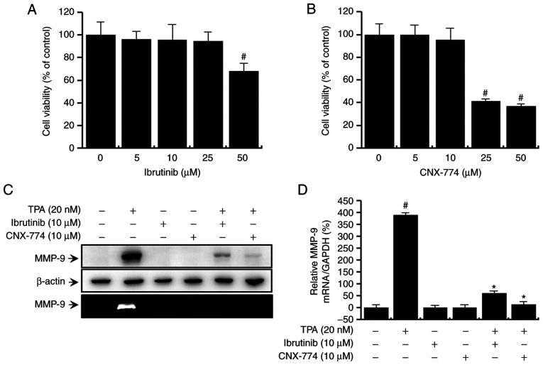
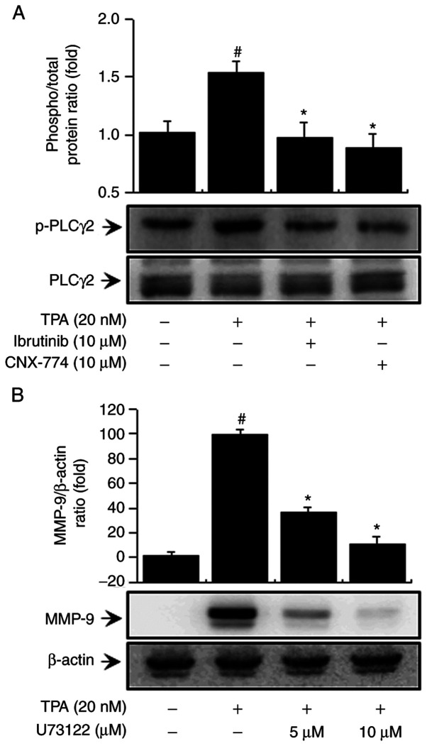
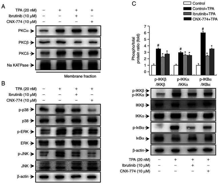
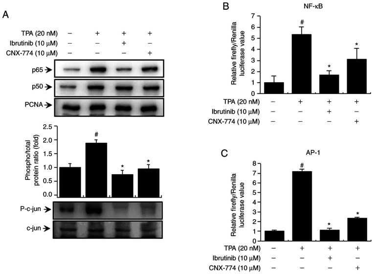
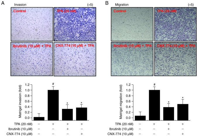
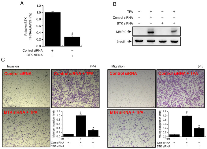
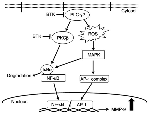
Bruton's agammaglobulinemia tyrosine kinase (BTK) is an important cytoplasmic tyrosine kinase involved in B‑lymphocyte development, differentiation, and signaling. Activated protein kinase C (PKC), in turn, induces the activation of mitogen‑activated protein kinase (MAPK) signaling, which promotes cell proliferation, viability, apoptosis, and metastasis. This effect is associated with nuclear factor‑κB (NF‑κB) activation, suggesting an anti‑metastatic effect of BTK inhibitors on MCF‑7 cells that leads to the downregulation of matrix metalloproteinase (MMP)‑9 expression. However, the effect of BTK on breast cancer metastasis is unknown. In this study, the anti‑metastatic activity of BTK inhibitors was examined in MCF‑7 cells focusing on MMP‑9 expression in 12‑O‑tetradecanoylphorbol‑13‑acetate (TPA)‑stimulated MCF‑7 cells. The expression and activity of MMP‑9 in MCF‑7 cells were investigated using quantitative polymerase chain reaction analysis, western blotting, and zymography. Cell invasion and migration were investigated using the Matrigel invasion and cell migration assays. BTK inhibitors [ibrutinib (10 µM), CNX‑774 (10 µM)] significantly attenuated TPA‑induced cell invasion and migration in MCF‑7 cells and inhibited the activation of the phospholipase Cγ2/PKCβ signaling pathways. In addition, small interfering RNA specific for BTK suppressed MMP‑9 expression and cell metastasis. Collectively, results of the present study indicated that BTK suppressed TPA‑induced MMP‑9 expression and cell invasion/migration by activating the MAPK or IκB kinase/NF‑κB/activator protein‑1 pathway. The results clarify the mechanism of action of BTK in cancer cell metastasis by regulating MMP‑9 expression in MCF‑7 cells.
Keywords: Bruton's agammaglobulinemia tyrosine kinase; matrix metalloproteinase‑9; protein kinase C; MCF‑7 cells.
Conflict of interest statement
The authors declare that they have no competing interests.
Figures
Similar articles
-
Role of NOX1 and NOX5 in protein kinase C/reactive oxygen species‑mediated MMP‑9 activation and invasion in MCF‑7 breast cancer cells.Mol Med Rep. 2024 Oct;30(4):188. doi: 10.3892/mmr.2024.13312. Epub 2024 Sep 2. Mol Med Rep. 2024. PMID: 39219290 Free PMC article.
-
Downregulation of matriptase suppresses the PAR‑2/PLCγ2/PKC‑mediated invasion and migration abilities of MCF‑7 breast cancer cells.Oncol Rep. 2021 Dec;46(6):247. doi: 10.3892/or.2021.8198. Epub 2021 Oct 5. Oncol Rep. 2021. PMID: 34608498 Free PMC article.
-
Noncatalytic Bruton's tyrosine kinase activates PLCγ2 variants mediating ibrutinib resistance in human chronic lymphocytic leukemia cells.J Biol Chem. 2020 Apr 24;295(17):5717-5736. doi: 10.1074/jbc.RA119.011946. Epub 2020 Mar 17. J Biol Chem. 2020. PMID: 32184360 Free PMC article.
-
Resistance Mutations to BTK Inhibitors Originate From the NF-κB but Not From the PI3K-RAS-MAPK Arm of the B Cell Receptor Signaling Pathway.Front Immunol. 2021 Jun 10;12:689472. doi: 10.3389/fimmu.2021.689472. eCollection 2021. Front Immunol. 2021. PMID: 34177947 Free PMC article. Review.
-
Review of the development of BTK inhibitors in overcoming the clinical limitations of ibrutinib.Eur J Med Chem. 2022 Feb 5;229:114009. doi: 10.1016/j.ejmech.2021.114009. Epub 2021 Nov 22. Eur J Med Chem. 2022. PMID: 34839996 Review.
Cited by
-
BTK Expression Level Prediction and the High-Grade Glioma Prognosis Using Radiomic Machine Learning Models.J Imaging Inform Med. 2024 Aug;37(4):1359-1374. doi: 10.1007/s10278-024-01026-9. Epub 2024 Feb 21. J Imaging Inform Med. 2024. PMID: 38381384 Free PMC article.
-
PLCG2, A Regulator of Lung Adenocarcinoma Proliferation and Migration Associated with Immune Infiltration.Curr Cancer Drug Targets. 2025;25(2):159-169. doi: 10.2174/0115680096307100240801095132. Curr Cancer Drug Targets. 2025. PMID: 39177130
-
Metronomic Chemotherapy for Metastatic Breast Cancer Treatment: Clinical and Preclinical Data between Lights and Shadows.J Clin Med. 2022 Aug 12;11(16):4710. doi: 10.3390/jcm11164710. J Clin Med. 2022. PMID: 36012949 Free PMC article. Review.
-
Bruton's Tyrosine Kinase Inhibitors (BTKIs): Review of Preclinical Studies and Evaluation of Clinical Trials.Molecules. 2023 Mar 6;28(5):2400. doi: 10.3390/molecules28052400. Molecules. 2023. PMID: 36903645 Free PMC article. Review.
-
Overexpression of Bruton Tyrosine Kinase Inhibits the Proliferation, Migration, and Invasion of Non-Small Cell Lung Cancer Cells.Anal Cell Pathol (Amst). 2023 Aug 18;2023:3377316. doi: 10.1155/2023/3377316. eCollection 2023. Anal Cell Pathol (Amst). 2023. PMID: 37638060 Free PMC article.
References
MeSH terms
Substances
LinkOut - more resources
Full Text Sources
Other Literature Sources
Medical
Research Materials
Miscellaneous
