Review
. 2021 Apr 27;15(4):5819-5837.
doi: 10.1021/acsnano.0c08510. Epub 2021 Mar 24.

From Protein Building Blocks to Functional Materials

Affiliations
Review

From Protein Building Blocks to Functional Materials

Yi Shen et al. ACS Nano. .

Abstract

Proteins are the fundamental building blocks for high-performance materials in nature. Such materials fulfill structural roles, as in the case of silk and collagen, and can generate active structures including the cytoskeleton. Attention is increasingly turning to this versatile class of molecules for the synthesis of next-generation green functional materials for a range of applications. Protein nanofibrils are a fundamental supramolecular unit from which many macroscopic protein materials are formed. In this Review, we focus on the multiscale assembly of such protein nanofibrils formed from naturally occurring proteins into new supramolecular architectures and discuss how they can form the basis of material systems ranging from bulk gels, films, fibers, micro/nanogels, condensates, and active materials. We review current and emerging approaches to process and assemble these building blocks in a manner which is different to their natural evolutionarily selected role but allows the generation of tailored functionality, with a focus on microfluidic approaches. We finally discuss opportunities and challenges for this class of materials, including applications that can be involved in this material system which consists of fully natural, biocompatible, and biodegradable feedstocks yet has the potential to generate materials with performance and versatility rivalling that of the best synthetic polymers.

Keywords: biomaterial; condensate; drug delivery; fiber; film; gel; microfluidic; protein; self-assembly.


Conflict of interest statement

The authors declare no competing financial interest.

Figures
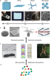
Figure 1
Figure 1
Assembly of proteins from the molecular level to functional materials. (a) Protein molecules can form bulk gels, films, fibers, and microgels.,,− The examples represent BLG fibrillar hydrogel, lysozyme fibrillar film, a fiber formed from fused in sarcoma (FUS) protein condensates, whey protein fibrillar fibers, and cyro-scanning electron microscopy (SEM) image of a lysozyme fibrillar microgel. The scale bars are 1 mm, 20 μm, 1 cm, and 20 μm, respectively. The figure is adapted with permissions from ref (2), Copyright 2017 Wiley-VCH; ref (5), ref (38), Copyright 2010 and 2020, Springer Nature; ref (39); ref (40), Copyright 2015, American Chemical Society. (b) Methods that can direct the assembly of the building blocks into materials. The examples include drop casting, 3D printing, microfluidics, and ultrasonication. The figure is adapted with permissions from ref (41), Copyright 2008, Wiley-VCH; ref (16), Copyright 2019, Springer Nature. (c) Nanoscale fibrils containing antiparallel and parallel β-sheets are the building blocks of the natural protein materials., The figure is adapted with permissions from ref (42), Copyright 2014, Springer Nature; ref (43), Copyright 2014, Wiley-VCH. (d) Nanofibrils’ formation is a result of protein molecule self-assembly.
Figure 2
Figure 2
Hydrogels and aerogels generated by protein nanofibrils and nanoparticles (NPs). (a) Gels made of β-lactoglobulin (BLG) fibrils and CaNPs with both Ca2+ and CaNPs working as cross-linkers to stabilize the network. The figure is adapted with permission from ref (2), Copyright 2017, Wiley-VCH. (b) Aerogels formed by BLG fibrils and Au crystals and AuNPs. Amyloid aerogel with imbedded gold crystals showed increasing conductivity when an increasing pressure is applied., (c) Aerogels made of BLG fibrils and AgNPs showed optical properties. Figures adapted with permissions from ref (6), Copyright 2015, Wiley-VCH; ref (7), Copyright 2017, Wiley-VCH.
Figure 3
Figure 3
Protein films and protein hybrid films. (a) Lysozyme was used to form the nanofibrils structured 2D film (top). This free-standing film with ordered nanostructures showed peaked birefringence signal at 45 deg under cross-polarized microscopy (bottom). The figure is adapted with permission from ref (5), Copyright 2010, Springer Nature. (b) Films made of silk and BLG fibrils showed transparent optical property and birefringence signal under cross-polarized microscopy. The figure is adapted with permission from ref (43), Copyright, 2014 Wiley-VCH. (c) Schematic illustration of purification process by amyloid–carbon film (left), and color change of Na2PdCl4 solution after filtration (middle). SEM image showing general and detailed structure of the amyloid-carbon film (right). The figure is adapted with permission from ref (3), Copyright 2016, Springer Nature.
Figure 4
Figure 4
Assembly of protein nanofibrils into microfibers. (a) Amyloid fibrils from lysozyme assembled into microfibers using the wet-spinning technique. An oppositely charged polysaccharide was used to generate hydrogel fibers. The figure is adapted with permission from ref (27), Copyright 2011, American Chemical Society. (b) Fibronectin fibers were generated by extrusion of proteins through nanopore membrane. The proteins undergo conformational changes during extrusion. The figure is adapted with permissions under Creative Commons CC BY license from ref (120), Copyright 2016, Oxford University Press (left); with permission from ref (119), Copyright 2015, American Chemical Society (right). (c) Microfluidic spinning of recombinant silk protein resulted in aligned and β-sheet rich fibers. Cross-polarized optical microscopy image shows birefringence in the microfiber (bottom). The figure is adapted with permission from ref (122), Copyright 2008, National Academy of Sciences, U.S.A. (d) Microfluidic device was used to generate fibers with aligned β-lactoglobulin nanofibrils. Left top panel shows the optical image of the microfluidic channel. Left bottom panels show the SEM image of the hydrogel fiber (left) and the confocal image of a fiber stained with Thioflavin T to visualize the protein fibrils (right). Right panels show the SEM image (left) and fluorescent image (right) of yarn-like fibers generated through microfluidic spinning. This figure is adapted with permission from, ref (48), Copyright 2019, Wiley-VCH.
Figure 5
Figure 5
(a,b) Protein microcapsules. Schematic representation of forming water-in-oil lysozyme-based microgels and oil-in-water microgels, respectively. Middle and right panels depict confocal and cryo-SEM micrographs of the corresponding microgel systems. Figure adapted with permission from ref (40), Copyright 2015, American Chemical Society. (c) A range of native silk microgel morphologies can be formed on the basis of the solution flow rates applied. From left to right: spheres, cylinders, short fibers, and thin fibers. Scale bar, 5 μm. Figure adapted with permission under a Creative Commons CC BY license from ref (9), Copyright 2017, Springer Nature. (d) SEM micrographs of regenerated silk fibroin microgels formed by varying the ethanol content from 20 to 40%. The panels on the right are magnified micrographs of the corresponding images on the left panels. The scale bars are 50 and 10 μm from left to right. Reprinted with permission from ref (138), Copyright 2019, Wiley-VCH. (e) Schematic showing the production of the hybrid inorganic/organic microgels and their subsequent use as antibacterial agents for a surgical site on a murine model. Reprinted with permission from ref (139), Copyright 2020, American Chemical Society. (f) Spatially inhomogeneous gelatin microgels. i, Microfluidic setup with heating accessories. ii, Gelatin microgels with different radial density. Green and red (magenta) nanospheres were premixed in the gelatin and enzyme (transglutaminase) solutions, respectively. Scale bar, 100 μm. iii, Gelatin microgels through versatile cross-linking regimes. Figure adapted with permission under a Creative Commons CC BY license from ref (47), Copyright 2020, Wiley-VCH.
Figure 6
Figure 6
(a) Schematic representation of the hybrid nano/microfluidic device used to generate water-in-oil nanodroplets and their subsequent formation into nanogels. (b) SEM micrographs of the silk nanoparticles and the corresponding size distribution histogram. (c) Confocal microscopy images of ovarian cancer cells (red) in the presence of silk nanoparticles (green). (d) 3D reconstruction of a single cancer cell which was imaged at different angles with respect to the z axis in order to show that the nanoparticles have penetrated the membrane and are well within the cell. Figures a–d adapted with permission under a Creative Commons CC BY license from ref (49), Copyright 2020, American Association for the Advancement of Science. (e–g) Schematic diagram of the capillary device used to form hierarchical emulsions with two different types of aqueous droplets and corresponding brightfield microscopy images of these double emulsions. Figure adapted with permission from ref (153), Copyright 2012, Royal Society of Chemistry. (h) Schematic representation of the 3-D devices used to generate hierarchical emulsions. (i) Collected droplets consisting of two, three, or four internal droplets. (h, i) Figure adapted with permission from ref (154), Copyright 2017, American Chemical Society.
Figure 7
Figure 7
(a) 3D construction of collagen to rebuild the components of human hearts. i, Schematic of printing acidified collagen solution into neutral support bath (pH 7.4). ii, Schematic of dual-material printing using a collagen ink and a high-concentration cell ink. iii, Cross-sectional view of the collagen heart, showing left and right ventricles and interior structures. Adapted with permission from ref (46), Copyright 2019, The American Association for the Advancement of Science. (b) Transendothelial migration of cancer cells in a microfluidic system at the collagen-channel barrier. i, Brightfield image of collagen-channel system. ii, Fluorescent images of the segmentation of the projected cancer cell areas (red contours) identified by cell number in the microvessels. Adapted with permission under a Creative Commons CC BY license from ref (167), Copyright 2018, Springer Nature. (c) i, Native tissues demonstrate a wide range of stiffness. ii, Stem cells interact with extracellular matrices, neighboring cells, and soluble factors. iii, Stem cells differentiate into various cell lineages when cultured on collagen-coating substrates with varying elasticity. Adapted with permission from ref (170), Copyright 2012, American Chemical Society. (d) i, Schematic of a triple-helical structure of self-assembling collagen protein. ii, Schematic of a mineralized collagen hydrogel. iii, SEM image of a collagen-based hydrogel. iv, Transmission electron microscopy (TEM) image of a collagen hydrogel network containing gold nanoparticles. Adapted with permission from ref (160), Copyright 2016, Wiley-VCH.
Figure 8
Figure 8
(a) Schematic diagram showing the introduction of preformed spherulites into the microfluidic device (top). To monitor the cantilever deflection, images were acquired through the glass slide at the bottom of the device (bottom). Reprinted with permission from ref (173). (b) Schematics of fibril growth within supercritical droplets and their buckling. Fibrils may pierce through the droplet because of an increase in their cross-section or through shrinkage of the droplet diameter (top). Reprinted with permission from ref (179). Copyright 2016, Springer Nature. Bright-field time-lapse microscopy of FF tube self-assembly and unbuckling due to droplet shrinkage (bottom). Reprinted with permission from ref (53), Copyright 2018, with permission from the Royal Society of Chemistry. (c) Scheme displaying the jet-like release of spheres through droplets and phase diagram indicating the condition for jetting (top). High magnification images of a single microdroplet releasing its nanosphere content during evaporation. Scale bar represents 30 μm. When the nanosphere assembly inside the droplet has reached a critical mass, nanosphere release could be detected within seconds (bottom). Reprinted with permission from ref (180), Copyright 2018, American Chemical Society. (d) Mechanism of budding-like division of w/w emulsion droplets mediated by protein nanofibrils. Schematic diagram (top) and fluorescence microscope images (bottom) describing the mechanistic steps in the budding-like division of w/w droplets. The fibril network (stained green) contracts and phase-separates from the remaining liquid phase through a dewetting transition. In this transition, the as-formed protrusions coalesce (as pinpointed by the white arrows) until a sufficient amount of fibrils adsorbs at the w/w interface to stabilize daughter droplets. Complete fission of dextran-rich subdroplets (faked red color) is observed after total decomposition of the fibril networks in the PEG-rich continuous phase. Scale bars, 100 μm. Figure reprinted with permission under a Creative Commons CC BY license from ref (156), Copyright 2018, Springer Nature.

References

    1. Teng Z.; Xu R.; Wang Q. RSC Advances Beta-Lactoglobulin-Based Encapsulating Systems as Emerging Bioavailability Enhancers for Nutraceuticals : A Review. RSC Adv. 2015, 5 (2013), 35138–35154. 10.1039/C5RA01814E. - DOI
    1. Shen Y.; Nyström G.; Mezzenga R. Amyloid Fibrils Form Hybrid Colloidal Gels and Aerogels with Dispersed CaCO 3 Nanoparticles. Adv. Funct. Mater. 2017, 27 (45), 1700897.10.1002/adfm.201700897. - DOI
    1. Bolisetty S.; Mezzenga R. Amyloid–Carbon Hybrid Membranes for Universal Water Purification. Nat. Nanotechnol. 2016, 11 (4), 365–371. 10.1038/nnano.2015.310. - DOI - PubMed
    1. Shen Y.; Posavec L.; Bolisetty S.; Hilty F. M.; Nyström G.; Kohlbrecher J.; Hilbe M.; Rossi A.; Baumgartner J.; Zimmermann M. B.; Mezzenga R. Amyloid Fibril Systems Reduce, Stabilize and Deliver Bioavailable Nanosized Iron. Nat. Nanotechnol. 2017, 12 (7), 642–647. 10.1038/nnano.2017.58. - DOI - PubMed
    1. Knowles T. P. J.; Oppenheim T. W.; Buell A. K.; Chirgadze D. Y.; Welland M. E. Nanostructured Films from Hierarchical Self-Assembly of Amyloidogenic Proteins. Nat. Nanotechnol. 2010, 5 (3), 204–207. 10.1038/nnano.2010.26. - DOI - PMC - PubMed

Publication types