. 2021 Mar 24;15(3):e0008352.
doi: 10.1371/journal.pntd.0008352. eCollection 2021 Mar.

Distinct features of the Leishmania cap-binding protein LeishIF4E2 revealed by CRISPR-Cas9 mediated hemizygous deletion

Affiliations

Distinct features of the Leishmania cap-binding protein LeishIF4E2 revealed by CRISPR-Cas9 mediated hemizygous deletion

Nofar Baron et al. PLoS Negl Trop Dis. .

Abstract

Leishmania parasites cycle between sand-fly vectors and mammalian hosts adapting to alternating environments by stage-differentiation accompanied by changes in the proteome profiles. Translation regulation plays a central role in driving the differential program of gene expression since control of gene regulation in Leishmania is mostly post-transcriptional. The Leishmania genome encodes six eIF4E paralogs, some of which bind a dedicated eIF4G candidate, and each eIF4E is assumed to have specific functions with perhaps some overlaps. However, LeishIF4E2 does not bind any known eIF4G ortholog and was previously shown to comigrate with the polysomal fractions of sucrose gradients in contrast to the other initiation factors that usually comigrate with pre-initiation and initiation complexes. Here we deleted one of the two LeishIF4E2 gene copies using the CRISPR-Cas9 methodology. The deletion caused severe alterations in the morphology of the mutant cells that became round, small, and equipped with a very short flagellum that did not protrude from its pocket. Reduced expression of LeishIF4E2 had no global effect on translation and growth, unlike other LeishIF4Es; however, there was a change in the proteome profile of the LeishIF4E2(+/-) cells. Upregulated proteins were related mainly to general metabolic processes including enzymes involved in fatty acid metabolism, DNA repair and replication, signaling, and cellular motor activity. The downregulated proteins included flagellar rod and cytoskeletal proteins, as well as surface antigens involved in virulence. Moreover, the LeishIF4E2(+/-) cells were impaired in their ability to infect cultured macrophages. Overall, LeishIF4E2 does not behave like a general translation factor and its function remains elusive. Our results also suggest that the individual LeishIF4Es perform unique functions.


Conflict of interest statement

The authors have declared that no competing interests exist.

Figures

Fig 1
Fig 1. Multiple sequence alignment of the LeishIF4E2.
(A)The ORFs of LeishIF4E2 from different Leishmania and Trypanosoma specis along with mammalian eIF4E1 were aligned using Jalview (2.10.5). The sequences were retrieved from L. mexicana (L. mex, LmxM.19.1480), L. major (L. maj, LmjF.19.1500); L. donovani, (L. don, LdBPK_191520.1), T. brucei (T. bruc, Tb927.10.16070) and T. congolense (T. cong, TcIL3000_0_03820). The alignment file was saved in FASTA format. The final alignment showing the predicted secondary structure of LeishIF4E2 was developed using the downloaded PDB file (5EHC, DOI: 10.2210/pdb5EHC/pdb) of Homo sapiens eIF4E1 (https://www.rcsb.org/), along with the FASTA alignment file, using the online ESPript 3 tool [60]. Secondary structure elements of the aligned sequences are (α: alpha helices, ƞ: 310-helix, β: beta-strands, TT: strict β-turns). White letters over a red background indicate identical residues and red letters over a white background indicate sequence similarity.
Fig 2
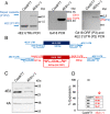
Fig 2. CRISPR-Cas9 mediated hemizygous deletion of LeishIF4E2.
(A) Diagnostic PCR was performed to confirm the deletion of single allele of LeishIF4E2. Genomic DNA was extracted from L. mexicana Cas9/T7 cells and from the LeishIF4E2(+/-) mutant. PCR was performed using different combinations of primers derived from the LeishIF4E2 5’ UTR (Forward) and 3’ UTR (Reverse) (P1/P2 –left panel); G418 ORF (Forward) and Reverse (P3/P4, middle panel); and a primer set derived from the G418 resistance gene, forward and the 3’ UTR (Reverse) (P3/P2). (B) Schematic representation of LeishIF4E2 locus and primers (represented by arrows). The PCR was applied to test the presence or absence of the LeishIF4E2 gene and the G418 resistance marker. Primers derived from the LeishIF4E2 UTRs are shown in blue and primers derived from the ORF of G418 are shown in red. (C) Western analysis monitoring the protein level of LeishIF4E2 in the LeishIF4E2(+/-) mutant and in Cas9/T7 control was performed using LeishIF4E2 specific antibodies. LeishIF4A-1 served as a loading control. (D) Dot plot representing the densitometry analysis of the LeishIF4E2 protein levels in the LeishIF4E2(+/-) mutant and in the Cas9/T7 controls.
Fig 3
Fig 3. The LeishIF4E2(+/-) mutant shows altered promastigote morphology.
(A) Mid-log phase (Day 2) promastigotes of WT, Cas9/T7 expressers, LeishIF4E2(+/-) cells, and LeishIF4E2 add-back cells were fixed with 2% paraformaldehyde and visualized by phase contrast microscopy at 100x magnification. WT, Cas9/T7 expressing cells showed normal promastigote morphology while LeishIF4E2(+/-) were round with reduced flagellar length. (B) All the cell lines were stained with 20 μg/mL propidium iodide (PI) for 30 min and cell viability was analyzed using the ImageStream X Mark II Imaging flow cytometer (Millipore). 20,000 cells were analyzed for each sample and percent viable cells were determined. (C). The circularity of single, viable and focused cells from each of the cell lines was quantified using flow cytometry, and is shown as percentage of the total number of cells measured. Data from three independent experiments are shown.
Fig 4
Fig 4. Global translation and growth are not altered in the LeishIF4E2(+/-) mutant cells.
(A) LeishIF4E2(+/-) cells, WT, Cas9/T7 expressing cells and transgenic parasites expressing the full length LeishIF4E21-281 and its truncated form devoid of the C-terminus, LeishIF4E21-217 were incubated with 1 μg/mL puromycin for 1 hr. Cycloheximide treated cells were used as a negative control for complete inhibition of translation. Puromycin treated cells were lysed and resolved over 12% SDS-PAGE and subjected to western analysis using antibodies against puromycin. (B) Ponceau staining was used to indicate comparable protein loads. (C) Densitometry analysis of puromycin incorporation in the different cells lines, was compared to WT cells (considered as 100%). Data from all three independent experiments are represented. (D) All cells were cultured at 25 oC in M199 containing essential nutrients. Cell counts were monitored daily during 4 consecutive days. The curves were obtained from three independent assays, error bars are also marked. Growth curves of WT cells are shown in yellow, Cas9/T7 are in brown, the LeishIF4E2(+/-) mutant cells are in green, cells expressing the full length LeishIF4E2 1–281 are in purple, and the truncated LeishIF4E21-217 cells are in blue.
Fig 5
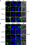
Fig 5. The LeishIF4E2(+/-) mutant cells show reduced infectivity to macrophages.
Stationary phase (Day 5) L. mexicana WT, Cas9/T7 expressing cells, LeishIF4E2(+/-) mutant cells and the add-back LeishIF4E2 line were pre-stained with CFSE (green), counted, washed and used to infected RAW 264.7 macrophages, at a ratio of 10:1 for one hour. The cells were then washed to remove unattached parasites, and the macrophages were cultured for 1h (A) or 24h (B) at 37°C. Macrophage nuclei were stained with DAPI and the infected macrophage slides were processed for confocal microscopy, showing a Z-projection produced by Image J software. Fields containing 100 cells were further evaluated to quantify the infection. The scale bar represents 5 μm.
Fig 6
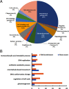
Fig 6. The categorized proteome of the upregulated proteins in LeishIF4E2(+/-) mutant cells as compared to Cas9/T7 control cells.
The proteomic content of LeishIF4E2(+/-) and Cas9/T7 cells was determined by LC-MS/MS analysis, in triplicates. Raw mass spectrometric data were analyzed and quantified using the MaxQuant software and the peptide data were searched against the annotated L. mexicana proteins listed in TriTrypDB. The summed intensities of the peptides that served to identify the individual proteins were used to quantify changes in the proteomic content of specific proteins. Statistical analysis was done using the Perseus software. Proteins that were upregulated in the LeishIF4E2(+/-) mutant by 1.7 fold as compared to Cas9/T7 cell extracts, with p<0.05 are shown. (A) Proteins in LeishIF4E2(+/-) that were upregulated (>1.7 fold) as compared to Cas9/T7 extracts were clustered manually into functional categories. The pie chart represents the summed intensities of upregulated proteins in each category, in the LeishIF4E2(+/-) mutant. Numbers in brackets indicate the number of proteins in each category, % represent their summed relative intensity in the analysis. (B) Enriched proteins were classified by the GO enrichment tool in TriTrypDB, based on Biological Function. The threshold for the calculated enrichment of proteins based on their GO terms was set for 2.5 fold, with p<0.05. This threshold eliminated most of the general groups that represented parental GO terms. GO terms for which only a single protein was annotated were filtered out as well.
Fig 7
Fig 7. LeishIF4E2(+/-) mutant cells easily transform to axenic amastigote-like cells.
(A) Promastigotes of the LeishIF4E2(+/-) mutant, control WT, and Cas9/T7 expressing cells grown under normal conditions are shown. (B) Morphology of cells transferred to conditions that induce differentiation to axenic-amastigotes (33°C/pH 5.5) during four days. Images were captured at 100x magnification with a Zeiss Axiovert 200M microscope equipped with AxioCam HRm CCD camera. The scale bar is 10 μm. (C) Shared upregulated proteins in the mutant LeishIF4E2(+/-) promastigotes (compared to Cas9/T7 cells) and in published amastigotes proteome. The total protein of the LeishIF4E2(+/-) mutant promastigotes was compared to the proteome of Cas9/T7 cells. The list of upregulated proteins was further compared with the proteins enriched in the amastigote proteome of the virulent L. amazonensis PH8 strain (de Rezende et al, PLOS NTD 2017) and of L. mexicana amastigotes (Paape et al, Mol Cell Prot 2008). L amazonensis gene IDs were converted to L. mexicana, for the sake of comparison.

Similar articles

Cited by

References

    1. Sacks DL, Perkins PV. Identification of an infective stage of Leishmania promastigotes. Science. 1984;223(4643):1417–9. 10.1126/science.6701528 - DOI - PubMed
    1. Zilberstein D, Shapira M. The role of pH and temperature in the development of Leishmania parasites. Ann Rev Microbiol. 1994;48(1):449–70. 10.1146/annurev.mi.48.100194.002313 - DOI - PubMed
    1. Haile S, Papadopoulou B. Developmental regulation of gene expression in trypanosomatid parasitic protozoa. Currt Opin Microbiol. 2007;10(6):569–77. 10.1016/j.mib.2007.10.001 - DOI - PubMed
    1. Sonenberg N, Hinnebusch AGJC. Regulation of translation initiation in eukaryotes: mechanisms and biological targets. Cell. 2009;136(4):731–45. 10.1016/j.cell.2009.01.042 - DOI - PMC - PubMed
    1. Peter D, Igreja C, Weber R, Wohlbold L, Weiler C, Ebertsch L, et al.. Molecular architecture of 4E-BP translational inhibitors bound to eIF4E. Molecular Cell. 2015;57(6):1074–87. 10.1016/j.molcel.2015.01.017 - DOI - PubMed

Publication types

MeSH terms

Substances

LinkOut - more resources