. 2021 Sep;259(9):2559-2571.
doi: 10.1007/s00417-021-05156-6. Epub 2021 Mar 24.

The extracellular matrix complexity of idiopathic epiretinal membranes and the bilaminar arrangement of the associated internal limiting membrane in the posterior retina

Affiliations

The extracellular matrix complexity of idiopathic epiretinal membranes and the bilaminar arrangement of the associated internal limiting membrane in the posterior retina

Annalisa Altera et al. Graefes Arch Clin Exp Ophthalmol. 2021 Sep.

Abstract

Purpose: To study the composition of the internal limiting membrane (ILM) of the retina, the extracellular matrix (ECM) of idiopathic epiretinal membranes (iERMs), and the relationships occurring between the two membranes.

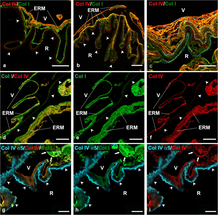
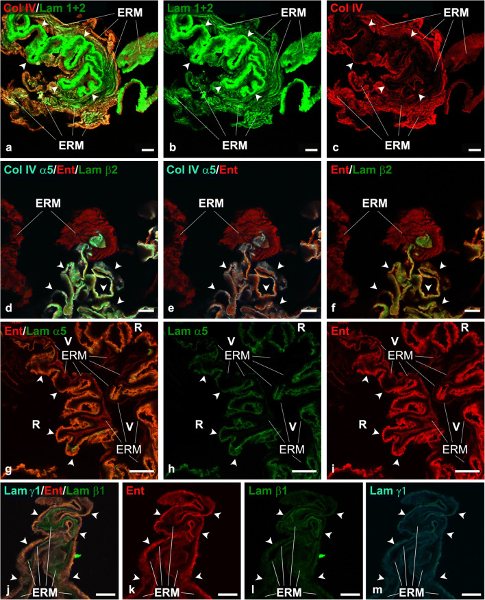
Methods: Forty-six iERMs, 24 of them associated with the ILM, were collected and included in this study. The investigation has been carried out by immunofluorescence and confocal microscopy on glutaraldehyde- and osmium-fixed epon-embedded samples and on frozen samples. Sections were double or triple labelled with antibodies against vimentin; collagens I, III, IV, α5(IV), and VI; laminin 1 + 2; laminin α2-, α4-, α5-, β1-, β2-, β3-, γ1-, and γ2-chains; entactin; and fibronectin.

Results: iERM thickness was not uniform. Almost 14% of iERMs showed thickenings due to folding of their ECM component under the cell layer. The vitreal side of iERMs was often shorter than the attached ILM. In this case, the ILM resulted folded under the iERM. ILMs contained laminin 111; laminin α2-, α5-, β1-, β2-, and γ1-chains; entactin; collagens I; α5(IV); [α1(IV)]2α2(IV); and VI. Laminins, entactin, and α5(IV) were gathered on the retinal half of the ILM, whereas collagens [α1(IV)]2α2(IV) and I were restricted to the vitreal side. Collagen VI was detected on both sides of the ILM. iERMs expressed laminin 111, collagens III, [α1(IV)]2α2(IV) and VI, entactin, and fibronectin. Entactin co-localized with laminins and collagen IV.

Conclusions: Analysis of laminins and collagen chain expression indicates that ILM contains laminin 111 (former laminin 1), laminin 521 (former laminin 11), laminin 211 (former laminin 2), collagen [α1(IV)]2α2(IV), and collagen α3(IV)α4(IV)α5. In contrast, iERMs express only collagen [α1(IV)]2α2(IV) and laminin 111. In addition, both iERMs and ILMs contain entactin. The presence of three major constituents of the basement membranes co-localized together in iERMs is suggestive for a deranged process of basement membrane formation which fails to assemble properly. In view of the many interactions occurring among its proteins, the ECM of either the iERMs or the ILMs can account for their reciprocal adhesiveness. In addition, the peculiar deposition of the ECM observed in some samples of iERM is suggestive for its involvement in the formation of macular puckers.

Keywords: Collagen IV; Epiretinal membrane; Extracellular matrix; Internal limiting membrane; Laminin; Vitreoretinal interface.


Conflict of interest statement

The authors declare that they have no conflict of interest

Figures

Fig. 1
Fig. 1
General arrangement of the ECM in iERMs. a–f Two iERMs, one of them associated with the ILM, double-labelled with anti-collagen I (green) and anti-collagen IV (red). a–c Idiopathic ERM provided with a very thick layer of ECM proteins. d–f Idiopathic ERM provided with a very thin layer of ECM proteins. The iERM has a closely associated ILM which is visible only for a very faint reddish background shadow (arrowheads). g–l Two iERMs triple-labelled with anti-vimentin (cyan), anti-collagen I (green) and anti-collagen IV (red) antibodies. g–i Knot-like local thickenings of the iERM, resulting from its folding under the cell layer, can be observed with a certain frequency. j–l Less pronounced foldings under the cell layer are likely knots-like accumulations in the process of formation. V vitreal side, R retinal side; magnification bars = 20 μm
Fig. 2
Fig. 2
Relationships between iERMs and ILMs and differential distribution of collagen IV isoforms. a–f Four iERMs, three of them associated with the ILM, double-labelled with anti-collagen I (green) and anti-collagen IV (red). g–i One iERM associated with the ILM triple-labelled with the anti-α5(IV) (cyan), anti-collagen I (green) and anti-collagen IV (red). In addition to a perfectly parallel and adherent course, iERMs can display a more complex pattern of association with the ILM: a Idiopathic ERMs can be seen partially detached from the ILM (arrowheads), bridging different areas of the ILM. The result is the folding of the ILM under the iERM; b Idiopathic ERMs can adhere to the ILM (arrowheads) though their thickness is not uniform. The result is again the folding of the ILM. On the right side of b, the pattern of iERM/ILM association is similar to the sample shown in a, with a partially detached iERM. c In other cases, iERMs can have a lamellar and looser arrangement. In such circumstances, it is possible to observe the decreasing length of the collagen lamellae from the retinal side of the membrane towards the vitreal side. The progressive shortening of the lamellae is highlighted by the dotted line. d–f Idiopathic ERM partially associated with the ILM (arrowheads). The ILM is double-labelled on its vitreal rim (the one facing the iERM). g–i The iERM (arrows) and the vitreal rim of the ILM are double-labelled with anti-collagen IV and anti-collagen I antibodies. In contrast, the antibody against the α5-chain of collagen IV labels exclusively the retinal half of the ILM (arrowheads). The absence of co-localization between anti-α5(IV) and anti-collagen IV antibodies indicates that the latter antibody recognizes the ([α1(IV)]2α2(IV) isoform, the only isoform that does not contain the α5-chain. Where optimally cross-sectioned, ILM also shows an unlabelled stripe between the anti-collagen I/IV double labelled vitreal rim and the anti-α5(IV) immunoreactive retinal half ERM = Epiretinal membrane; V vitreal side; R retinal side. a-f) Magnification bars = 20 μm. g-i) Magnification bars = 10 μm.
Fig. 3
Fig. 3
Chain-specific laminin expression in ILM and iERMs. a–c Idiopathic ERM/ILM double labelled with anti-type IV collagen (red) and anti laminin 1+2 (green) antibodies. The iERM expresses both antigens. The ILM (arrowheads) shows an exuberant immunoreactivity for the anti-laminin 1+2 antibody. d–f Idiopathic ERM/ILM triple labelled with anti-α5(IV) (cyan), anti-entactin (red), and anti-laminin β2 (green) antibodies. The α5 chain of collagen IV and the laminin β2 are expressed exclusively in the ILM (arrowheads). Entactin, in contrast, is expressed either in the iERM or in the ILM. g–i Idiopathic ERM/ILM double labelled with anti-entactin (red) and anti-laminin α5 (green) antibodies. Whereas laminin α5 is present almost exclusively in the ILM (arrowheads), entactin is expressed in both membranes. V vitreal side of the iERM, R retinal side of the ILM. j–m Idiopathic ERM/ILM triple labelled with anti-laminin γ1 (cyan), anti-entactin (red), and anti-laminin β1 (green) antibodies. Though with a different degree of intensity, all antigens are expressed either in the ILM (arrowheads) or in the iERM. Entactin and the laminin γ1 are prevalently located in the ILM, whereas the laminin β1 fluorescence is more evident in the iERM. ERM epiretinal membrane. a–f Magnification bars = 10 μm. gm Magnification bars = 20 μm
Fig. 4
Fig. 4
Expression of fibronectin, collagen III, and collagen IV in iERM and ILM. a–c Idiopathic ERM/ILM triple labelled with anti-α5(IV) (cyan), anti-collagen IV ([α1(IV)]2α2(IV) isoform) (red), and anti-fibronectin (green) antibodies. The iERM expresses both fibronectin and the [α1(IV)]2α2(IV) isoform of collagen IV though their distribution co-localizes only focally. The α5 chain of collagen IV is restricted to the ILM. Note the different localization of the [α1(IV)]2α2(IV) isoform of collagen IV and the α5 chain of same collagen in the ILM. d-f) Idiopathic ERM/ILM triple labelled with vimentin (cyan), anti-collagen IV ([α1(IV)]2α2(IV) isoform) (red), and anti-collagen III (green) antibodies. Collagens III and IV co-localize perfectly in the iERM. In contrast, the ILM, which is well visible where the iERM is detached, shows only the usual weak immunoreactivity for the [α1(IV)]2α2(IV) isoform of collagen IV on its vitreal rim. Note how cells are gathered on the vitreal side of the ERM and the relationships occurring between the ILM and the ERM with the latter which is shorter and bridges more distant points of the ILM. R retinal side, V vitreal side. g–l Idiopathic ERM/ILM triple labelled with vimentin (cyan), anti-collagen IV ([α1(IV)]2α2(IV) isoform) (red), and anti-collagen VI (green) antibodies. The [α1(IV)]2α2(IV) isoform of collagen IV and collagen VI co-localizes in the iERM though there are areas labelled exclusively anti-collagen IV or anti-collagen VI antibodies. In general, however, collagen VI seems more represented (particularly in jl). The ILM is labelled on both the retinal and the vitreal edges. Where the iERM is detached from the ILM, collagen VI fluorescence is visible on the retinal edge of the ILM co-localizing perfectly with the [α1(IV)]2α2(IV) isoform of collagen IV. R retinal side, V vitreal side. ILM inner limiting membrane, ERM epiretinal membrane. ai Magnification bars = 20 μm. jl Magnification bars = 10 μm

Similar articles

Cited by

References

    1. Bertelli E, Kondova I, Lagermann JAM. The Retina. In: Bertelli E, editor. Anatomy of the eye and human visual system. Padua: Piccin Nuova Libraria; 2019. pp. 187–247.
    1. Kohno T, Sorgente N, Ishibashi T, Goodnight R, Ryan SJ. Immunofluorescence studies of fibronectin and laminin in the human eye. Invest Ophthalmol Vis Sci. 1987;28:506–514. - PubMed
    1. Ponsioen TL, van Luyn MJA, van der Worp RJ, van Meurs JC, Hooymans JMM, Los LI. Collagen distribution in the human vitreoretinal interface. Invest Ophthalmol Vis Sci. 2008;49:4089–4095. doi: 10.1167/iovs.07-1456. - DOI - PubMed
    1. Clark SJ, Keenan TD, Fielder HL, Collinson LJ, Holley RJ, van Kuppevelt TH MCL, Day AJ, Bishop PN. Mapping the differential distribution of glycosaminoglycans in the adult human retina, choroid, and sclera. Invest Ophthalmol Vis Sci. 2011;52:6511–6521. doi: 10.1167/iovs.11-7909. - DOI - PMC - PubMed
    1. Keenan TDL, Clark SJ, Unwin RD, Ridge LA, Day AJ, Bishop PN. Mapping the differential distribution of proteoglycan core proteins in the adult human retina, choroid, and sclera. Invest Ophthalmol Vis Sci. 2012;53:7528–7538. doi: 10.1167/iovs.12-10797. - DOI - PMC - PubMed

LinkOut - more resources