Review
. 2021 Mar 24;23(4):50.
doi: 10.1007/s11886-021-01478-3.

PET Stress Testing with Coronary Flow Capacity in the Evaluation of Patients with Coronary Artery Disease and Left Ventricular Dysfunction: Rethinking the Current Paradigm

Affiliations
Review

PET Stress Testing with Coronary Flow Capacity in the Evaluation of Patients with Coronary Artery Disease and Left Ventricular Dysfunction: Rethinking the Current Paradigm

Robert M Bober et al. Curr Cardiol Rep. .

Abstract

Purpose of review: Cardiomyopathy with underlying left ventricular (LV) dysfunction is a heterogenous group of disorders that may be present with, and/or secondary to, coronary artery disease (CAD). The purpose of this review is to demonstrate, via case illustrations, the benefits offered by cardiac positron-emission tomography (PET) stress testing with coronary flow capacity (CFC) in the evaluation and treatment of patients with left ventricular (LV) dysfunction and CAD.

Recent findings: CFC, a metric that is increasing in prominence, represents the integration of several absolute perfusion metrics into clinical strata of CAD severity. Our prior work has demonstrated improvement in regional perfusion metrics as a result of revascularization to territories with severe reduction in CFC. Conversely, when CFC is adequate, there is no change in regional perfusion metrics following revascularization, despite angiographically severe stenosis. Furthermore, Gould et al. demonstrated decreased rates of myocardial infarction and death following revascularization of myocardium with severely reduced CFC, with no clinical benefit observed following revascularization of patients with preserved CFC. In a series of cases, we present pre-revascularization and post-revascularization PET scans with perfusion metrics in patients with LV dysfunction and CAD. In these examples, we demonstrate improvement in LV function and perfusion metrics following revascularization only in cases where baseline CFC is severely reduced. PET with CFC offers unique guidance regarding revascularization in patients with reduced LV function and CAD.

Keywords: Cardiac PET; Coronary flow capacity; Ischemic cardiomyopathy; Left ventricular dysfunction; Myocardial blood flow.


Conflict of interest statement

Robert M. Bober reports grants from Bracco Diagnostics, Inc. (ClinicalTrials.gov Identifier: NCT02931331) during the conduct of the study; and personal fees from Bracco Diagnostics, Inc., outside of the submitted work. Selim R. Krim reports personal fees from Abbott, outside the submitted work. The other authors declare that they have no conflict of interest.

Figures

Fig. 1
Fig. 1
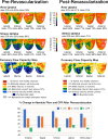
Pre-revascularization and post-revascularization rest and stress relative perfusion images (1st and 2nd rows, respectively) and coronary flow capacity maps (3rd row) of a patient with cardiomyopathy and associated perfusion abnormalities secondary to severe CAD. The patient history and revascularization specifics are located within the text. The graphs demonstrate the percent change from the baseline of regional resting MBF (blue bars), stress MBF (orange bars) and CFR (gray bars). The black horizontal lines denote 20% day-to-day normal variability of absolute perfusion. A change in absolute flow metrics outside of the black horizontal lines likely represents procedural or medical therapy influence on absolute perfusion. There is marked reduction of the “blue” regions in revascularized territories. Absolute rest and stress MBF improved in all territories that were “blue” and underwent revascularization. Regional and whole-heart average stress MBF increased ~50% which is well above normal day-to-day variability. LV function improved following revascularization as a result of increased MBF and improvement in CFC
Fig. 2
Fig. 2
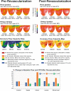
Pre-revascularization and post-revascularization rest and stress relative perfusion images (1st and 2nd rows, respectively) and coronary flow capacity maps (3rd row) of a patient with cardiomyopathy without significant perfusion abnormalities but with severely reduced “blue” CFC secondary to severe CAD. The patient history and revascularization specifics are located within the text. The graphs demonstrate the percent change from the baseline of regional resting MBF (blue bars), stress MBF (orange bars), and CFR (gray bars). The black horizontal lines denote 20% day-to-day normal variability of absolute perfusion. A change in absolute flow metrics outside of the black horizontal lines likely represents procedural or medical therapy influence on absolute perfusion. There is marked improvement in “blue” CFC with revascularization from 39 to 0% of the LV myocardium. Whole-heart stress MBF improved ~80% with regions improving ~40–120%. All revascularized regions demonstrated an increase in sMBF beyond expected 20% day-to-day variability
Fig. 3
Fig. 3
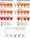
Pre-revascularization and post-revascularization rest and stress relative perfusion images (1st and 2nd rows, respectively) and coronary flow capacity maps (3rd row) of a patient with cardiomyopathy secondary to a combination of prior myocardial infarctions and a primary “non-ischemic” cardiomyopathy. The patient received a LIMA-to-LAD graft because of disease deemed significant based on visual assessment in the LAD and global cardiomyopathy. The patient history is located within the text. The graphs demonstrate the percent change from the baseline of regional resting MBF (blue bars), stress MBF (orange bars), and CFR (gray bars). The black horizontal lines denote 20% day-to-day normal variability of absolute perfusion. A change in absolute flow metrics outside of the black horizontal lines likely represents procedural or medical therapy influence on absolute perfusion. After revascularization, there was neither significant change in absolute flow metrics nor in CFC in the LAD distribution (revascularized territory) or the non-revascularized territory. All absolute PET flow metrics were within 20% of preoperative values
Fig. 4
Fig. 4
Pre-revascularization and post-revascularization rest and stress relative perfusion images (1st and 2nd rows, respectively) and coronary flow capacity maps (3rd row) of a patient with a primary “non-ischemic” cardiomyopathy with concomitant left main disease visually determined as “significant.” The patient received CABG (LIMA to the LAD and SVG to the OM). The patient history is located within the text. The graphs demonstrate the percent change from the baseline of regional resting MBF (blue bars), stress MBF (orange bars), and CFR (gray bars). The black horizontal lines denote 20% day-to-day normal variability of absolute perfusion. A change in absolute flow metrics outside of the black horizontal lines likely represents procedural or medical therapy influence on absolute perfusion. Repeat PET stress testing approximately 3 years after CABG demonstrated nearly identical findings as the pre-revascularization PET, with unchanged CFC within the revascularized territories (anterior, septal, and lateral walls). There was a slight decrease in CFC in the RCA territory, consistent with progression of mild nonobstructive RCA disease or perhaps progression of a primary myocardial process

Similar articles

Cited by

References

    1. Anderson JL, Bittl JA, Bridges CR, et al. PRACTICE GUIDELINE 2011 ACCF/AHA guideline for coronary artery bypass graft surgery. J Am Coll Cardiol. 2011;58:e123–e210. doi: 10.1016/j.jacc.2011.08.009. - DOI - PubMed
    1. Petrie MC, Jhund PS, She L, Adlbrecht C, Doenst T, Panza JA, Hill JA, Lee KL, Rouleau JL, Prior DL, Ali IS, Maddury J, Golba KS, White HD, Carson P, Chrzanowski L, Romanov A, Miller AB, Velazquez EJ, STICH Trial Investigators Ten-year outcomes after coronary artery bypass grafting according to age in patients with heart failure and left ventricular systolic dysfunction: an analysis of the extended follow-up of the STICH Trial (Surgical Treatment for Ischemic Heart Failure) Circulation. 2016;134:1314–1324. doi: 10.1161/CIRCULATIONAHA.116.024800. - DOI - PMC - PubMed
    1. Maron BJ, Towbin JA, Thiene G, Antzelevitch C, Corrado D, Arnett D, Moss AJ, Seidman CE, Young JB. Contemporary definitions and classification of the cardiomyopathies: an American Heart Association Scientific Statement from the Council on Clinical Cardiology, Heart Failure and Transplantation Committee; Quality of Care and Outcomes Research and Functional Genomics and Translational Biology Interdisciplinary Working Groups; and Council on Epidemiology and Prevention. Circulation. 2006;113:1807–1816. doi: 10.1161/CIRCULATIONAHA.106.174287. - DOI - PubMed
    1. Elliott P, Andersson B, Arbustini E, Bilinska Z, Cecchi F, Charron P, Dubourg O, Kühl U, Maisch B, McKenna W, Monserrat L, Pankuweit S, Rapezzi C, Seferovic P, Tavazzi L, Keren A. Classification of the cardiomyopathies: a position statement from the European Society of Cardiology Working Group on Myocardial and Pericardial Diseases. Eur Heart J. 2008;29:270–276. doi: 10.1093/eurheartj/ehm342. - DOI - PubMed
    1. Gould KL, Johnson NP, Bateman TM, Beanlands RS, Bengel FM, Bober R, Camici PG, Cerqueira MD, Chow BJW, di Carli MF, Dorbala S, Gewirtz H, Gropler RJ, Kaufmann PA, Knaapen P, Knuuti J, Merhige ME, Rentrop KP, Ruddy TD, Schelbert HR, Schindler TH, Schwaiger M, Sdringola S, Vitarello J, Williams KA, Sr, Gordon D, Dilsizian V, Narula J. Anatomic versus physiologic assessment of coronary artery disease: role of coronary flow reserve, fractional flow reserve, and positron emission tomography imaging in revascularization decision-making. J Am Coll Cardiol. 2013;62:1639–1653. doi: 10.1016/j.jacc.2013.07.076. - DOI - PubMed

Publication types

MeSH terms