. 2021 May 6;81(9):1951-1969.e6.
doi: 10.1016/j.molcel.2021.03.003. Epub 2021 Mar 23.

Multiple, short protein binding motifs in ORC1 and CDC6 control the initiation of DNA replication

Affiliations

Multiple, short protein binding motifs in ORC1 and CDC6 control the initiation of DNA replication

Manzar Hossain et al. Mol Cell. .

Abstract

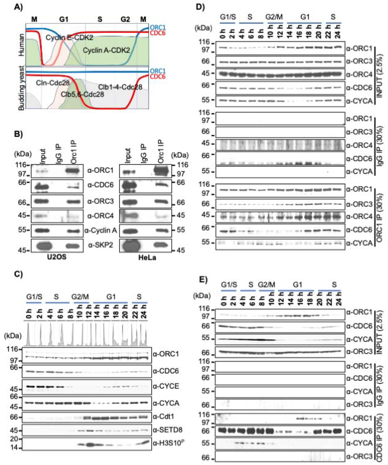
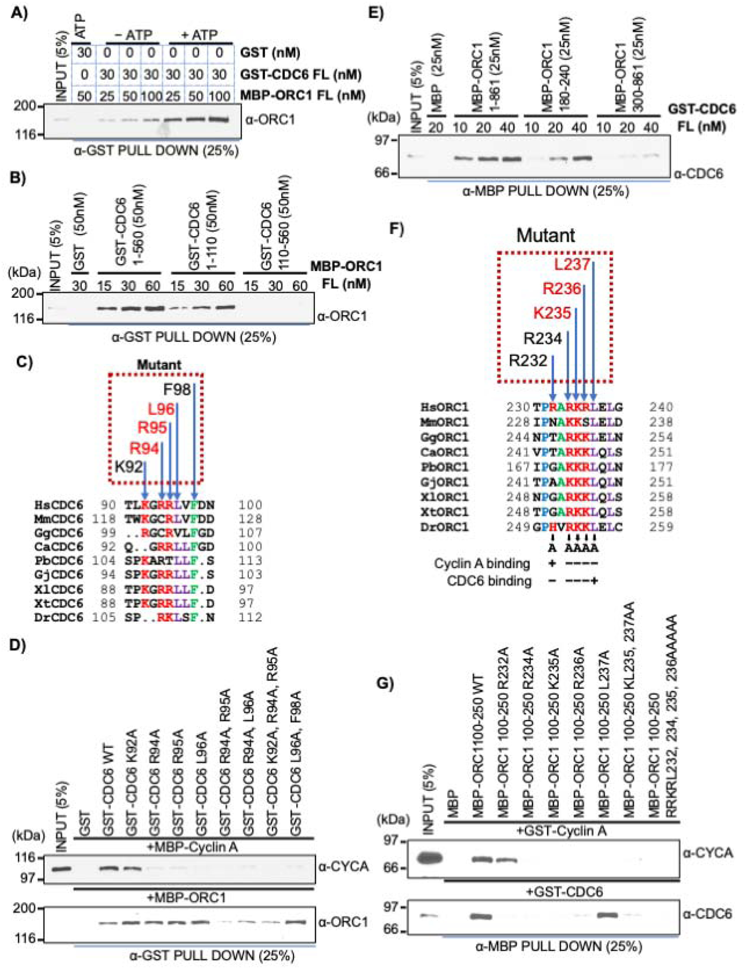
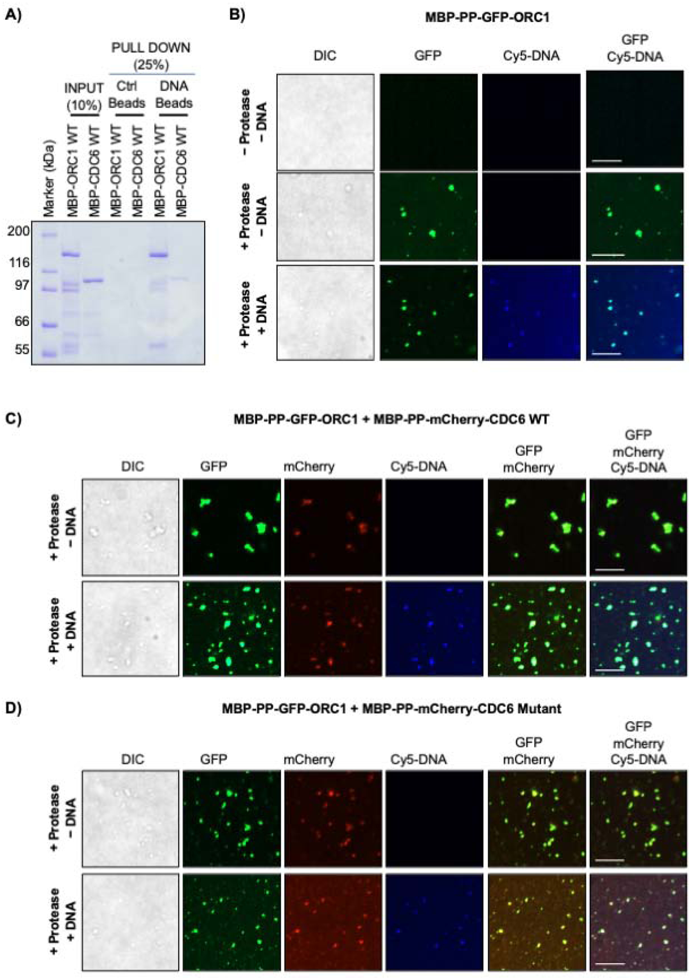
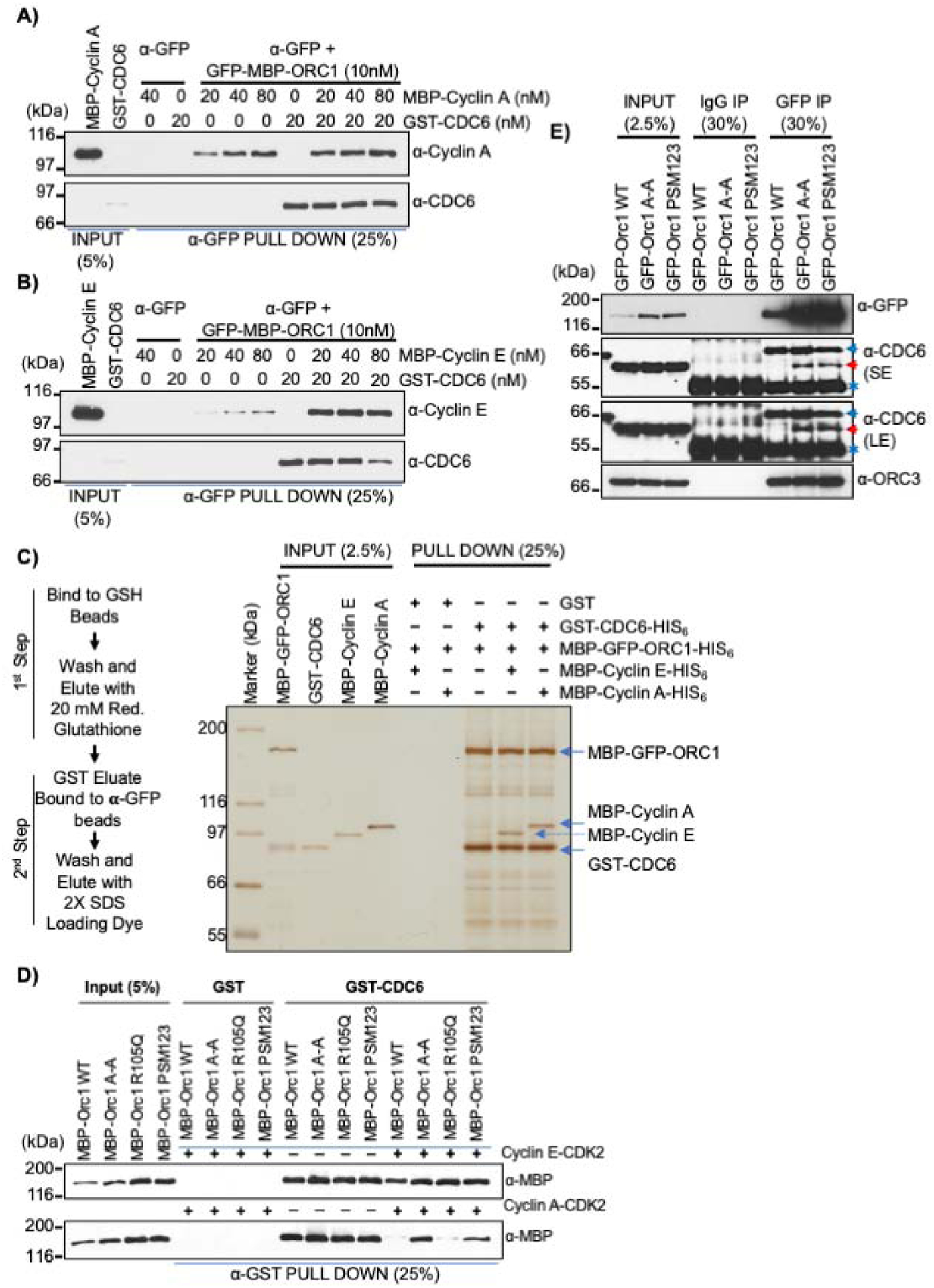
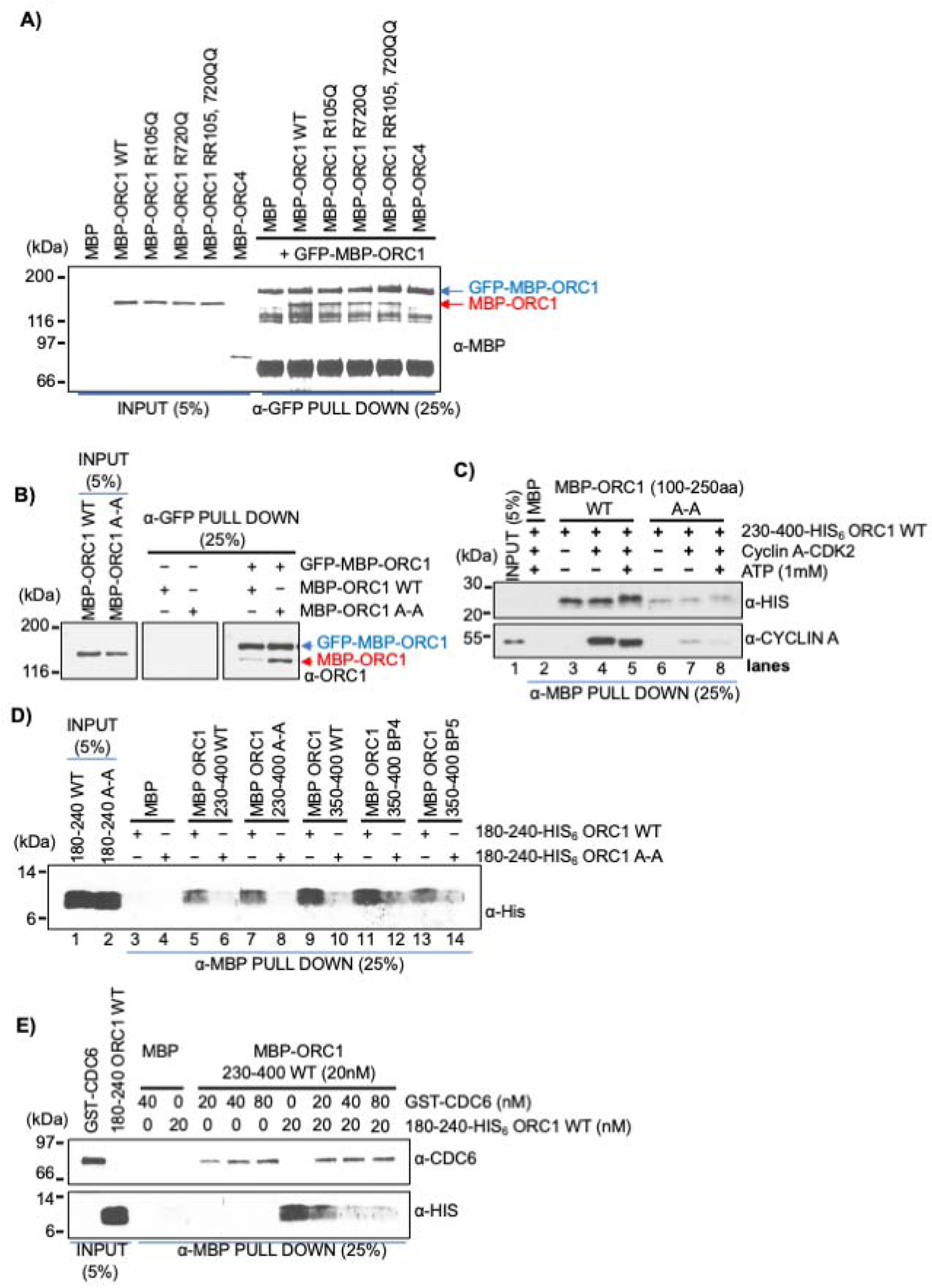
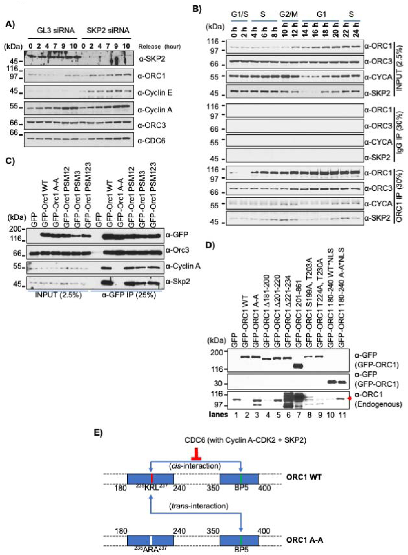
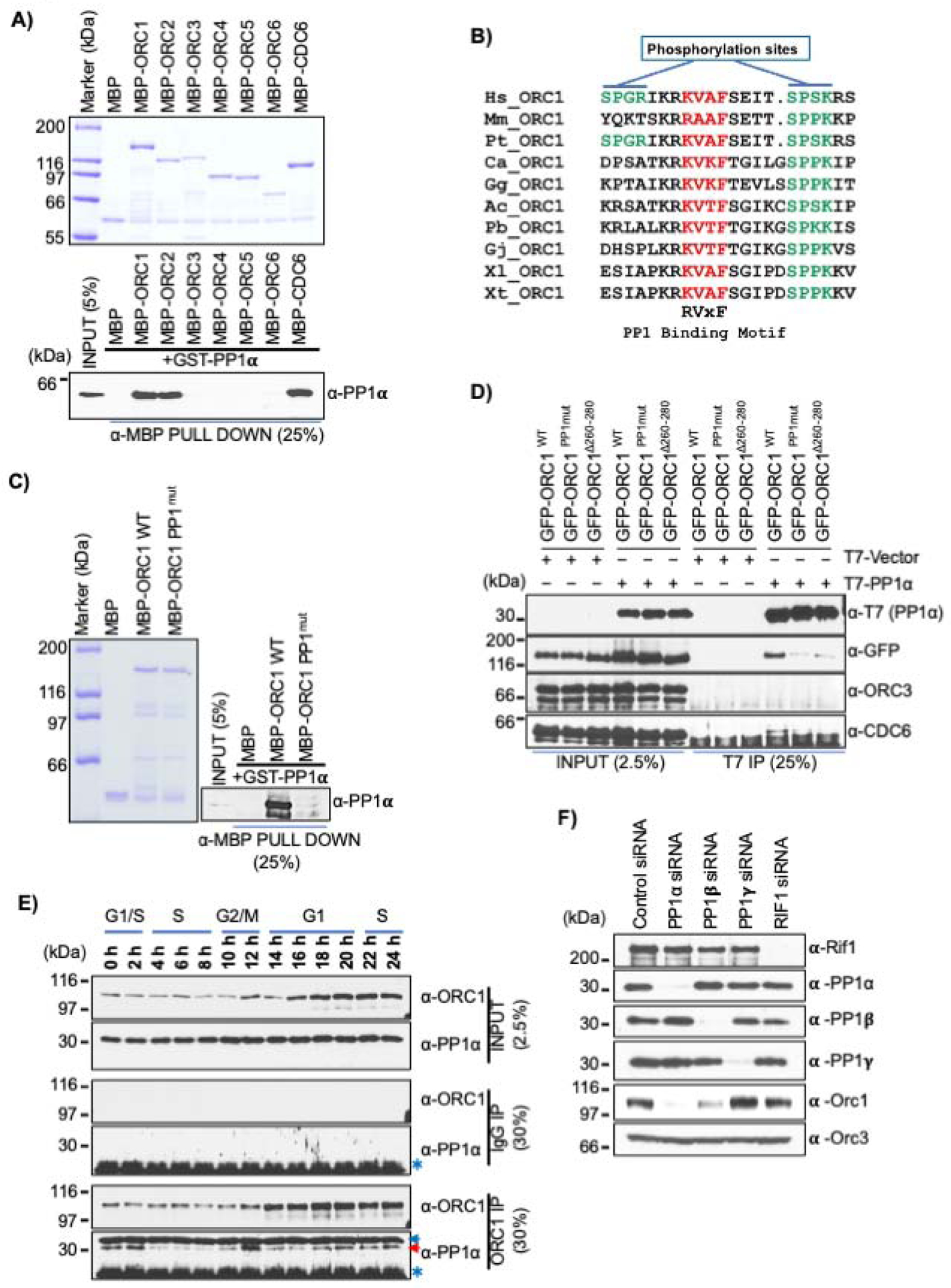
The initiation of DNA replication involves cell cycle-dependent assembly and disassembly of protein complexes, including the origin recognition complex (ORC) and CDC6 AAA+ ATPases. We report that multiple short linear protein motifs (SLiMs) within intrinsically disordered regions (IDRs) in ORC1 and CDC6 mediate cyclin-CDK-dependent and independent protein-protein interactions, conditional on the cell cycle phase. A domain within the ORC1 IDR is required for interaction between the ORC1 and CDC6 AAA+ domains in G1, whereas the same domain prevents CDC6-ORC1 interaction during mitosis. Then, during late G1, this domain facilitates ORC1 destruction by a SKP2-cyclin A-CDK2-dependent mechanism. During G1, the CDC6 Cy motif cooperates with cyclin E-CDK2 to promote ORC1-CDC6 interactions. The CDC6 IDR regulates self-interaction by ORC1, thereby controlling ORC1 protein levels. Protein phosphatase 1 binds directly to a SLiM in the ORC1 IDR, causing ORC1 de-phosphorylation upon mitotic exit, increasing ORC1 protein, and promoting pre-RC assembly.

Keywords: CDC6; DNA replication; PP1 phosphatase; cell division cycle; cyclin-dependent protein kinases; initiation; liquid-liquid phase transition; origin recognition complex; protein degradation; short linear protein motifs.


Conflict of interest statement

Declaration of interests B.S. is a member of the science advisory board of Circle Pharma and is an advisor to EnGeneIC and Pfizer. The authors declare no competing interests.

Figures

Figure 1.
Figure 1.. Dynamic interaction between ORC1 and CDC6 proteins during the human cell cycle.
(A) Schematic of dynamic expression pattern of human and the yeast S. cerevisiae ORC1, CDC6 and Cyclin-CDK kinases across the cell division cycle. (B) Immunoprecipitation of ORC1 from asynchronous U2OS (left panel) and HeLa (right panel) cell lysates showing interactions with CDC6, ORC3, ORC4, Cyclin A and SKP2 proteins. Input and IP are 5% and 30%, respectively. Molecular weight markers, kDa. (C) Dynamic expression profile of pre-RC and cell cycle proteins detected by immunoblotting of extracts from double thymidine block and released synchronized HeLa cells. DNA content is indicated. CYCE and CYCA denotes Cyclin E and Cyclin A, respectively. (D-E) Double thymidine synchronized and released HeLa cell lysate prepared at different time points were immunoprecipitated either with an ORC1 antibody (D) or a CDC6 antibody (E) and immunoblotted as indicated. The input and IgG IP denote loading control and mock IP in the experiment, respectively.
Figure 2.
Figure 2.. ATP-dependent ORC1 and CDC6 interaction region overlaps with their Cy motifs.
(A) Constant amounts of GST-CDC6 protein bound beads were incubated with varying concentrations of MBP-ORC1 protein in absence or presence of 1mM ATP and blotted with anti-ORC1 antibody. FL, full length protein. GST protein served as negative control. See also Figure S2B. (B) Equimolar amounts of GST-CDC6 wild type and N- and C-truncation fragments (1–110 and 110–560) were incubated with full length MBP-ORC1 protein, precipitated with anti-GST antibody and blotted with anti-ORC1 antibody. See also Figure S2E. (C) Multiple sequence alignment of the 90–100 amino acid region of CDC6 from vertebrate species (Homo sapiens, Hs; Mus musculus, Mm; Calypte anna, Ca; Gallus gallus, Gg; Python bivittatus, Pb; Gekko japonicus, Gj; Xenopus laevis, Xl; Xenopus tropicalis, Xt; Danio rerio, Dr). The dotted box above the alignment indicates the conserved residues of human CDC6 protein selected for making recombinant mutant proteins used in binding studies. The residues colored red in the dotted box represent Cy motif of human CDC6 protein. (D) Single (K92A, R94A, R95A, and L96A), double (R94A, R95A; R94A, L96A; L96A, F98A) and triple (K92A, R94A and R95A) mutants of GST-CDC6 full length protein, along with wild type protein, were incubated with either MBP-Cyclin A (top) or MBP-ORC1 (bottom) in a GST pull down assay. The proteins were detected by western blot using their respective antibodies as indicated. See also Figure S2G. (E) Increasing amounts of GST-CDC6 full-length protein binding to MBP-ORC1 wild (1–861) or fragments 180–240 aa or 300–861 aa in α-MBP antibody immunoprecipitate and blotted with anti-CDC6 antibody. See also Figure S2O. (F) Multiple alignment showing sequence conservation the 230–240 aa region of vertebrate ORC1 sequences containing the Cy motif (Homo sapiens, Hs; Mus musculus, Mm; Calypte anna, Ca; Gallus gallus, Gg; Python bivittatus, Pb; Gekko japonicus, Gj; Xenopus laevis, Xl; Xenopus tropicalis, Xt; Danio rerio, Dr). The dotted box above the alignment indicates the conserved residues of human ORC1 protein selected for making recombinant MBP-ORC1 mutants. The residues colored red in the dotted box represent the Cy motif of human ORC1 protein. A summary of binding to CDC6 and Cyclin A is shown below. (G) Human ORC1 single (R232A, R234A, K235A, R236A and L237A), double (K235A, L237A) or quadruple (R232A, R234A, K235A, R236A) mutants in the 100–250 aa region of recombinant MBP-ORC1 protein were incubated with either GST-Cyclin A (top) or GST-CDC6 (bottom) in an anti-MBP antibody pull down assay, then blotted with their respective antibodies as indicated. See also Figure S2P.
Figure 3.
Figure 3.. Phase separation of ORC1 and CDC6 proteins on DNA.
(A) MBP-ORC1 and MBP-CDC6 proteins were incubated with biotinylated dsDNA streptavidin beads in presence of 1mM ATP. Streptavidin beads alone served as control. (B) MBP-PP-GFP-ORC1 (4 μM) do not undergo a liquid-liquid phase separation without protease, but upon addition of protease in the absence or presence of 4 μM Cy5-dsDNA cleaved full length GFP-ORC1 forms liquid droplets. (C-D) MBP-PP-GFP-ORC1 (2 μM) protein was mixed with either 2 μM of MBP-PP-mCherry-CDC6 WT (C) or CDC6 Cy mutant protein (D) in the presence of protease without or with Cy5-dsDNA (4 μM). Merged images shows co-recruitment of cleaved GFP-ORC1 and mCherry-CDC6 (WT or mutant) with or without Cy5-dsDNA. See Figures S3C and S3D. In figures B-D, the images are representative of three independent experiments. The liquid droplets formed were captured after digestion with protease at 4°C for 16 hours. Scale bars 10 μm. PP; PreScission protease site.
Figure 4.
Figure 4.. G1-S Cyclin-CDK kinases have differential effects on ORC1-CDC6 protein interactions.
(A-B) In a α-GFP antibody pull-down assay, a constant amount of recombinant GFP-MBP-ORC1 protein was incubated with GST-CDC6 alone or with increasing molar amounts of MBP-Cyclin A (A) or MBP-Cyclin E (B) and the levels of Cyclin or CDC6 co-precipitated were detected by immuno-blotting. See Figures S4C and S4D. (C) Schematic outline of a two-step affinity purification of protein complexes using purified recombinant proteins (Left panel). MBP-GFP-ORC1-HIS6 (50nM) and GST-CDC6-HIS6 (50nM) were incubated together or in combination with MBP-Cyclin E-HIS6 (50nM) or MBP-Cyclin A-HIS6 (50nM) and control GST (50nM) protein were incubated with either MBP-Cyclin E-HIS6 or MBP-Cyclin A-HIS6 and MBP-GFP-ORC1-HIS6 as indicated. After two step purification, the protein complexes were detected with silver stain (right panel). The reaction contained 1mM ATP. See Figure S4E and S4F. (D) In a GST pull-down assay, GST-CDC6 protein was incubated with MBP-ORC1 WT or its mutants in the presence or absence of Cyclin E-CDK2 (top panel) or Cyclin A-CDK2 (bottom panel) with 1mM ATP. The western blot is probed with anti-MBP antibody and GST protein served as negative control. (E) Mitotic cells from stable GFP-ORC1 WT or ORC1 mutant cell lines were collected after nocodazole treatment and cell lysates were used for immunoprecipitation with anti-GFP antibody. The samples were further immunoblotted with CDC6 antibody and ORC3 antibody. A red arrow indicates specific CDC6 band, while a blue arrow and an asterisk symbol indicate non-specific and cross-reactive heavy IgG bands, respectively. SE, short exposure and LE, long exposure.
Figure 5.
Figure 5.. Self-interaction between the Cy motif containing domain and the BP5 basic patch of human ORC1 is blocked by CDC6 protein.
(A) MBP-GFP-ORC1 wild type protein bound to α-GFP antibody magnetic beads was incubated with either MBP-ORC1 WT or its mutants (R105Q; R720Q; R105Q, R720Q) or MBP-ORC4 and blotted with anti-GFP antibody in a GFP pull-down assay. (B) MBP-GFP-ORC1 protein was incubated with either MBP-ORC1 wild type (WT) or MBP-ORC1 Cy motif mutant (A-A) in the presence of 1mM ATP. MBP-GFP-ORC1 protein was immunoprecipitated with α-GFP antibody magnetic beads, then immunoblotted with the indicated antibodies (right panel). (C) ORC1 fragments, MBP-ORC1 100–250 aa WT or its A-A mutant proteins bound to α-MBP antibody magnetic beads were incubated with ORC1 230–400-His6 protein either alone or in presence of Cyclin A-CDK2 with or without 1mM ATP in MBP pull-down assays. Reactions were blotted with anti-HIS and anti-Cyclin A antibodies. (D) MBP-ORC1-230-400 WT or its Cy motif mutant along with MBP-ORC1-350-400 WT or its basic patch mutants (BP4 and BP5) bound to α-MBP antibody magnetic beads were incubated with His-tagged ORC1 protein fragments, 180–240 WT and 180–240 A-A and probed with anti-His antibody in MBP pull-down assays. See Figure S5B. (E) CDC6 competitively inhibits inter-molecular ORC1 interaction. The constant amount of recombinant MBP-ORC1-230-400 protein fragment bound to α-MBP antibody magnetic beads was incubated with either GST-CDC6 or ORC1-180-240-His6 individually or both together in MBP pull-down assays and immunoblotted with anti-CDC6 and anti-His antibodies. See Figure S5C.
Figure 6.
Figure 6.. Cyclin A-CDK2 recruits SPK2 to promote ORC1 degradation.
(A) U2OS cells transfected with either SKP2 or control GL3 siRNAs were synchronized by double thymidine block and harvested at indicated time points after release from the block. Cell lysates were immunoblotted as indicated. (B) Double thymidine synchronized HeLa cells collected at different time points were lysed and ORC1 immunoprecipitation analyzed by western blot. (C) HEK293 cells were transfected with GFP-ORC1 WT or its Cy motif and CDK phosphorylation site mutants as indicated for overexpression and cells were lysed after 36 hours. Cell lysate immunoprecipitated with anti-GFP antibody were analyzed by immunoblotting with indicated antibodies. (D) Cell lysate from HEK293 cells overexpressing GFP-tagged wild type ORC1 or its mutants [Cy mutant (A-A); internal deletion mutants from full length (Δ181–200, Δ201–220, and Δ221–234); N-terminal deletion (200–861); predicted phosphorylation site double mutants (S199A, T203A and T224A, T230A); NLS fused minimal region 180–240 WT or A-A mutants] were used either for immunoprecipitation in figure S6D or blotted with ORC1 antibody to detect endogenous level of ORC1 proteins. The input of anti-GFP blots from figure S6D is used again for comparison. Red arrow denotes endogenous ORC1 protein band. See also Figure S6D. (E) Schematic showing self-interaction of two distinct regions of ORC1: 180–240 that binds CDC6 as well as contains the Cy motif and 350–400 containing the BP5 basic patch. The cis-interaction is competitively inhibited by wild type CDC6 protein. Upon overexpression of the cis-interaction defective ORC1 Cy mutant, the mutant protein defective in binding to Cyclin A-SKP2 protein establishes a trans-interaction with its wild type counterpart, thus providing protection of wild type protein by sequestering it from the action of SCFSKP2 ubiquitylation and protein degradation.
Figure 7.
Figure 7.. ORC1 directly binds to PP1 protein phosphatase.
(A) Purified MBP tagged ORC and CDC6 proteins bound to α-MBP antibody magnetic beads were incubated with purified GST-PP1α protein in an MBP pull-down assay and blotted with anti-PP1α antibody (bottom panel); MBP-fused recombinant proteins shown in top panel. (B) Alignment of PP1 binding motif in ORC1 vertebrate species. The PP1 binding RVxF motif is highlighted in red, while the adjacent CDK phosphorylation sites are indicated in green. (Homo sapiens, Hs; Mus musculus, Mm; Pan troglodytes, Pt), Aves (Calypte anna, Ca; Gallus gallus, Gg), Reptiles (Anolis carolinensis, Ac; Python bivittatus, Pb; Gekko japonicus, Gj) and Amphibia (Xenopus laevis, Xl; Xenopus tropicalis, Xt). (C) MBP-ORC1 WT protein or corresponding PP1 binding mutant, MBP-ORC1PP1mut bound to α-MBP antibody magnetic beads were incubated with purified GST-PP1α protein in MBP pull-down assay and blotted with anti-PP1α antibody (right panel). MBP-fused recombinant proteins are shown in left panel. (D) HEK293 cells were co-transfected with GFP-ORC1 WT or its PP1 binding mutants (GFP-ORC1PP1mut and GFP-ORC1Δ260−280) with either T7-PP1α or empty T7 vector. Anti-T7 antibody precipitates were immunoblotted with the indicated antibodies. (E) HeLa cells were synchronized using double thymidine block and collected at different time points after release. Cell extracts were immunoprecipitated with anti-ORC1 or control IgG antibody and further immunoblotted with anti-ORC1 and anti-PP1α antibodies. The red arrow indicates specific PP1 band, while the blue arrow and an asterisk symbol indicates non-specific and cross-reactive light IgG bands, respectively. (F) siRNAs targeting PP1 isoforms and RIF1 were used to deplete endogenous proteins from asynchronous growing U2OS cells. Total cell lysate of siRNA treated U2OS cells were prepared after 36 hours of knockdown and immunoblotted with indicated antibodies.

Comment in

Similar articles

Cited by

References

    1. Adams PD, Sellers WR, Sharma SK, Wu AD, Nalin CM, and Kaelin WG (1996). Identification of a cyclin-cdk2 recognition motif present in substrates and p21-like cyclin-dependent kinase inhibitors. Mol Cell Biol 16, 6623–6633. - PMC - PubMed
    1. Bashir T, Dorrello NV, Amador V, Guardavaccaro D, and Pagano M (2004). Control of the SCFSkp2–Cks1 ubiquitin ligase by the APC/CCdh1 ubiquitin ligase. Nature 428, 190–193. - PubMed
    1. Baum B, Nishitani H, Yanow S, and Nurse P (1998). Cdc18 transcription and proteolysis couple S phase to passage through mitosis. Embo J 17, 5689–5698. - PMC - PubMed
    1. Bell SP, and Labib K (2016). Chromosome Duplication in Saccharomyces cerevisiae. Genetics 203, 1027–1067. - PMC - PubMed
    1. Bell SP, and Stillman B (1992). ATP-dependent recognition of eukaryotic origins of DNA replication by a multiprotein complex. Nature 357, 128–134. - PubMed

Publication types

MeSH terms

LinkOut - more resources