. 2021 Mar 24;13(7):10312-10325.
doi: 10.18632/aging.202793. Epub 2021 Mar 24.

Membrane-cytoplasm translocation of annexin A4 is involved in the metastasis of colorectal carcinoma

Affiliations

Membrane-cytoplasm translocation of annexin A4 is involved in the metastasis of colorectal carcinoma

Ya Peng et al. Aging (Albany NY). .

Abstract

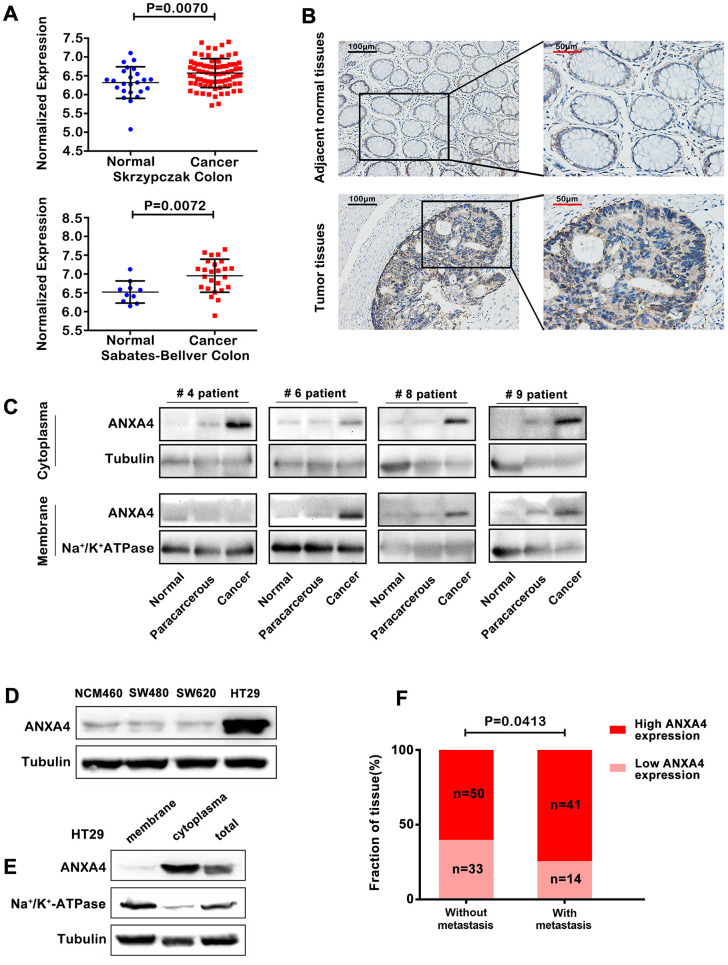
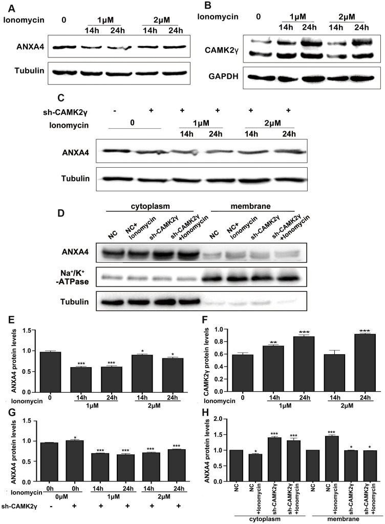
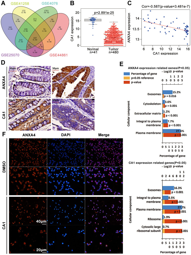
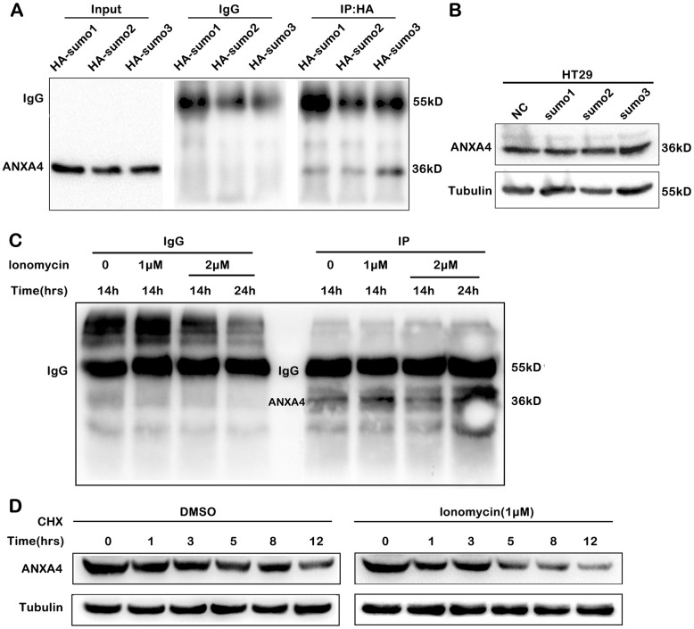
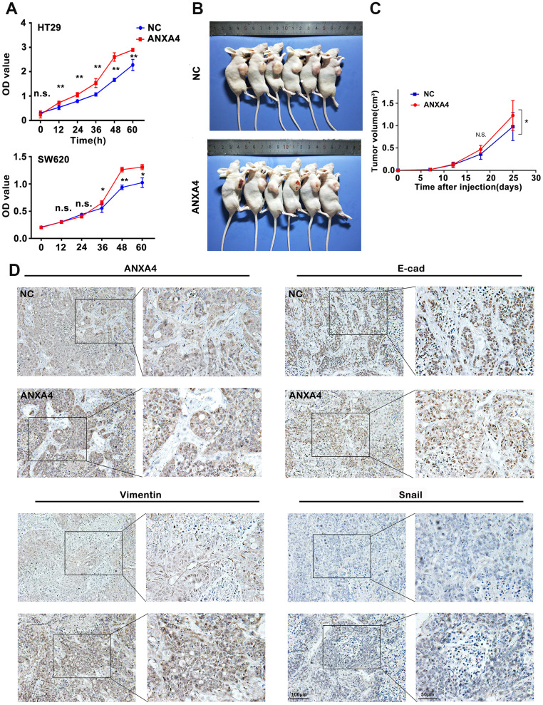
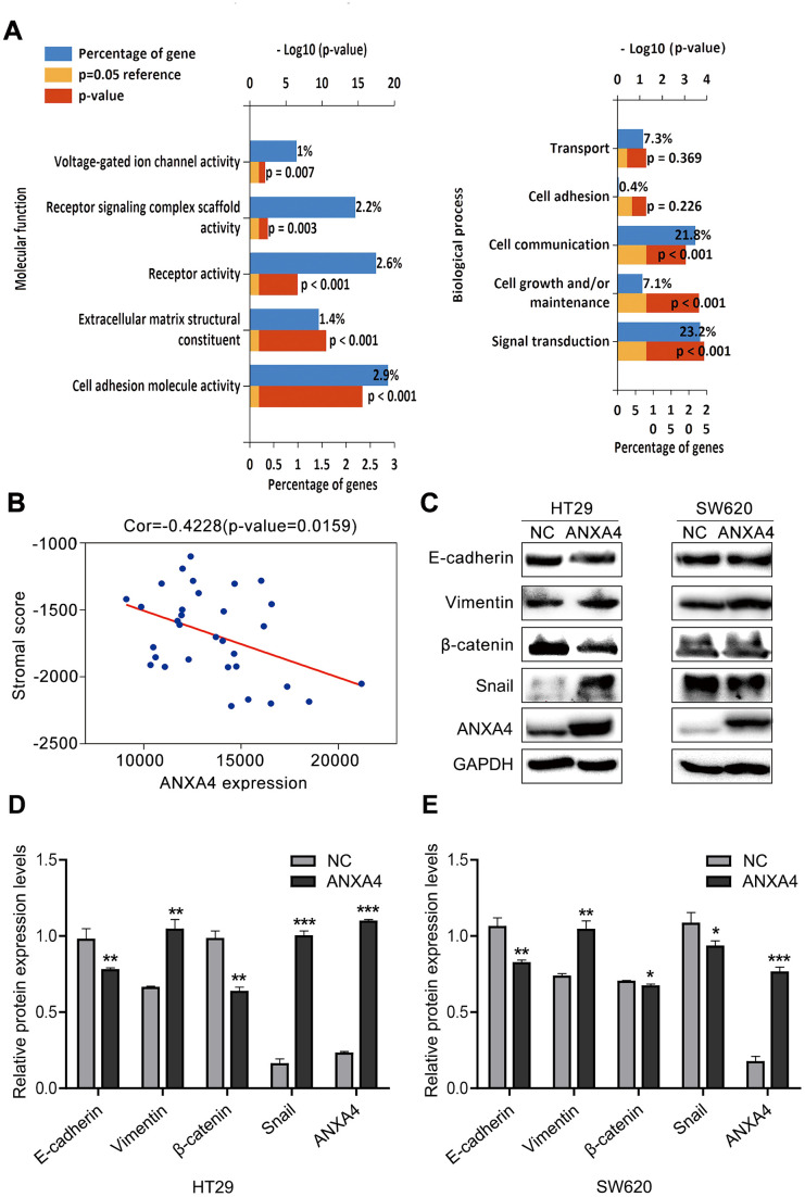
Annexin A4 (ANXA4) is a Ca2+- and phospholipid-binding protein that belongs to the annexin family, which is involved in the development of multiple tumour types via NF-κB signalling. In this study, we verified the high expression and membrane-cytoplasm translocation of ANXA4 in colorectal carcinoma (CRC). Calcium/calmodulin-dependent protein kinase II gamma (CAMK2γ) was found to be important for high ANXA4 expression in CRC, whereas carbonic anhydrase (CA1) promoted ANXA4 aggregation in the cell membrane. An increased Ca2+ concentration attenuated the small ubiquitin-like modifier (SUMO) modification of cytoplasmic ANXA4 and ANXA4 stabilization, and relatively high expression of ANXA4 promoted CRC tumorigenesis and epithelial-mesenchymal transition (EMT).

Keywords: SUMO; annexin A4; calcium/calmodulin-dependent protein kinase II gamma; carbonic anhydrase; membrane-cytoplasm translocation.


Conflict of interest statement

CONFLICTS OF INTEREST: The authors state no conflicts of interest.

Figures

Figure 1
Figure 1
Membrane-cytoplasm translocation of ANXA4 in CRCA. (A) ANXA4 mRNA expression levels were significantly higher in colorectal cancer tissue samples than in normal tissue samples, as indicated by analysis of the Oncomine colorectal cancer dataset, Skrzypczak Colorectal dataset and Sabates-Bellver Colon dataset. (B) Representative images of the protein expression of ANXA4 in CRC and tumour-adjacent tissue samples (n=138) generated by immunohistochemistry. (C) Representative blots of the membrane and cytoplasmic protein expression of ANXA4 in CRC and tumour-adjacent tissue samples (n=19) generated by western blotting. Tubulin was used as the loading control for total protein; Na+/K+-ATPase was used as the loading control for cell membrane protein. (D) The expression of ANXA4 in normal colon cells (NCM460) and colorectal cancer cells (SW480, SW620, and HT-29 cells) determined by western blotting. Tubulin was used as the loading control for total protein. (E) The membrane and cytoplasmic protein expression levels of ANXA4 in HT-29 cells determined by western blotting. Na/K-ATPase was used as the loading control for cell membrane protein; Tubulin was used as the loading control for total protein. (F) Correlation analysis between high ANXA4 expression and metastasis in CRC patients (n=138) by chi-square test analysis (p<0.05).
Figure 2
Figure 2
Calcium regulated the expression and location of ANXA4. (A) Time-course expression analysis of ANXA4 after the addition of the Ca2+ agonist ionomycin (1 μM or 2 μM) at 14 h and 24 h. Tubulin was used as the loading control for total protein. *p < 0.05; **p < 0.01; **p < 0.001. (B) Time-course expression analysis of CAMK2γ after the addition of the Ca2+ agonist ionomycin (1 μM or 2 μM) at 14 h and 24 h. Tubulin was used as the loading control for total protein. *p < 0.05; **p < 0.01; **p < 0.001. (C) Time-course expression analysis of ANXA4 in HT-29 cells transfected with CAMK2γ after the addition of the Ca2+ agonist ionomycin (1 μM or 2 μM) at 14 h and 24 h. Tubulin was used as the loading control for total protein. *p < 0.05; **p < 0.01; ***p < 0.001. (D) Membrane and cytoplasmic levels of ANXA4 in HT-29 cells transfected with CAMK2γ after the addition of the Ca2+ agonist ionomycin (1 μM) at 14 h. Na/K-ATPase was used as the loading control for cell membrane protein; Tubulin was used as the loading control for total protein. *p < 0.05; **p < 0.01; ***p < 0.001. (EH) Relative quantitative of comparison of WB analysis mentioned in (AD). *p < 0.05; **p < 0.01; ***p < 0.001.
Figure 3
Figure 3
CA1 promoted ANXA4 aggregation in the cell membrane. (A) 187 commonly changed genes in CRC samples from the GSE 25070 (USA), GSE 41258 (Israel), GSE 44076 (Spain) and GSE 44861 (USA) datasets. (B) The CA1 mRNA expression level was significantly lower in colorectal cancer tissues than in normal tissues in the TCGA database. (C) Negative correlation analysis between ANXA4 and CA1 in the GSE 20916 and GSE 8671 datasets. (D) Decreased expression of CA1 and increased expression of ANXA4 in Chinese CRC samples, as determined by IHC (n=103). (E) Common cellular component enrichment results for CA1 and ANXA4 determined by GO analysis. (F) CA1 promoted ANXA4 aggregation in the cell membrane, as demonstrated by immunofluorescence imaging.
Figure 4
Figure 4
SUMOylation of ANXA4 stabilized ANXA4 expression. (A) Immunoprecipitation analysis of ANXA4 SUMOylation after cells were cotransfected with flag-ANXA4 and HA-SUMO1, HA-SUMO2, or HA-SUMO3 and then subjected to western blotting with an anti-HA antibody. (B) Western blot analysis of ANXA4 proteins in HT-29 cells after transfection with HA-sumo1, HA-sumo2 or HA-sumo3. Tubulin was used as the loading control. (C) Immunoprecipitation analysis of ANXA4 SUMOylation after cells were cotransfected with flag-ANXA4 and HA-SUMO1, treated with the Ca2+ agonist ionomycin (1 μM or 2 μM) at 14 h and 24 h and then subjected to western blotting with an anti-HA antibody. (D) Western blot analysis of the expression of ANXA4 in HT-29 cells after treatment with cycloheximide with the addition of the Ca2+ agonist ionomycin (1 μM).
Figure 5
Figure 5
ANXA4 promotes tumorigenesis and EMT in a xenograft model. (A) CCK8 assay assessing the viability of HT-29 cells transfected with HA-ANXA4. The data are shown as the mean ± SEM of three independent experiments. *p < 0.05, **p<0.01. (B) HT-29 cells stably transfected with ANXA4 were subcutaneously injected into six nude mice (2×10⁶/120 μl per mouse). After 25 days, the mice were sacrificed, and the tumours were removed. (C) Tumour growth curves of the xenograft tumours (n=6). Tumour volume was calculated at different time points (7, 12, 18, and 25 days) using the formula V=(a×b2)/2. *p < 0.05. (D) Protein expression levels of ANXA4 and the EMT-related molecules E-cadherin, Vimentin, and Snail in tumours determined by IHC.
Figure 6
Figure 6
ANXA4 downregulated the stromal response in CRC. (A) Common molecular function and biological process enrichment terms of ANXA4 determined by GO analysis. (B) Negative correlation analysis between ANXA4 and the stromal score. (C) Effects of ANXA4 overexpression on the EMT-related molecules E-cadherin, Vimentin, β-catenin and Snail. GAPDH was used as the loading control for total protein. (D, E) Relative quantitative analysis of E-cadherin, Vimentin, β-catenin and Snail expression in HT-29 cells (D) and SW620 cells (E). *p < 0.05; **p < 0.01; ***p < 0.001.

Similar articles

Cited by

References

    1. Abakushina EV, Gelm YV, Pasova IA, Bazhin AV. Immunotherapeutic approaches for the treatment of colorectal cancer. Biochemistry (Mosc). 2019; 84:720–28. 10.1134/S0006297919070046 - DOI - PubMed
    1. Raskov H, Pommergaard HC, Burcharth J, Rosenberg J. Colorectal carcinogenesis—update and perspectives. World J Gastroenterol. 2014; 20:18151–64. 10.3748/wjg.v20.i48.18151 - DOI - PMC - PubMed
    1. Wei B, Guo C, Liu S, Sun MZ. Annexin A4 and cancer. Clin Chim Acta. 2015; 447:72–78. 10.1016/j.cca.2015.05.016 - DOI - PubMed
    1. Park TJ, Kim JH, Bae JS, Park BL, Cheong HS, Pasaje CF, Park JS, Uh ST, Kim MK, Choi IS, Park CS, Shin HD. Potential association between ANXA4 polymorphisms and aspirin-exacerbated respiratory disease. Diagn Mol Pathol. 2012; 21:164–71. 10.1097/PDM.0b013e3182461d0d - DOI - PubMed
    1. Boye TL, Maeda K, Pezeshkian W, Sønder SL, Haeger SC, Gerke V, Simonsen AC, Nylandsted J. Annexin A4 and A6 induce membrane curvature and constriction during cell membrane repair. Nat Commun. 2017; 8:1623. 10.1038/s41467-017-01743-6 - DOI - PMC - PubMed

Publication types

MeSH terms