Observational Study
. 2021 Mar 26;100(12):e25232.
doi: 10.1097/MD.0000000000025232.

Correlation of autopsy pathological findings and imaging features from 9 fatal cases of COVID-19 pneumonia

Affiliations
Observational Study

Correlation of autopsy pathological findings and imaging features from 9 fatal cases of COVID-19 pneumonia

Lingyun Zhao et al. Medicine (Baltimore). .

Abstract

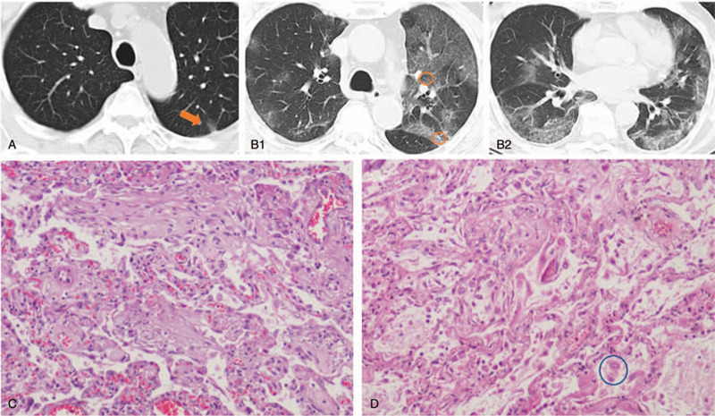
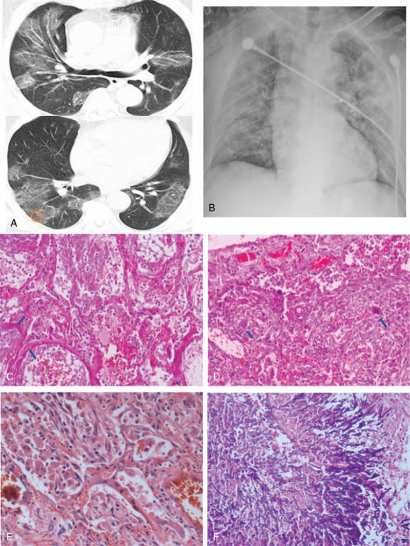
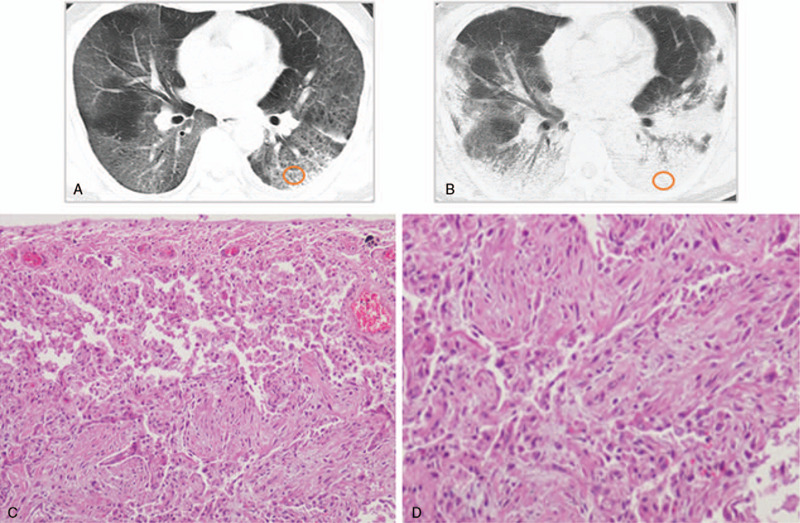
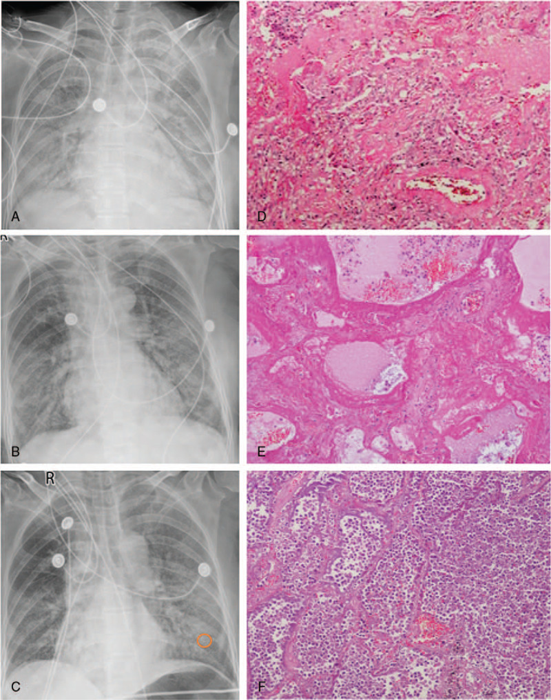
We aimed to investigate the relationship of radiological features and the corresponding pulmonary pathology of patients with Coronavirus Disease (COVID-19) pneumonia.In this multicenter study, serial chest CT and radiographic images from 9 patients (51-85 years old, 56% male) were reviewed and analyzed. Postmortem lungs were sampled and studied from these autopsies, with a special focus on several corresponding sites based on imaging features.The predominant pattern of pulmonary injury in these 9 cases was diffuse alveolar damage (DAD) and interstitial inflammation. Moreover, acute fibrinous exudates, organization, inflammatory cell infiltration, hyaline membranes, pulmonary edema, pneumocyte hyperplasia, and fibrosis were all observed. The histopathology features varied according to the site and severity of each lesion. In most of the 9 cases, opacities started from a subpleural area and peripheral structures were more severely damaged based on gross views and pathological examinations. Fibrosis could occur in early stages of infection and this was supported by radiological and pathological findings. The radiological features of COVID-19 pneumonia, at the critically ill stage, were diffuse ground-glass opacities with consolidation, interstitial thickening, and fibrous stripes, which was based in the fibrous tissue proliferation in the alveolar and interlobular septa, and filled alveoli with organizing exudation. Fungal and bacterial co-infections were also observed in 6 cases.Typical imaging features can be correlated with underlying pathological findings. Combining assessments of imaging features with pathological findings therefore can enhance our understanding of the histopathological mechanism of COVID-19 pneumonia, and facilitate early radiological diagnosis and prognosis estimation of COVID-19 pneumonia, which has important implications for the development of clinical targeted treatments and research related to COVID-19 pneumonia.


Conflict of interest statement

The authors have no conflicts of interests to disclose.

Figures

Figure 1
Figure 1
A-B: Large-scale patchy ground-glass opacities with consolidation and interstitial thickening was observed (the 5th day from onset), presenting as “white lungs” in radiographic images. C: The 10th day from onset. D: Exudative phase of diffuse alveolar damage showing hyaline membrane formation (arrows), serous and fibrinous exudate, pulmonary congestion and edema, and small amounts of inflammatory cell infiltration in the right upper lobe (×100). E: Pulmonary congestion, hemorrhaging, exudate organization in alveolar spaces, and fibrosis of the alveolar septa. Thickened alveolar walls and widened interstitial tissues were accompanied by lymphocytes and other inflammatory cell infiltration and fibroblast proliferation in the right upper lobe (×100). Yellow circles in figures represent pathological sampling sites.
Figure 2
Figure 2
A: In early stage of the disease, a very small ground-glass opacity originated from the subpleural area (arrow, the 3rd day from onset). B1: Several days later, multiple opacities were observed bilaterally, especially in the left upper lobe, combined with the “paving stone signs” (the 12th day from onset). B2: Ground-glass opacities with interlobular septal thickening and fibrosis stripes. C: In the subpleural portion of the left upper lobe, fibrous tissue proliferation in the alveolar septa, alveolar collapse, alveolar epithelium exfoliation and adenoid alveolar formation (×100). D: In the central part of the left upper lobe, the alveolar structure was roughly preserved, small foci fibrosis of alveolar septa with widened alveolar walls, type II pneumocytes hyperplasia, and serous, lymphocytes, monocytes and macrophage exudation (blue circle) in the alveolar spaces (×100). Yellow circles: subpleural and central pathological sampling sites.
Figure 3
Figure 3
A: At the 15th day, a large degree of patchy ground-glass opacities along the bronchus and periphery with superposed vasodilation, interstitial thickening were observed. B: “white lungs,” manifested as obvious ground-glass opacities and fibrosis (the 21st day from onset). C: In the central part of the right lower lobe, fibrin exudate, hyaline membrane formation (arrows) and inflammatory cell infiltration in alveoli. Hyperemia and thickening of the alveolar wall, part of which was replaced by fibrous tissue (×100). D: In the subpleural part of the right lower lobe, alveolar structure destruction was seen. The granuloma nodules were composed of epithelioid cells and multinucleated giant cells (arrows) with central suppuration (×40). E: Fibroblast proliferation and a large number of monocyte and lymphocytes infiltration. (×200) F: Secondary fungal infections with Aspergillus mycelium in the subpleural part of right lower lobe (×100). Yellow circle: subpleural pathological sampling site.
Figure 4
Figure 4
A: A large degree of patchy ground-glass opacities with interstitial thickening or reticulation. “paving stone signs” were seen (17th day after onset). B: 7 days later, lager scale and higher density of the lesions, and pleural thickening indicated a progression. Consolidation, fibrosis, bronchiectasis, bronchial traction and small air-sacs were presented, particularly in the periphery. C: Pulmonary fibrosis and alveolar destruction in the subpleural part of the dorsal side of the left upper lobe (×100). D: Fibrous tissue proliferation in the alveolar septa, alveolar collapse, organized exudate in alveoli, alveolar epithelium exfoliation and adenoid alveolar formation (×200). Yellow circle: subpleural pathological sampling site.
Figure 5
Figure 5
A: “White lungs” (23rd day after onset). B: The increased transparence of bilateral lungs indicated an improvement (26th day after onset). C: A large degree of patchy ground-glass opacities with interstitial thickening or reticulation were observed in the left upper and lower lobes (28th day from onset). CR image showed bronchopneumonia in the left lower lobe. D: Alveolar structure destruction, widened alveolar septum and focal hemorrhagic necrosis. Diffuse alveolar damage with hyaline membrane formation and alveolar septal fibrosis, pulmonary congestion and edema in the left lower lobe (×40). E: Hyaline membrane and alveolar exudation in the left lower lobe (×100). F: Secondary bacterial infection in the left lower lobe: a large number of neutrophil exudations in the alveolar spaces (suppurative inflammation). The alveoli structures were still preserved (×100). Yellow circle: pathological sampling site for suppurative infection.

Similar articles

Cited by

  • Risk factors for the development of interstitial lung disease following severe COVID-19 pneumonia and outcomes of systemic corticosteroid therapy: 3-month follow-up.
    Günay S, Parlak IS, Hezer H, Şeref Parlak EŞ, Umut MS, Hancıoğlu Z, Çelenk Ergüden H, Kocaman Y, Dalkıran A, Sertçelik Ü, Şerifoğlu İ, Akpınar E, Göktaş MF, Fidan M, Babahanoğlu B, Cander FS, Çıvgın E, Er M, Kılıç H, Argüder E, Tuğ T, Ünsal E, Hasanoğlu C, Günay İ, Babayiğit M, Ağca B, Karalezli A. Günay S, et al. Sarcoidosis Vasc Diffuse Lung Dis. 2023 Sep 13;40(3):e2023029. doi: 10.36141/svdld.v40i3.14418. Sarcoidosis Vasc Diffuse Lung Dis. 2023. PMID: 37712369 Free PMC article.
  • Residual Lung Abnormalities after COVID-19 Hospitalization: Interim Analysis of the UKILD Post-COVID-19 Study.
    Stewart I, Jacob J, George PM, Molyneaux PL, Porter JC, Allen RJ, Aslani S, Baillie JK, Barratt SL, Beirne P, Bianchi SM, Blaikley JF, Chalmers JD, Chambers RC, Chadhuri N, Coleman C, Collier G, Denneny EK, Docherty A, Elneima O, Evans RA, Fabbri L, Gibbons MA, Gleeson FV, Gooptu B, Greening NJ, Guio BG, Hall IP, Hanley NA, Harris V, Harrison EM, Heightman M, Hillman TE, Horsley A, Houchen-Wolloff L, Jarrold I, Johnson SR, Jones MG, Khan F, Lawson R, Leavy O, Lone N, Marks M, McAuley H, Mehta P, Parekh D, Hanley KP, Platé M, Pearl J, Poinasamy K, Quint JK, Raman B, Richardson M, Rivera-Ortega P, Saunders L, Saunders R, Semple MG, Sereno M, Shikotra A, Simpson AJ, Singapuri A, Smith DJF, Spears M, Spencer LG, Stanel S, Thickett DR, Thompson AAR, Thorpe M, Walsh SLF, Walker S, Weatherley ND, Weeks ME, Wild JM, Wootton DG, Brightling CE, Ho LP, Wain LV, Jenkins GR. Stewart I, et al. Am J Respir Crit Care Med. 2023 Mar 15;207(6):693-703. doi: 10.1164/rccm.202203-0564OC. Am J Respir Crit Care Med. 2023. PMID: 36457159 Free PMC article.
  • Pentoxifylline as a Potential Adjuvant Therapy for COVID-19: Impeding the Burden of the Cytokine Storm.
    Feret W, Nalewajska M, Wojczyński Ł, Witkiewicz W, Kłos P, Dziedziejko V, Pawlik A. Feret W, et al. J Clin Med. 2021 Nov 15;10(22):5305. doi: 10.3390/jcm10225305. J Clin Med. 2021. PMID: 34830588 Free PMC article. Review.
  • Post-COVID Interstitial Lung Disease-The Tip of the Iceberg.
    Kewalramani N, Heenan KM, McKeegan D, Chaudhuri N. Kewalramani N, et al. Immunol Allergy Clin North Am. 2023 May;43(2):389-410. doi: 10.1016/j.iac.2023.01.004. Epub 2023 Mar 3. Immunol Allergy Clin North Am. 2023. PMID: 37055095 Free PMC article. Review.
  • Autopsy findings of pediatric COVID-19: a systematic review.
    Octavius GS, Wijaya JH, Tan AO, Muljono MP, Chandra S, Juliansen A. Octavius GS, et al. Egypt J Forensic Sci. 2022;12(1):32. doi: 10.1186/s41935-022-00288-0. Epub 2022 Jul 14. Egypt J Forensic Sci. 2022. PMID: 35855892 Free PMC article.

References

    1. International Committee on Taxonomy of Viruses. Naming the 2019 Coronavirus. Available at: https://talk.ictvonline.org/.[accessed February 11, 2020]
    1. Zhu N, Zhang D, Wang W, et al. . A novel coronavirus from patients with pneumonia in China, 2019. N Engl J Med 2020;382:727–33. - PMC - PubMed
    1. World Health Organization. Weekly epidemiological update - December 29, 2020. Available at: https://www.who.int/publications/m/item/weekly-epidemiological-update---...
    1. Huang C, Wang Y, Li X, et al. . Clinical features of patients infected with 2019 novel coronavirus in Wuhan, China. Lancet 2020;395:497–506. - PMC - PubMed
    1. Chen N, Zhou M, Dong X, et al. . Epidemiological and clinical characteristics of 99 cases of 2019 novel coronavirus pneumonia in Wuhan, China: a descriptive study. Lancet 2020;395:507–13. - PMC - PubMed

Publication types