. 2021 Apr 7;143(13):4979-4992.
doi: 10.1021/jacs.0c10644. Epub 2021 Mar 24.

Tunable Methacrylamides for Covalent Ligand Directed Release Chemistry

Affiliations

Tunable Methacrylamides for Covalent Ligand Directed Release Chemistry

Rambabu N Reddi et al. J Am Chem Soc. .

Abstract

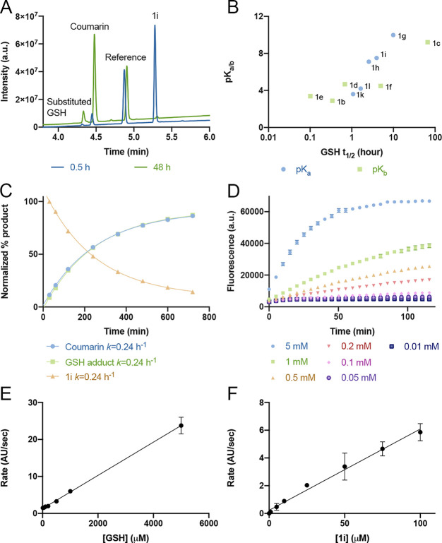
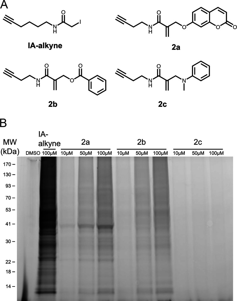
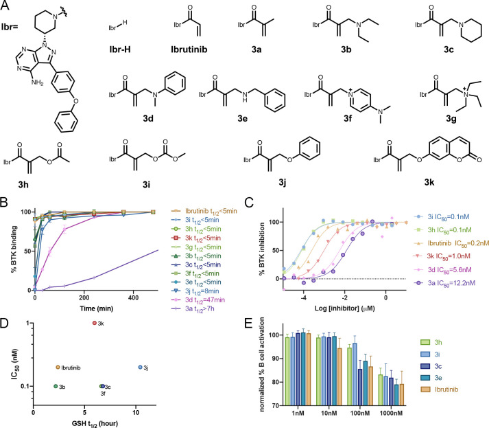
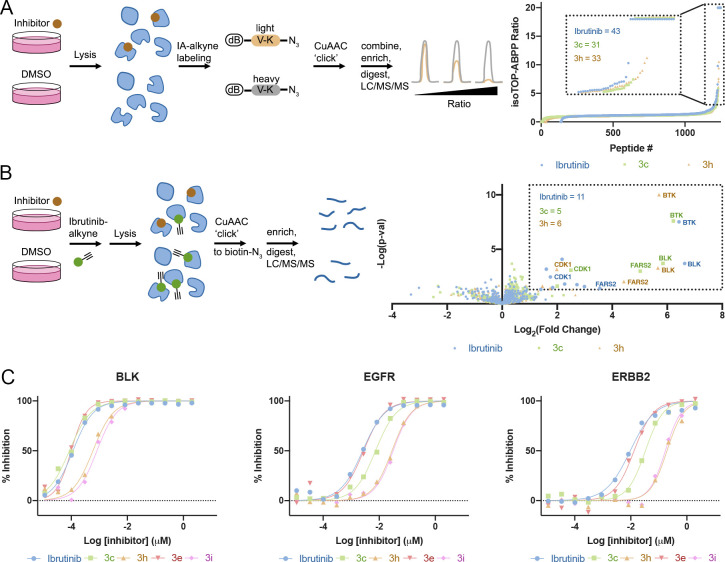
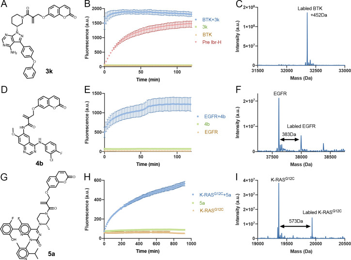
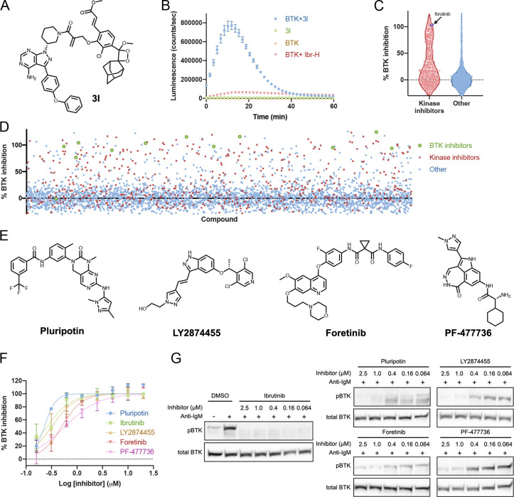
Targeted covalent inhibitors are an important class of drugs and chemical probes. However, relatively few electrophiles meet the criteria for successful covalent inhibitor design. Here we describe α-substituted methacrylamides as a new class of electrophiles suitable for targeted covalent inhibitors. While typically α-substitutions inactivate acrylamides, we show that hetero α-substituted methacrylamides have higher thiol reactivity and undergo a conjugated addition-elimination reaction ultimately releasing the substituent. Their reactivity toward thiols is tunable and correlates with the pKa/pKb of the leaving group. In the context of the BTK inhibitor ibrutinib, these electrophiles showed lower intrinsic thiol reactivity than the unsubstituted ibrutinib acrylamide. This translated to comparable potency in protein labeling, in vitro kinase assays, and functional cellular assays, with improved selectivity. The conjugate addition-elimination reaction upon covalent binding to their target cysteine allows functionalizing α-substituted methacrylamides as turn-on probes. To demonstrate this, we prepared covalent ligand directed release (CoLDR) turn-on fluorescent probes for BTK, EGFR, and K-RasG12C. We further demonstrate a BTK CoLDR chemiluminescent probe that enabled a high-throughput screen for BTK inhibitors. Altogether we show that α-substituted methacrylamides represent a new and versatile addition to the toolbox of targeted covalent inhibitor design.


Conflict of interest statement

The authors declare the following competing financial interest(s): R.N.R., E.R., A.R., and N.L. are inventors on a patent application describing CoLDR chemistry.

Figures

Figure 1
Figure 1
New type of acrylamide-based electrophiles for covalent inhibitors. (A) Various acrylamide substitutions can modify its properties, including both intrinsic reactivity and reversibility. (B) Schematic representation of the reaction of a target cysteine with a substituted α-methacrylamide through CoLDR (covalent ligand directed release) chemistry.
Figure 2
Figure 2
GSH reactivity correlates to the pKa/b of the leaving group. (A) Example LC chromatogram showing monitoring of the reaction of 1i (100 μM) with GSH (5 mM) at 30 min (blue) and 48 h (green). GSH adduct: retention time (RT) = 4.3 min, m/z = 480; coumarin: RT = 4.5 min; reference: RT = 4.8 min; 1i: RT = 5.3 min; m/z = 332. UV absorption measured between 220 and 400 nm. (B) GSH t1/2 vs pKa of the protonated leaving group (pKb for amines). (C) Rates of formation in LC-MS (absorption 220–400 nm) of coumarin and GSH adduct and depletion of 1i in a reaction between 100 μM 1i and 5 mM GSH in PBS buffer, pH 8, 37 °C (n = 3). (D) Fluorescence intensity of 1i (100 μM, n = 4) as a function of incubation time with different GSH concentrations (PBS buffer pH 8, 37 °C, Ex/Em = 385/435 nm). (E) Rates of the turn-on fluorescence reaction of 1i (100 μM; n = 4) as a function of GSH concentration presented in D. (F) Rates of the turn-on fluorescence reaction of GSH (5 mM; PBS buffer pH 8, 37 °C, Ex/Em = 385/435 nm; n = 4) as a function of 1i concentration. The linearity in E and F indicates that the first step of the reaction (thiol addition) is the rate-limiting step.
Figure 3
Figure 3
α-Methacrylamides show varied proteomic reactivity. (A) Chemical structures of model electrophilic alkyne probes. (B) In situ proteomic labeling with the alkyne probes. Mino cells were treated for 2 h with either DMSO, IA-alkyne, or 2a2c, then lysed, reacted with TAMRA-azide using Cu-AAC, and imaged via in-gel fluorescence (532 nm).
Figure 4
Figure 4
α-Substituted derivatives of ibrutinib as potential inhibitors. (A) Chemical structures of the ibrutinib derivatives. (B) Time course LC-MS binding assay (2 μM compound, ibrutinib or 3a3k, and 2 μM BTK at room temperature; n = 3; error bars indicate standard deviation). (C) In vitro kinase activity assay using wild-type BTK (0.6 nM BTK, 5 μM ATP) for selected analogues (see Figure S11 for all). (D) GSH half-life (t1/2) of ibrutinib derivatives does not correlate to measured IC50 values. Note that 3d and 3e are not presented since their GSH t1/2 > 100 h (E) Dose-dependent inhibition of B cell response after anti-IgM-induced activation and treatment with ibrutinib analogues for 24 h (n = 6; error bars indicate standard deviation).
Figure 5
Figure 5
Selectivity of ibrutinib derivatives. (A) isoTOP ABPP using desthiobiotin–valine–azidolysine light or heavy peptides, schematic description, and result summary. Mino cells treated with 1 μM compound for 2 h (n = 4). Proteins in the box have a heavy to light (H/L) ratio ≥ 3. (B) Pull-down proteomics schematic description and result summary. Mino cells treated with 1 μM compound for 1 h and 10 μM ibrutinib-alkyne for an additional 1 h (n = 4). Proteins in the box show significant change (fold change > 2; p < 0.05). (C) In vitro kinase activity assays with selected kinases; see additional plots for BTK, BMX, and ITK in Figure S15.
Figure 6
Figure 6
Turn-on fluorescent probes using CoLDR chemistry. (A, D, G) Structures of turn-on fluorescent probes for BTK, EGFR, and K-RasG12C, respectively. (B, E, H) Time dependence of fluorescence intensity (representing the release of the coumarin moiety) measured at Ex/Em = 385/435 nm (n = 3). Green curves show that the compounds in and of themselves (2 μM) are not fluorescent. Orange curves show that the proteins themselves (2 μM) are also not fluorescent. Only upon mixing of probe and target (blue curves) do we see an increase in fluorescence. (C, F, I) Deconvoluted LC/MS spectra for BTK, EGFR, and K-RasG12C incubated with 3k, 4b, and 5a at the end of each plate reader measurement. The adduct mass corresponds to a labeling event in which the coumarin moiety was released, validating the proposed mechanism. For BTK (D) we completed a reversible version of ibrutinib Ibr-H (2 μM; 0.5 h preincubation; Figure 4A) with 3k (red curve). This considerably slowed the release of coumarin and the corresponding increase in fluorescence. All error bars indicate standard deviation.
Figure 7
Figure 7
Chemiluminescent BTK probe allows high-throughput screening for BTK inhibitors. (A) Structure of the chemiluminescent probe 3l. (B) Time dependence of the luminescence signal (representing the release of the chemiluminescent moiety, n = 3). The compound in and of itself (2 μM; green) is not luminescent. The protein itself (2 μM; orange) is also not luminescent. Only upon mixing of probe and target (blue) do we see an increase in luminescence. Preincubation of BTK with a reversible version of ibrutinib Ibr-H (2 μM; 0.5 h; red) inhibits luminescence (100 ms integration). (C) Schematic summary of %BTK binding inhibition in HTS using 3l showing an enrichment of known kinase inhibitors in the library to bind BTK compared to nonkinase inhibitors. (D) Overall view of %BTK binding inhibition in the HTS. Known kinase inhibitors in red and known BTK inhibitors in green. (E) Structures of selected hits from HTS with 3l. (F) Dose response (n = 2) of %BTK binding inhibition of selected hits from HTS with 3l. (G) Inhibition of BTK phosphorylation in Mino cells with hit compounds. Cells were incubated for 1 h with inhibitors followed by 10 min activation with anti-IgM (full gels Figure S24). All error bars indicate standard deviation.

References

    1. Zhao Z.; Bourne P. E. Progress with Covalent Small-Molecule Kinase Inhibitors. Drug Discovery Today 2018, 23 (3), 727–735. 10.1016/j.drudis.2018.01.035. - DOI - PubMed
    1. Liu Q.; Sabnis Y.; Zhao Z.; Zhang T.; Buhrlage S. J.; Jones L. H.; Gray N. S. Developing Irreversible Inhibitors of the Protein Kinase Cysteinome. Chem. Biol. 2013, 20 (2), 146–159. 10.1016/j.chembiol.2012.12.006. - DOI - PMC - PubMed
    1. Baillie T. A. Targeted Covalent Inhibitors for Drug Design. Angew. Chem., Int. Ed. 2016, 55 (43), 13408–13421. 10.1002/anie.201601091. - DOI - PubMed
    1. Singh J.; Petter R. C.; Baillie T. A.; Whitty A. The Resurgence of Covalent Drugs. Nat. Rev. Drug Discovery 2011, 10 (4), 307–317. 10.1038/nrd3410. - DOI - PubMed
    1. Shannon D. A.; Weerapana E. Covalent Protein Modification: The Current Landscape of Residue-Specific Electrophiles. Curr. Opin. Chem. Biol. 2015, 24, 18–26. 10.1016/j.cbpa.2014.10.021. - DOI - PubMed

Publication types