

Attenuated TGFB signalling in macrophages decreases susceptibility to DMBA-induced mammary cancer in mice
- PMID: 33761981
- PMCID: PMC7992865
- DOI: 10.1186/s13058-021-01417-8
Attenuated TGFB signalling in macrophages decreases susceptibility to DMBA-induced mammary cancer in mice
Abstract
Background: Transforming growth factor beta1 (TGFB1) is a multi-functional cytokine that regulates mammary gland development and cancer progression through endocrine, paracrine and autocrine mechanisms. TGFB1 also plays roles in tumour development and progression, and its increased expression is associated with an increased breast cancer risk. Macrophages are key target cells for TGFB1 action, also playing crucial roles in tumourigenesis. However, the precise role of TGFB-regulated macrophages in the mammary gland is unclear. This study investigated the effect of attenuated TGFB signalling in macrophages on mammary gland development and mammary cancer susceptibility in mice.
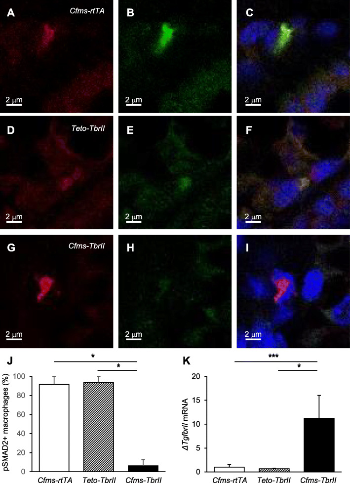
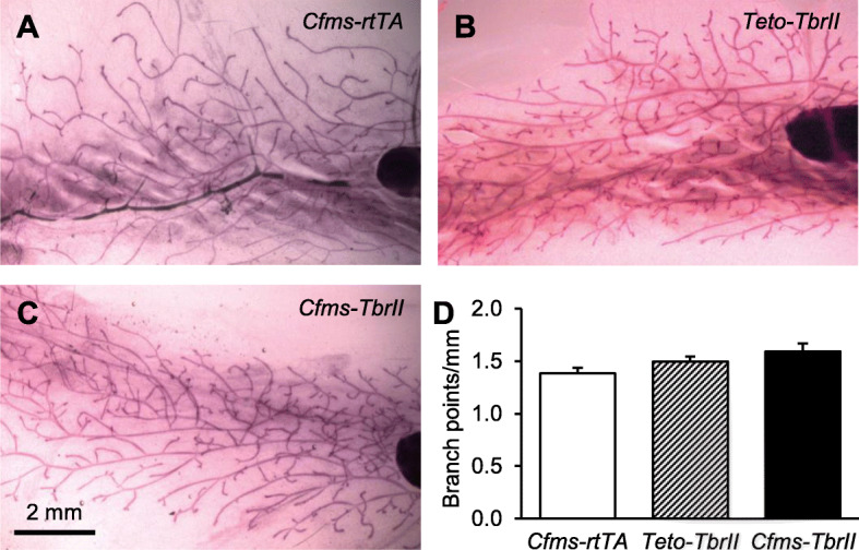
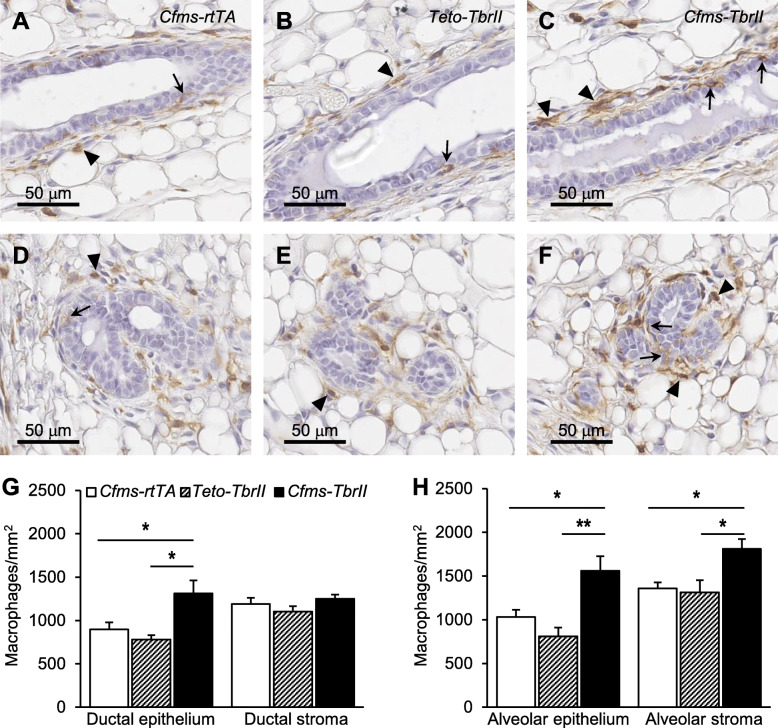
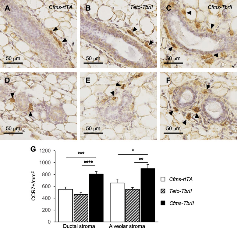
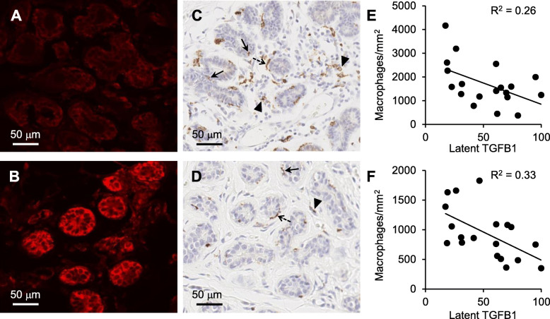
Methods: A transgenic mouse model was generated, wherein a dominant negative TGFB receptor is activated in macrophages, in turn attenuating the TGFB signalling pathway specifically in the macrophage population. The mammary glands were assessed for morphological changes through wholemount and H&E analysis, and the abundance and phenotype of macrophages were analysed through immunohistochemistry. Another cohort of mice received carcinogen 7,12-dimethylbenz(a)anthracene (DMBA), and tumour development was monitored weekly. Human non-neoplastic breast tissue was also immunohistochemically assessed for latent TGFB1 and macrophage marker CD68.
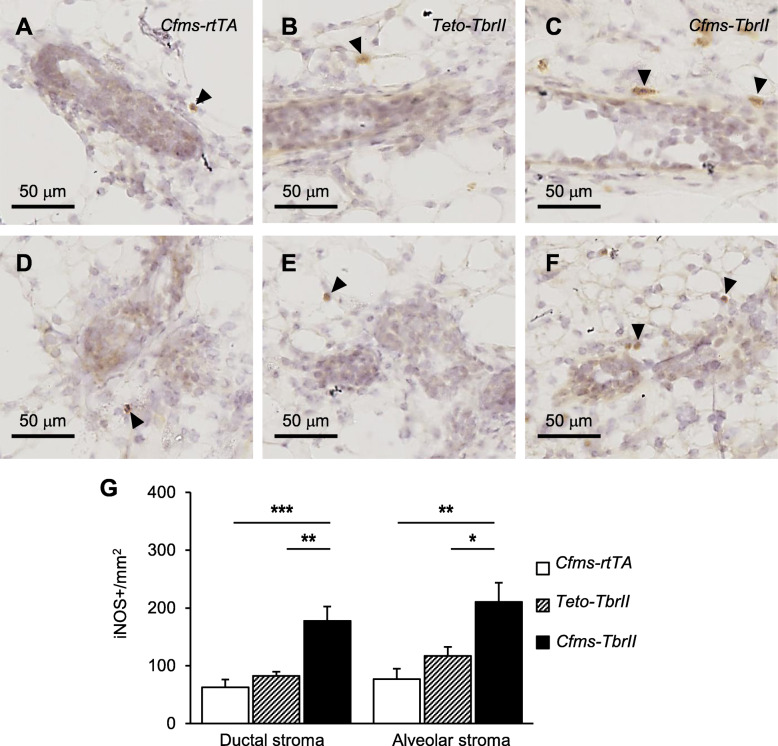
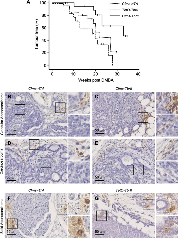
Results: Attenuation of TGFB signalling resulted in an increase in the percentage of alveolar epithelium in the mammary gland at dioestrus and an increase in macrophage abundance. The phenotype of macrophages was also altered, with inflammatory macrophage markers iNOS and CCR7 increased by 110% and 40%, respectively. A significant decrease in DMBA-induced mammary tumour incidence and prolonged tumour-free survival in mice with attenuated TGFB signalling were observed. In human non-neoplastic breast tissue, there was a significant inverse relationship between latent TGFB1 protein and CD68-positive macrophages.
Conclusions: TGFB acts on macrophage populations in the mammary gland to reduce their abundance and dampen the inflammatory phenotype. TGFB signalling in macrophages increases mammary cancer susceptibility potentially through suppression of immune surveillance activities of macrophages.
Keywords: Cancer; Macrophage; Mammary gland; Transforming growth factor beta.
Conflict of interest statement
The authors declare that they have no competing interests.
Figures

Similar articles
-
CCL2-driven inflammation increases mammary gland stromal density and cancer susceptibility in a transgenic mouse model.Breast Cancer Res. 2017 Jan 11;19(1):4. doi: 10.1186/s13058-016-0796-z. Breast Cancer Res. 2017. PMID: 28077158 Free PMC article.
-
Regulation of epithelial cell turnover and macrophage phenotype by epithelial cell-derived transforming growth factor beta1 in the mammary gland.Cytokine. 2013 Feb;61(2):377-88. doi: 10.1016/j.cyto.2012.12.002. Epub 2013 Jan 3. Cytokine. 2013. PMID: 23290315
-
Cytokine networks that mediate epithelial cell-macrophage crosstalk in the mammary gland: implications for development and cancer.J Mammary Gland Biol Neoplasia. 2014 Jul;19(2):191-201. doi: 10.1007/s10911-014-9319-7. Epub 2014 Jun 13. J Mammary Gland Biol Neoplasia. 2014. PMID: 24924120 Review.
-
Opposing roles for mammary epithelial-specific PPARγ signaling and activation during breast tumour progression.Mol Cancer. 2015 Apr 15;14:85. doi: 10.1186/s12943-015-0347-8. Mol Cancer. 2015. PMID: 25889730 Free PMC article.
-
The role of activin in mammary gland development and oncogenesis.J Mammary Gland Biol Neoplasia. 2011 Jun;16(2):117-26. doi: 10.1007/s10911-011-9214-4. Epub 2011 Apr 8. J Mammary Gland Biol Neoplasia. 2011. PMID: 21475961 Review.
Cited by
-
TGFβ control of immune responses in cancer: a holistic immuno-oncology perspective.Nat Rev Immunol. 2023 Jun;23(6):346-362. doi: 10.1038/s41577-022-00796-z. Epub 2022 Nov 15. Nat Rev Immunol. 2023. PMID: 36380023 Free PMC article. Review.
-
Advancing osteoarthritis therapy with GMOCS hydrogel-loaded BMSCs-exos.J Nanobiotechnology. 2024 Aug 19;22(1):493. doi: 10.1186/s12951-024-02713-z. J Nanobiotechnology. 2024. PMID: 39160590 Free PMC article.
-
Pistacia lentiscus L. revealed in vitro anti-proliferative activity on MCF-7 breast cancer cells and in vivo anti-mammary cancer effect on C57BL/6 mice through necrosis, anti-inflammatory and antioxidant enhancements.PLoS One. 2024 Apr 18;19(4):e0301524. doi: 10.1371/journal.pone.0301524. eCollection 2024. PLoS One. 2024. PMID: 38635559 Free PMC article.
-
A role for platelets in metabolic reprogramming of tumor-associated macrophages.Front Physiol. 2023 Aug 24;14:1250982. doi: 10.3389/fphys.2023.1250982. eCollection 2023. Front Physiol. 2023. PMID: 37693009 Free PMC article. Review.
-
Obesity contributes to hepatocellular carcinoma development via immunosuppressive microenvironment remodeling.Front Immunol. 2023 May 17;14:1166440. doi: 10.3389/fimmu.2023.1166440. eCollection 2023. Front Immunol. 2023. PMID: 37266440 Free PMC article. Review.
References
Publication types
MeSH terms
Substances
Grants and funding
LinkOut - more resources
Full Text Sources
Other Literature Sources
Molecular Biology Databases
Miscellaneous
