GLUT5 (SLC2A5) enables fructose-mediated proliferation independent of ketohexokinase
- PMID: 33762003
- PMCID: PMC7992954
- DOI: 10.1186/s40170-021-00246-9
GLUT5 (SLC2A5) enables fructose-mediated proliferation independent of ketohexokinase
Abstract
Background: Fructose is an abundant source of carbon and energy for cells to use for metabolism, but only certain cell types use fructose to proliferate. Tumor cells that acquire the ability to metabolize fructose have a fitness advantage over their neighboring cells, but the proteins that mediate fructose metabolism in this context are unknown. Here, we investigated the determinants of fructose-mediated cell proliferation.
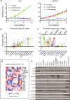
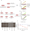
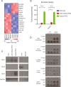
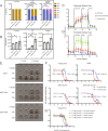
Methods: Live cell imaging and crystal violet assays were used to characterize the ability of several cell lines (RKO, H508, HepG2, Huh7, HEK293T (293T), A172, U118-MG, U87, MCF-7, MDA-MB-468, PC3, DLD1 HCT116, and 22RV1) to proliferate in fructose (i.e., the fructolytic ability). Fructose metabolism gene expression was determined by RT-qPCR and western blot for each cell line. A positive selection approach was used to "train" non-fructolytic PC3 cells to utilize fructose for proliferation. RNA-seq was performed on parental and trained PC3 cells to find key transcripts associated with fructolytic ability. A CRISPR-cas9 plasmid containing KHK-specific sgRNA was transfected in 293T cells to generate KHK-/- cells. Lentiviral transduction was used to overexpress empty vector, KHK, or GLUT5 in cells. Metabolic profiling was done with seahorse metabolic flux analysis as well as LC/MS metabolomics. Cell Titer Glo was used to determine cell sensitivity to 2-deoxyglucose in media containing either fructose or glucose.
Results: We found that neither the tissue of origin nor expression level of any single gene related to fructose catabolism determine the fructolytic ability. However, cells cultured chronically in fructose can develop fructolytic ability. SLC2A5, encoding the fructose transporter, GLUT5, was specifically upregulated in these cells. Overexpression of GLUT5 in non-fructolytic cells enabled growth in fructose-containing media across cells of different origins. GLUT5 permitted fructose to flux through glycolysis using hexokinase (HK) and not ketohexokinase (KHK).
Conclusions: We show that GLUT5 is a robust and generalizable driver of fructose-dependent cell proliferation. This indicates that fructose uptake is the limiting factor for fructose-mediated cell proliferation. We further demonstrate that cellular proliferation with fructose is independent of KHK.
Keywords: Fructose; GLUT5 (SLC2A5); Hexokinase; Ketohexokinase; Metabolism.
Conflict of interest statement
L.C.C. is a founder and member of the board of directors of Agios Pharmaceuticals and is a founder and receives research support from Petra Pharmaceuticals. M.D.G. reports personal fees from Novartis, Petra Pharmaceuticals, and Bayer. He receives research support from Pfizer. L.C.C. and M.D.G. are inventors on patents (pending) for Combination Therapy for PI3K-associated Disease or Disorder, The Identification of Therapeutic Interventions to Improve Response to PI3K Inhibitors for Cancer Treatment, and Anti-Fructose Therapy for Colorectal and Small Intestine Cancers. L.C.C. and M.D.G. are co-founders and shareholders in Faeth Therapeutics. All other authors report no competing interests.
Figures


Similar articles
-
Fructose Metabolism in Cancer: Molecular Mechanisms and Therapeutic Implications.Int J Med Sci. 2025 Jun 9;22(11):2852-2876. doi: 10.7150/ijms.108549. eCollection 2025. Int J Med Sci. 2025. PMID: 40520908 Free PMC article. Review.
-
Fructose-induced increases in expression of intestinal fructolytic and gluconeogenic genes are regulated by GLUT5 and KHK.Am J Physiol Regul Integr Comp Physiol. 2015 Sep;309(5):R499-509. doi: 10.1152/ajpregu.00128.2015. Epub 2015 Jun 17. Am J Physiol Regul Integr Comp Physiol. 2015. PMID: 26084694 Free PMC article.
-
GLUT5-KHK axis-mediated fructose metabolism drives proliferation and chemotherapy resistance of colorectal cancer.Cancer Lett. 2022 May 28;534:215617. doi: 10.1016/j.canlet.2022.215617. Epub 2022 Mar 4. Cancer Lett. 2022. PMID: 35257833
-
PFKFB3 Inhibits Fructose Metabolism in Pulmonary Microvascular Endothelial Cells.Am J Respir Cell Mol Biol. 2023 Sep;69(3):340-354. doi: 10.1165/rcmb.2022-0443OC. Am J Respir Cell Mol Biol. 2023. PMID: 37201952 Free PMC article.
-
Recent Progress on Fructose Metabolism-Chrebp, Fructolysis, and Polyol Pathway.Nutrients. 2023 Apr 5;15(7):1778. doi: 10.3390/nu15071778. Nutrients. 2023. PMID: 37049617 Free PMC article. Review.
Cited by
-
Establishing mammalian GLUT kinetics and lipid composition influences in a reconstituted-liposome system.Nat Commun. 2023 Jul 10;14(1):4070. doi: 10.1038/s41467-023-39711-y. Nat Commun. 2023. PMID: 37429918 Free PMC article.
-
Fructose Metabolism in Cancer: Molecular Mechanisms and Therapeutic Implications.Int J Med Sci. 2025 Jun 9;22(11):2852-2876. doi: 10.7150/ijms.108549. eCollection 2025. Int J Med Sci. 2025. PMID: 40520908 Free PMC article. Review.
-
GLUT5 armouring enhances adoptive T-cell therapy anti-tumour activity under glucose-limiting conditions.Immunother Adv. 2025 Apr 30;5(1):ltaf018. doi: 10.1093/immadv/ltaf018. eCollection 2025. Immunother Adv. 2025. PMID: 40575013 Free PMC article.
-
The Impact of Fructose Consumption on Human Health: Effects on Obesity, Hyperglycemia, Diabetes, Uric Acid, and Oxidative Stress With a Focus on the Liver.Cureus. 2024 Sep 24;16(9):e70095. doi: 10.7759/cureus.70095. eCollection 2024 Sep. Cureus. 2024. PMID: 39355469 Free PMC article. Review.
-
Fructose Induced KHK-C Increases ER Stress and Modulates Hepatic Transcriptome to Drive Liver Disease in Diet-Induced and Genetic Models of NAFLD.bioRxiv [Preprint]. 2023 Jan 27:2023.01.27.525605. doi: 10.1101/2023.01.27.525605. bioRxiv. 2023. Update in: Metabolism. 2023 Aug;145:155591. doi: 10.1016/j.metabol.2023.155591. PMID: 36747758 Free PMC article. Updated. Preprint.
References
-
- United States Department of Agriculture, Economic Research Service . USDA Sugar Supply: Table 50: US Consumption of Caloric Sweeteners. 2019.
-
- Khitan Z, Kim DH. Fructose: a key factor in the development of metabolic syndrome and hypertension. J Nutr Metab [Internet]. 2013;2013 [cited 2020 May 31]. Available from: https://www.ncbi.nlm.nih.gov/pmc/articles/PMC3677638/. - PMC - PubMed
Grants and funding
LinkOut - more resources
Full Text Sources
Other Literature Sources
Miscellaneous

