

Chronically altered NMDAR signaling in epilepsy mediates comorbid depression
- PMID: 33762011
- PMCID: PMC7992813
- DOI: 10.1186/s40478-021-01153-2
Chronically altered NMDAR signaling in epilepsy mediates comorbid depression
Abstract
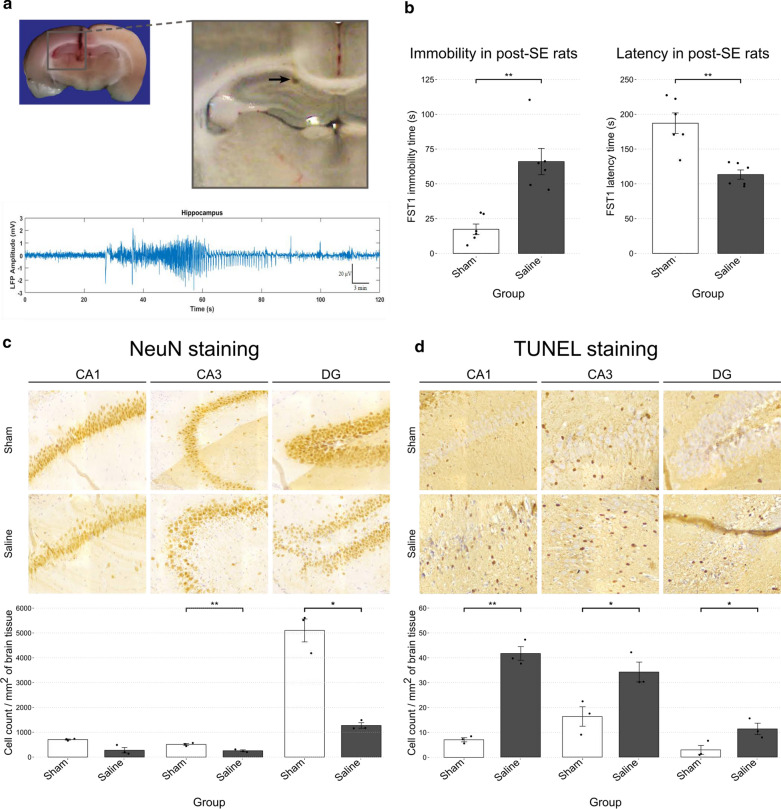
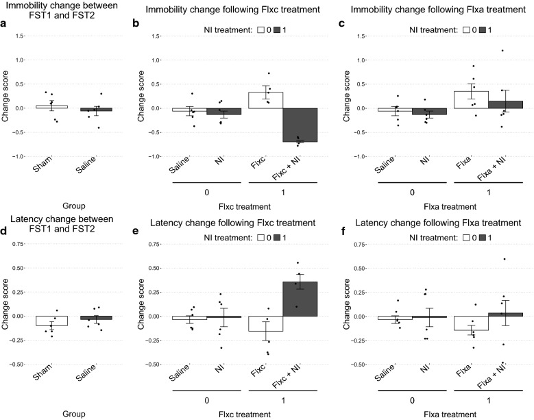
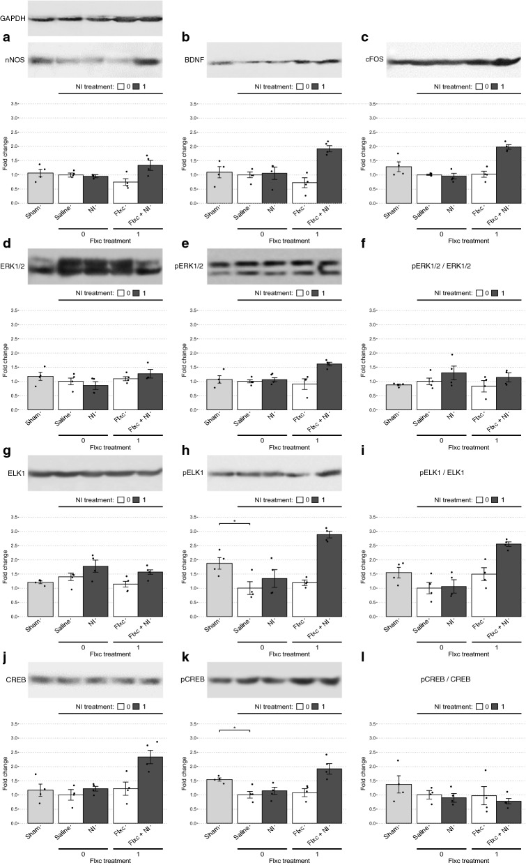
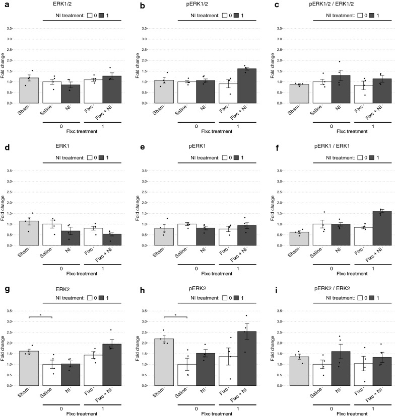
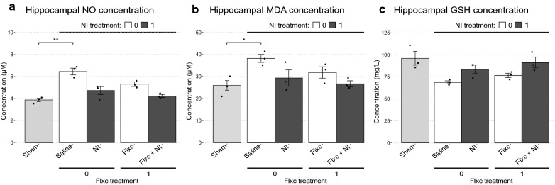
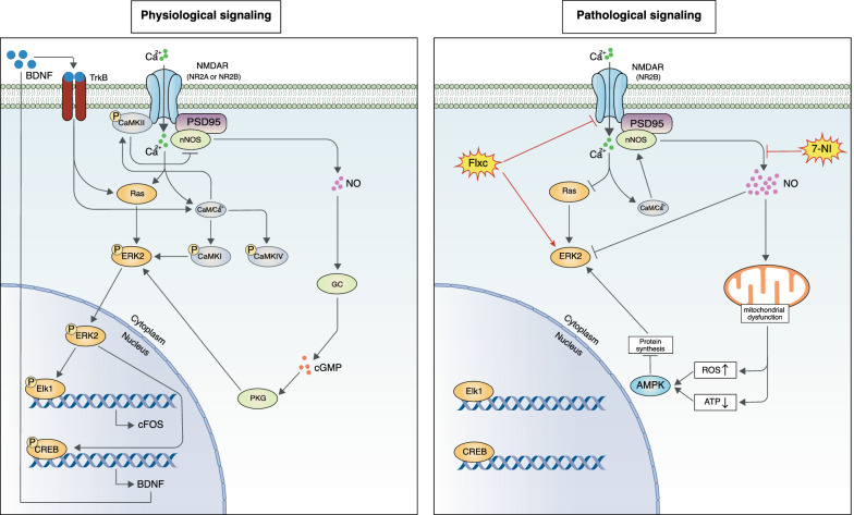
Depression is the most common psychiatric comorbidity of epilepsy. However, the molecular pathways underlying this association remain unclear. The NMDA receptor (NMDAR) may play a role in this association, as its downstream signaling has been shown to undergo long-term changes following excitotoxic neuronal damage. To study this pathway, we used an animal model of fluoxetine-resistant epilepsy-associated depression (EAD). We determined the molecular changes associated with the development of depressive symptoms and examined their response to various combinations of fluoxetine and a selective neuronal nitric oxide synthase inhibitor, 7-nitroindazole (NI). Depressive symptoms were determined using the forced swim test. Furthermore, expression and phosphorylation levels of markers in the ERK/CREB/ELK1/BDNF/cFOS pathway were measured to determine the molecular changes associated with these symptoms. Finally, oxidative stress markers were measured to more clearly determine the individual contributions of each treatment. While chronic fluoxetine (Flxc) and NI were ineffective alone, their combination had a statistically significant synergistic effect in reducing depressive symptoms. The development of depressive symptoms in epileptic rats was associated with the downregulation of ERK2 expression and ELK1 and CREB phosphorylation. These changes were exactly reversed upon Flxc + NI treatment, which led to increased BDNF and cFOS expression as well. Interestingly, ERK1 did not seem to play a role in these experiments. NI seemed to have augmented Flxc's antidepressant activity by reducing oxidative stress. Our findings suggest NMDAR signaling alterations are a major contributor to EAD development and a potential target for treating conditions associated with underlying excitotoxic neuronal damage.
Keywords: Brain-derived neurotrophic factor; Depression; Epilepsy; Immediate early genes; Intracellular signaling; MAP kinase signaling system; N-methyl-D-aspartate receptors; Nitric oxide; Nitric oxide synthase type I; Nitrosative stress.
Conflict of interest statement
The authors have no conflicts of interest to declare.
Figures
Similar articles
-
Effects of nitric oxide synthesis inhibitor or fluoxetine treatment on depression-like state and cardiovascular changes induced by chronic variable stress in rats.Stress. 2015;18(4):462-74. doi: 10.3109/10253890.2015.1038993. Epub 2015 Jun 11. Stress. 2015. PMID: 26068517
-
Antidepressant activity of fluoxetine in the zinc deficiency model in rats involves the NMDA receptor complex.Behav Brain Res. 2015;287:323-30. doi: 10.1016/j.bbr.2015.03.064. Epub 2015 Apr 4. Behav Brain Res. 2015. PMID: 25845739
-
Neural Plasticity Associated with Hippocampal PKA-CREB and NMDA Signaling Is Involved in the Antidepressant Effect of Repeated Low Dose of Yueju Pill on Chronic Mouse Model of Learned Helplessness.Neural Plast. 2017;2017:9160515. doi: 10.1155/2017/9160515. Epub 2017 Sep 17. Neural Plast. 2017. PMID: 29075536 Free PMC article.
-
The PSD-95/nNOS complex: new drugs for depression?Pharmacol Ther. 2012 Feb;133(2):218-29. doi: 10.1016/j.pharmthera.2011.11.005. Epub 2011 Nov 23. Pharmacol Ther. 2012. PMID: 22133842 Review.
-
The WAG/Rij strain: a genetic animal model of absence epilepsy with comorbidity of depression [corrected].Prog Neuropsychopharmacol Biol Psychiatry. 2011 Jun 1;35(4):854-76. doi: 10.1016/j.pnpbp.2010.11.010. Epub 2010 Nov 17. Prog Neuropsychopharmacol Biol Psychiatry. 2011. PMID: 21093520 Review.
Cited by
-
Effects of β-sitosterol on anxiety in migraine-induced rats: The role of oxidative/nitrosative stress and mitochondrial function.CNS Neurosci Ther. 2024 Sep;30(9):e14892. doi: 10.1111/cns.14892. CNS Neurosci Ther. 2024. PMID: 39301958 Free PMC article.
-
Targeting neuronal nitric oxide synthase and the nitrergic system in post-traumatic stress disorder.Psychopharmacology (Berl). 2022 Oct;239(10):3057-3082. doi: 10.1007/s00213-022-06212-7. Epub 2022 Aug 27. Psychopharmacology (Berl). 2022. PMID: 36029333 Review.
-
Therapeutic potential of N-methyl-D-aspartate receptor modulators in psychiatry.Neuropsychopharmacology. 2024 Jan;49(1):51-66. doi: 10.1038/s41386-023-01614-3. Epub 2023 Jun 27. Neuropsychopharmacology. 2024. PMID: 37369776 Free PMC article. Review.
-
CPEB3 can regulate seizure susceptibility by inhibiting the transcriptional activity of STAT3 on NMDARs expression.Mol Med. 2025 Feb 24;31(1):77. doi: 10.1186/s10020-025-01136-2. Mol Med. 2025. PMID: 39994587 Free PMC article.
-
The preventive effects of Saccharomyces boulardii against oxidative stress induced by lipopolysaccharide in rat brain.Heliyon. 2024 Apr 26;10(9):e30426. doi: 10.1016/j.heliyon.2024.e30426. eCollection 2024 May 15. Heliyon. 2024. PMID: 38720760 Free PMC article.
References
Publication types
MeSH terms
Substances
LinkOut - more resources
Full Text Sources
Other Literature Sources
Medical
Miscellaneous
