Case Reports
. 2021 Mar 24;14(3):e236097.
doi: 10.1136/bcr-2020-236097.

Bone mineral density changes in a free vascularised fibular graft in the distal femoral bone after osteosarcoma in a 10-year-old boy: a 7-year follow-up

Affiliations
Case Reports

Bone mineral density changes in a free vascularised fibular graft in the distal femoral bone after osteosarcoma in a 10-year-old boy: a 7-year follow-up

Andrea René Jørgensen et al. BMJ Case Rep. .

Abstract

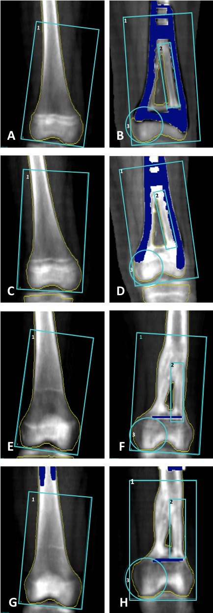
A 10-year-old boy presented with continuous reports of pain located to the left knee. Imaging revealed a sclerotic process in the left distal femur, and biopsies were consistent with chondroblastic osteosarcoma. As part of standard treatment the patient underwent neoadjuvant chemotherapy followed by limb sparring surgery and adjuvant chemotherapy. The entire tumour was excised and femoral bone reconstruction was performed with a double barrel free vascularised fibular graft. Bone mineral density (BMD) can be decreased in childhood survivors of cancer. The patient was followed for 7 years with dual-energy X-ray absorptiometry scans in order to assess BMD and graft adaption. Despite two accidental fractures to the graft region local and global BMD underwent an overall increase. Approximately 7 years after tumour resection the patient had a global Z-score of 0.2, which is considered within normal range.

Keywords: cancer intervention; orthopaedics; osteoporosis; paediatric oncology.


Conflict of interest statement

Competing interests: None declared.

Figures

Figure 1
Figure 1
Timeline. MAP-regimen, high-dose methotrexate, doxorubicin and cisplatin; LISS, Less invasive stabilization system; D-FVFG, double barrel free vascularised fibular graft; DXA, dual-energy X-ray absorptiometry; LCP, locking compression plate.
Figure 2
Figure 2
(A) Anterior–posterior radiograph showing a sclerotic process with periosteal spiculation in the distal lateral part of the femoral bone. (B) Diagnostic MRI showing a bone tumour measuring 55 mm in diameter with soft tissue extension but no involvement of neurovascular structures.
Figure 3
Figure 3
(A) Internal fixation of the femoral bone with a less invasive stabilization system (LISS) plate prior to excision of the tumour. (B) The free fibula graft measuring 225 mm. (C) The double-barrel free vascularised graft.
Figure 4
Figure 4
Radiographs 2 days postoperative. (A) Lateral view and (B) anterior–posterior view of the left femoral bone showing correct positioning of the doublebarrel free vascularised fibular graft and preservation of the epiphysial plate.
Figure 5
Figure 5
(A) Full body dual-energy X-ray absorptiometry (DXA)-scan 60 months after tumour resection showing signs of compensatory lumbar scoliosis due to leg length discrepancy. Boxes 1 and 2 (height 32 cm, width 12.5 cm) are positioned from the knee joint and 32 cm proximal, just below trochanter minor. They are used to estimate net bone mineral density for the left and right leg as seen in table 3 and figure 10. (B) Full body DXA-scan 81 months after tumour resection showing an almost realigned spinal scoliosis after a leg-lengthening procedure.
Figure 6
Figure 6
(A, B) Position of patient during dual-energy X-ray absorptiometry scans of the left distal femur.
Figure 7
Figure 7
Dual-energy X-ray absorptiometry scans of the right and left distal femoral bone. For the right distal femoral bone (A, C, E and G) the blue box outlines the region of interest (ROI) 1 (height 8.73 cm, width 16.32 cm) which is positioned with the lower horizontal line along the most distal part of the femoral condyles. For the left distal femoral bone the blue boxes outline ROI 1 (height 16.35 cm, width 9.53 cm), ROI 2 (height 8.16 cm, width 1.49 cm) of the lateral half of the double barrel free vascularised fibular graft positioned with the lower horizontal line along the distal end of the graft. ROI 3 (height 5.28 cm, width 4.08 cm) is positioned as a circle including the medial femoral condyle (non-bone transplant). The yellow line marks the bone border. The dark blue colour marks metal artefacts not included in the bone mineral density estimation. (A) and (B) 10 months, (C) and (D) 13 months, (E) and (F) 60 months and (G) and (H) 81 months.
Figure 8
Figure 8
BMD % change of the left distal femoral bone in ROI 1 (blue), ROI 2 (orange) and ROI 3 (green) in months after tumour resection. BMD, bone mineral density; ROI, regions of interest.
Figure 9
Figure 9
BMD % change in ROI 1 (blue) of the unaffected right distal femoral bone. BMD % change measured on total body scans: right leg (orange), left leg (grey) and the full body (yellow) in months after tumour resection. BMD, bone mineral density; ROI, regions of interest.
Figure 10
Figure 10
Total Z-score in months after tumour resection. DXA, dual-energy X-ray absorptiometry.

References

    1. Moore DD, Luu HH. Osteosarcoma. Cancer Treat Res 2014;162:65–92. 10.1007/978-3-319-07323-1_4 - DOI - PubMed
    1. Bacci G, Ferrari S, Bertoni F, et al. . Long-Term outcome for patients with nonmetastatic osteosarcoma of the extremity treated at the Istituto ortopedico rizzoli according to the Istituto ortopedico rizzoli/osteosarcoma-2 protocol: an updated report. J Clin Oncol 2000;18:4016–27. 10.1200/JCO.2000.18.24.4016 - DOI - PubMed
    1. Bumbasirevic M, Stevanovic M, Bumbasirevic V, et al. . Free vascularised fibular grafts in orthopaedics. Int Orthop 2014;38:1277–82. 10.1007/s00264-014-2281-6 - DOI - PMC - PubMed
    1. Bachrach LK, Gordon CM. Bone densitometry in children and adolescents. Pediatrics 2016;138:e20162398. 10.1542/peds.2016-2398 - DOI - PubMed
    1. Kaste SC, Ahn H, Liu T, et al. . Bone mineral density deficits in pediatric patients treated for sarcoma. Pediatr Blood Cancer 2008;50:1032–8. 10.1002/pbc.21281 - DOI - PubMed

Publication types