Review
. 2021 Mar 24;41(12):2541-2553.
doi: 10.1523/JNEUROSCI.0675-20.2020.

Evidences for Adult Hippocampal Neurogenesis in Humans

Affiliations
Review

Evidences for Adult Hippocampal Neurogenesis in Humans

Elena P Moreno-Jiménez et al. J Neurosci. .

Abstract

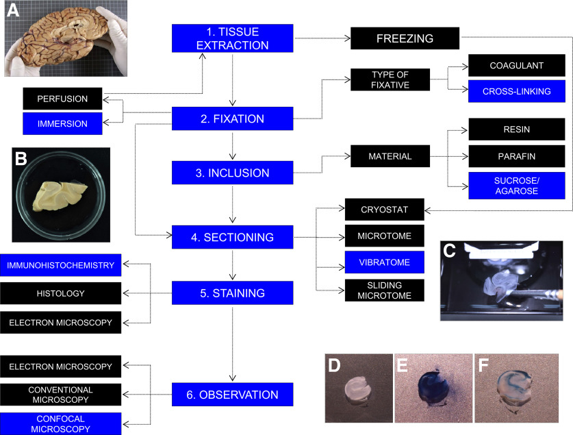
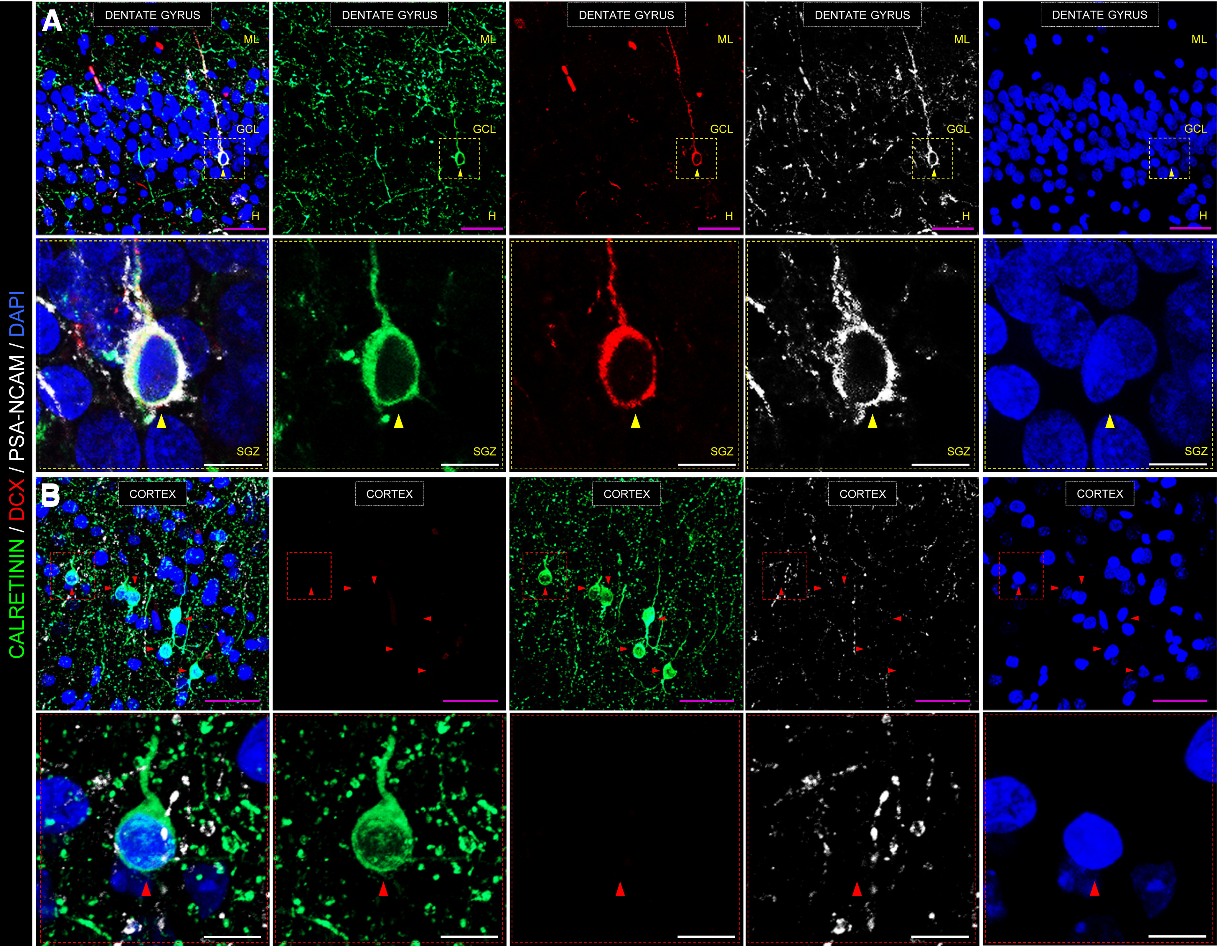
The rodent hippocampus generates new neurons throughout life. This process, named adult hippocampal neurogenesis (AHN), is a striking form of neural plasticity that occurs in the brains of numerous mammalian species. Direct evidence of adult neurogenesis in humans has remained elusive, although the occurrence of this phenomenon in the human dentate gyrus has been demonstrated in seminal studies and recent research that have applied distinct approaches to birthdate newly generated neurons and to validate markers of adult-born neurons. Our data point to the persistence of AHN until the 10th decade of human life, as well as to marked impairments in this process in patients with Alzheimer's disease. Moreover, our work demonstrates that the methods used to process and analyze postmortem human brain samples can limit the detection of various markers of AHN to the point of making them undetectable. In this Dual Perspectives article, we highlight the critical methodological aspects that should be strictly controlled in human studies and the robust evidence that supports the occurrence of AHN in humans. We also put forward reasons that may account for current discrepancies on this topic. Finally, the unresolved questions and future challenges awaiting the field are highlighted.

Keywords: adult neurogenesis; controversy; hippocampus; human; immature neuron; methodology.


Figures

Figure 1.
Figure 1.
Variety of methods routinely used to process postmortem human brain samples. During autopsy, the brain is removed from the skull (A). After dissecting the area of interest, tissue can be either frozen or fixed. Various chemical fixatives can be used. After fixation, samples can be embedded in resin, paraffin, or a mixture of agarose/sucrose (B) to favor tissue preservation and sectioning. Depending on the embedding support, a cryostat, microtome, or vibratome (C) sectioning method should be used. Staining is performed following previously established immunohistochemical, histologic, or electron immunohistochemical protocols. Human hippocampal sections subjected to immunohistochemistry staining before (D), during (E), and after (F) the final autofluorescence elimination step are shown. Stained sections can be observed under conventional epifluorescence, electron, or confocal microscopes. Blue represents the protocol followed in our laboratory. A complete description of this protocol can be found in Moreno-Jiménez et al. (2019) and Flor-García et al. (2020).
Figure 2.
Figure 2.
Representative images showing an immunohistochemical staining performed on postmortem human hippocampal samples. A, Low- and high-power magnification images show the anatomic organization of the human DG. In high-power magnification images, a triple-labeled DCX+/calretinin+/PSA-NCAM+ immature dentate granule cell is shown. B, Representative image of the human cortex is shown as a negative control for DCX staining. Note the abundance of calretinin+ interneurons, which is in sharp contrast to the absence of DCX+ cells in this structure. GCL, Granule cell layer; ML, molecular layer; H, hilus; SGZ, subgranular zone. Scale bars: magenta, 50 μm; white, 10 μm. Yellow triangles represent DCX+/calretinin+/PSA-NCAM+ immature dentate granule cell. Red triangles represent DCX/calretinin+/PSA-NCAM cortical interneuron. Immunohistochemistry was performed following a previously published protocol (Moreno-Jiménez et al., 2019; Flor-García et al., 2020), which included sodium borohydride (NaBH4), heat-mediated citrate antigen retrieval (HC-AR), and autofluorescence elimination steps. Low-power confocal stacks of images were obtained in a LSM800 Carl Zeiss confocal microscope, equipped with three GaAsP detectors and using a 40× immersion oil objective. A, 0.8 zoom, XY dimensions: 199.66 µm, Z interval: 1.5 µm, pinhole dimensions: 0.9 Airy units. B, 1.1 zoom, XY dimensions: 145.2 µm, Z interval: 1.5 µm, pinhole dimensions: 0.9 Airy units. High-power magnification images were obtained in the same confocal microscope using a 63× immersion oil objective (2.2 zoom, XY dimensions: 46 µm, Z interval: 0.5 µm, pinhole dimensions: 0.9 Airy units). All the images shown in this figure correspond to postmortem human samples obtained from a neurologically healthy control subject. Fixation time was limited to 24 h in freshly prepared 4% PFA at 4°C. The epidemiological data of this subject are the following: gender, male; age, 43; postmortem delay, 5 h; Braak-Tau stage, 0; CERAD stage, 0; and Braak α-synuclein stage, 0. Brain tissue donation, processing, and use for research were in compliance with published protocols (Martinez-Martin et al., 2010), which include the obtaining of informed consent for brain tissue donation from living donors, and approval of the whole donation process by the Ethical Committee of the Spanish Research Council (Committee Approval Reference 025-2020#).

References

    1. Akers KG, Martinez-Canabal A, Restivo L, Yiu AP, De Cristofaro A, Hsiang HL, Wheeler AL, Guskjolen A, Niibori Y, Shoji H, Ohira K, Richards BA, Miyakawa T, Josselyn SA, Frankland PW (2014) Hippocampal neurogenesis regulates forgetting during adulthood and infancy. Science 344:598–602. 10.1126/science.1248903 - DOI - PubMed
    1. Allen KM, Fung SJ, Weickert CS (2016) Cell proliferation is reduced in the hippocampus in schizophrenia. Aust N Z J Psychiatry 50:473–480. 10.1177/0004867415589793 - DOI - PMC - PubMed
    1. Altman J (1963) Autoradiographic investigation of cell proliferation in the brains of rats and cats. Anat Rec 145:573–591. 10.1002/ar.1091450409 - DOI - PubMed
    1. Altman J, Das GD (1965) Autoradiographic and histological evidence of postnatal hippocampal neurogenesis in rats. J Comp Neurol 124:319–335. 10.1002/cne.901240303 - DOI - PubMed
    1. Alvarez-Buylla A, Theelen M, Nottebohm F (1988) Birth of projection neurons in the higher vocal center of the canary forebrain before, during, and after song learning. Proc Natl Acad Sci USA 85:8722–8726. 10.1073/pnas.85.22.8722 - DOI - PMC - PubMed

Publication types