Review
. 2021 Mar 24;41(12):2554-2565.
doi: 10.1523/JNEUROSCI.0676-20.2020.

Positive Controls in Adults and Children Support That Very Few, If Any, New Neurons Are Born in the Adult Human Hippocampus

Affiliations
Review

Positive Controls in Adults and Children Support That Very Few, If Any, New Neurons Are Born in the Adult Human Hippocampus

Shawn F Sorrells et al. J Neurosci. .

Abstract

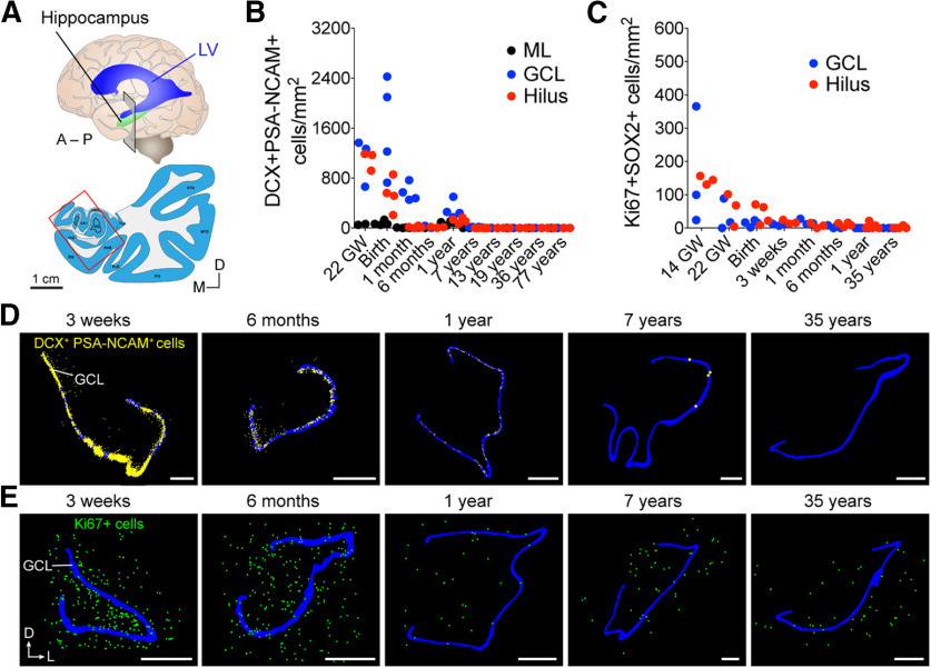
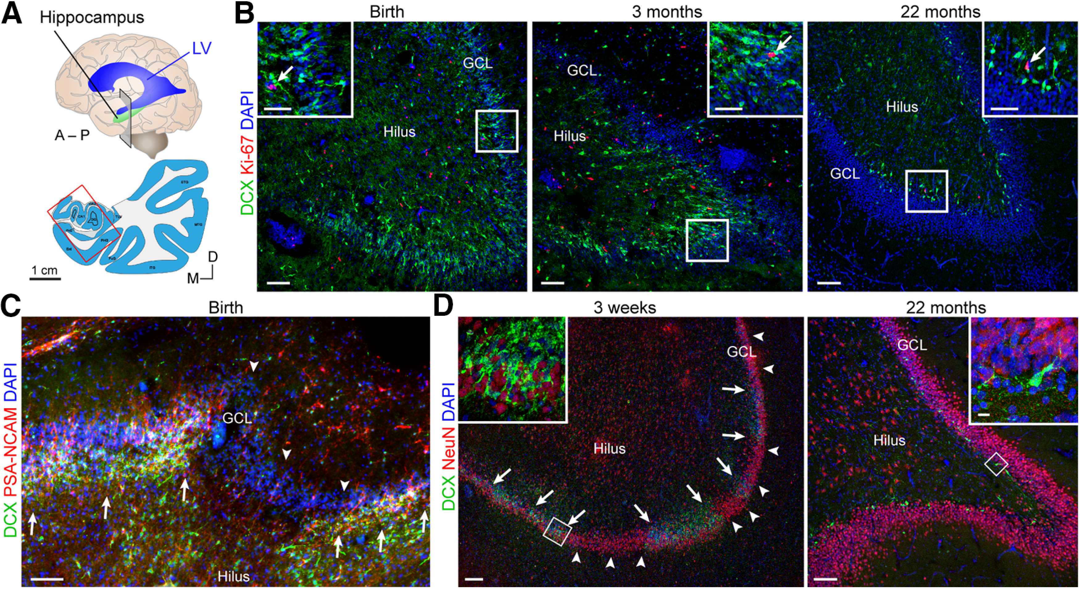
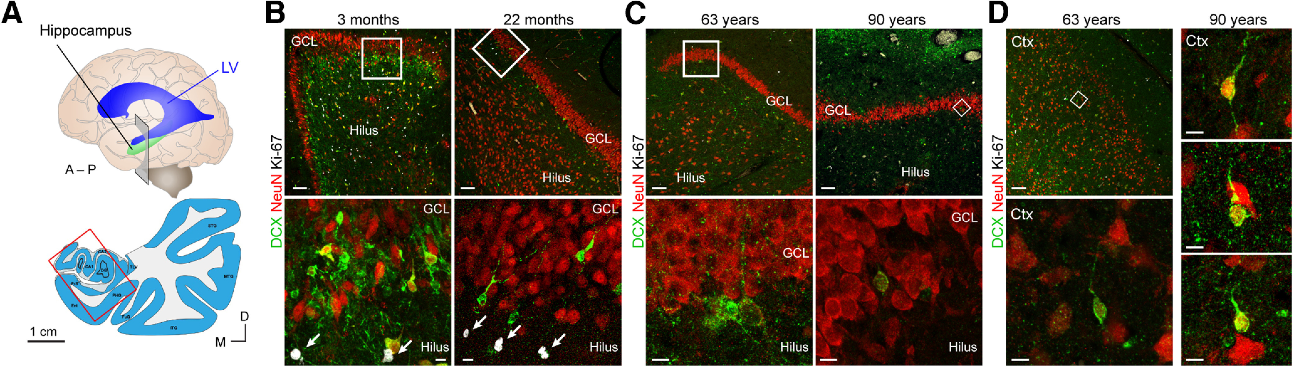
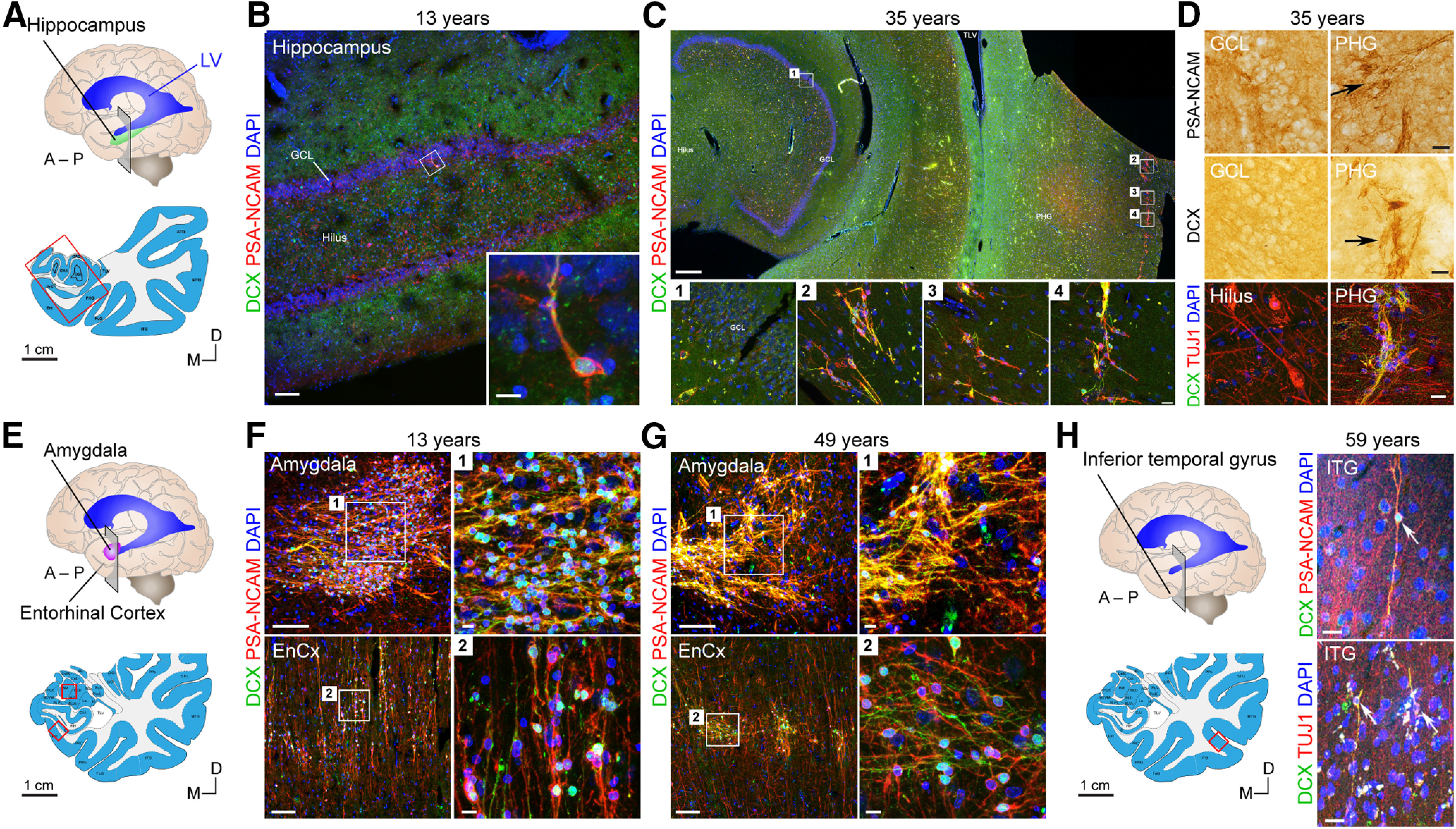
Adult hippocampal neurogenesis was originally discovered in rodents. Subsequent studies identified the adult neural stem cells and found important links between adult neurogenesis and plasticity, behavior, and disease. However, whether new neurons are produced in the human dentate gyrus (DG) during healthy aging is still debated. We and others readily observe proliferating neural progenitors in the infant hippocampus near immature cells expressing doublecortin (DCX), but the number of such cells decreases in children and few, if any, are present in adults. Recent investigations using dual antigen retrieval find many cells stained by DCX antibodies in adult human DG. This has been interpreted as evidence for high rates of adult neurogenesis, even at older ages. However, most of these DCX-labeled cells have mature morphology. Furthermore, studies in the adult human DG have not found a germinal region containing dividing progenitor cells. In this Dual Perspectives article, we show that dual antigen retrieval is not required for the detection of DCX in multiple human brain regions of infants or adults. We review prior studies and present new data showing that DCX is not uniquely expressed by newly born neurons: DCX is present in adult amygdala, entorhinal and parahippocampal cortex neurons despite being absent in the neighboring DG. Analysis of available RNA-sequencing datasets supports the view that DG neurogenesis is rare or absent in the adult human brain. To resolve the conflicting interpretations in humans, it is necessary to identify and visualize dividing neuronal precursors or develop new methods to evaluate the age of a neuron at the single-cell level.

Keywords: aging; dentate gyrus; doublecortin; human hippocampus; neural progenitors; neurogenesis; new neurons.


Figures

Figure 1.
Figure 1.
Early postnatal decline of dividing cells and immature neurons in the human hippocampus. A, Schematic of coronal section in the anterior hippocampus corresponding to maps in D, E. LV, Lateral ventricle; A, anterior; P, posterior; D, dorsal; L, lateral; M, medial. B, Quantification of DCX+PSA-NCAM+ cells in the human molecular layer (ML), GCL, and hilus between 22 gestational weeks (GW) and 77 years. C, Quantification of Ki-67+SOX2+ cells in the GCL and hilus between 14 GW and 35 years. D, Neurolucida maps of the location of DCX+PSA-NCAM+ cells and (E) Ki-67+ cells (bottom) in the human hippocampus between 3 weeks and 35 years. B–E, Reproduced in part from Sorrells et al. (2018). Scale bars: D, E, 1 mm.
Figure 2.
Figure 2.
Cells with morphology and marker expression of young immature neurons are readily detected at young ages in humans. A, Schematic of coronal section in the anterior hippocampus corresponding to images in B–D. B, DCX+ cells (green) and Ki-67+ cells (red) (insets, arrows) in the human DG at birth, 3 months, and 22 months. Although there is not a clear layer of proliferating cells forming a SGZ, there is a close correlation between proliferation and the presence of immature neurons. C, Already at birth, there are patches of the DG with (arrows) and without (arrowheads) DCX+PSA-NCAM+ cells. D, At 3 weeks, regions with many DCX+ cells (arrows) are separated by large regions where DCX+ cells are absent (arrowheads). Scale bars: B–D, 100 µm; B, Insets, 50 µm; D, Insets,10 µm.
Figure 3.
Figure 3.
Dual antigen retrieval in young children reveals DG DCX+ neurons with different morphology than those in the adult, as well as many neurons outside of the DG. A, Schematic of coronal section in the anterior hippocampus corresponding to images in B–D. B, Between 3 and 22 months, small DCX+ neurons (some also expressing NeuN) in the GCL are near Ki-67+ cells (arrows). C, In adults 63 and 90 years of age, large DCX+ neurons expressing NeuN are not near Ki-67+ cells. D, In adults 63 and 90 years of age, many DCX+ cells expressing NeuN can be found in the cortex of the same sections as in C. Scale bars: A, 1 cm; B–D, Top, 100 µm; B, C, Bottom, D, Bottom, Right, 10 µm.
Figure 4.
Figure 4.
Immature DCX+ neurons are readily detectable in 4% PFA-fixed material outside of the DG without dual antigen retrieval. A, Schematic of coronal section in the anterior hippocampus corresponding to images in B–D. B, Low- and high-power images of one individual DCX+PSA-NCAM+ neuron detected in the GCL in the same 13-year-old sample from Sorrells et al. (2018). C, In a 35-year-old adult, DCX+PSA-NCAM+ cells are not detected in the DG (C1); but within the same section, cells are readily detected in the cortical parahippocampal gyrus (PHG) (C2-C4). D, Cells in the PHG stain for DCX, PSA-NCAM, and TUJ1, but similar cells are not present in the DG, and TUJ1 labels cells throughout the hilus. E, Schematic of coronal section in the amygdala corresponding to images in F, G. F, DCX+PSA-NCAM+ cells in the amygdala and entorhinal cortex (EnCx) in the same 13-year-old individual as in B. G, DCX+PSA-NCAM+ cells in the amygdala and entorhinal cortex at 49 years of age. H, Schematic and examples of DCX+PSA-NCAM+ and DCX+TUJ1+ cells in the inferior temporal gyrus at 59 years of age. Scale bars: A, E, H, Left, 1 cm; C, Top, 1 mm; B, F, Left, G, Left, 100 µm; C, Insets, D, H, Right, 20 µm; B, Inset, F–H, Right, 10 µm.
Figure 5.
Figure 5.
Lack of evidence of adult human hippocampal neural stem cells in both single nucleus and bulk RNA-seq data. A, UMAP plot of data from Habib et al. (2017), representing 14,137 nuclei from adult human PFC and hippocampus. UMI count matrix was obtained from the GTEx portal (https://www.gtexportal.org/home/datasets) and processed using default settings in Seurat (filtering for nuclei expressing a minimum of 200 genes and genes expressed in a minimum of 3 cells). Nuclei (dots) are labeled and colored by cluster membership labels from the original study: exPFC, Glutamatergic neurons from the PFC; GABA, GABAergic interneurons; exCA1/3, pyramidal neurons from the hip CA region; exDG, granule neurons from the hip DG region; ASC, astrocytes; MG, microglia; ODC, oligodendrocytes; OPC, oligodendrocyte precursor cells; SMC, smooth muscle cells; END, endothelial cells. Cluster 14 is equivalent to the cluster that Habib et al. (2017) identified and labeled as NSCs. B, Region of origin of nuclei from A. CTX, PFC; HIP, hippocampus. C, Enrichment of ependymal cell type signatures in clusters from A. Ependymal gene sets consist of 440 genes from either mouse (Zeisel et al., 2015) or human (Kelley et al., 2018) data. Enrichment analysis was performed using a one-sided Fisher's exact test using differentially expressed genes of all clusters shown in A. Cluster 14, previously labeled as NSC, preferentially expresses ependymal genes. D, Feature plot of DCX expression in all nuclei. E, Quantification of UMI counts of DCX in all nuclei, grouped by region. Quantification of cell count per cluster, grouped by region. F, Bulk mRNA expression (TPM) of genes related to astrocytes and RGCs (GFAP, vimentin, SOX2), neurons (RBFOX3, DCX), and cell proliferation (MCM2, MKI67) separated in different age groups (20-30 years; 30-40 years; 40-50 years; 50-60 years; 60-70 years). The data used for the analysis were obtained from the GTEx Portal (GTEx Consortium, 2017). The donor eligibility requirements and sample collection process have been previously described (Carithers et al., 2015). We focused our analyses on the hippocampus, excluding the samples positive for Alzheimer disease (n = 4) for a total 117 samples; n = 4 in the 20-30 age group; n = 3 in the 30-40 age group; n = 13 in the 40-50 age group; n = 32 in the 50-60 age group; and n = 64 in the 60-70 age group. The data were normalized using 7 different housekeeping genes (PSMB4, GPI, RAB7A, VCP, C1orf43, CHMP2A, REEP5) previously described to be nonvariable in human tissue (Eisenberg and Levanon, 2013). The change along time is not significant in any of the genes, except for vimentin, which significantly increases from the 50-60 age range to the 60-70 age range (p = 0.016). The statistical analysis was done by one-way ANOVA followed by all pairwise comparisons by Holm-Sidak post hoc test. Data are mean ± SD.

Similar articles

Cited by

References

    1. Akter M, Kaneko N, Herranz-Pérez V, Nakamura S, Oishi H, García-Verdugo JM, Sawamoto K (2020) Dynamic changes in the neurogenic potential in the ventricular–subventricular zone of common marmoset during postnatal brain development. Cereb Cortex 30:4092–4109. 10.1093/cercor/bhaa031 - DOI - PubMed
    1. Altman J (1969) Autoradiographic and histological studies of postnatal neurogenesis: IV. Cell proliferation and migration in the anterior forebrain, with special reference to persisting neurogenesis in the olfactory bulb. J Comp Neurol 137:433–457. 10.1002/cne.901370404 - DOI - PubMed
    1. Altman J, Das GD (1965) Autoradiographic and histological evidence of postnatal hippocampal neurogenesis in rats. J Comp Neurol 124:319–335. 10.1002/cne.901240303 - DOI - PubMed
    1. Ambrogini P, Lattanzi D, Ciuffoli S, Agostini D, Bertini L, Stocchi V, Santi S, Cuppini R (2004) Morpho-functional characterization of neuronal cells at different stages of maturation in granule cell layer of adult rat dentate gyrus. Brain Res 1017:21–31. 10.1016/j.brainres.2004.05.039 - DOI - PubMed
    1. Bai J, Ramos RL, Ackman JB, Thomas AM, Lee RV, LoTurco JJ (2003) RNAi reveals doublecortin is required for radial migration in rat neocortex. Nat Neurosci 6:1277–1283. 10.1038/nn1153 - DOI - PubMed

Publication types