. 2021 Jun 10;95(13):e0228820.
doi: 10.1128/JVI.02288-20. Epub 2021 Jun 10.

Newcastle Disease Virus Entry into Chicken Macrophages via a pH-Dependent, Dynamin and Caveola-Mediated Endocytic Pathway That Requires Rab5

Affiliations

Newcastle Disease Virus Entry into Chicken Macrophages via a pH-Dependent, Dynamin and Caveola-Mediated Endocytic Pathway That Requires Rab5

Ran Zhao et al. J Virol. .

Abstract

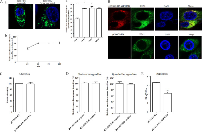
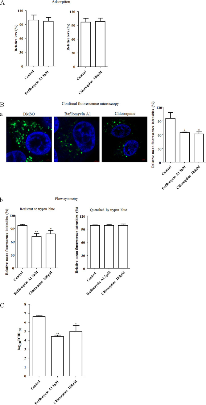
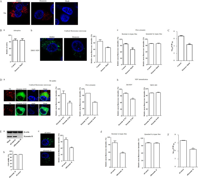
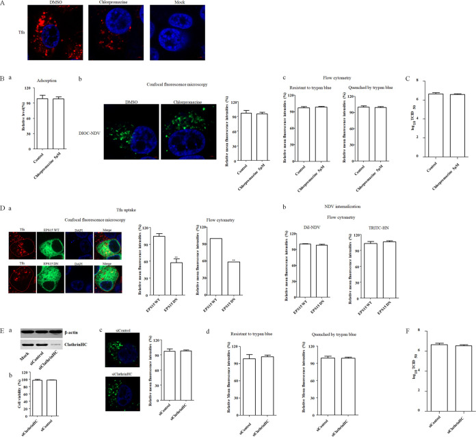
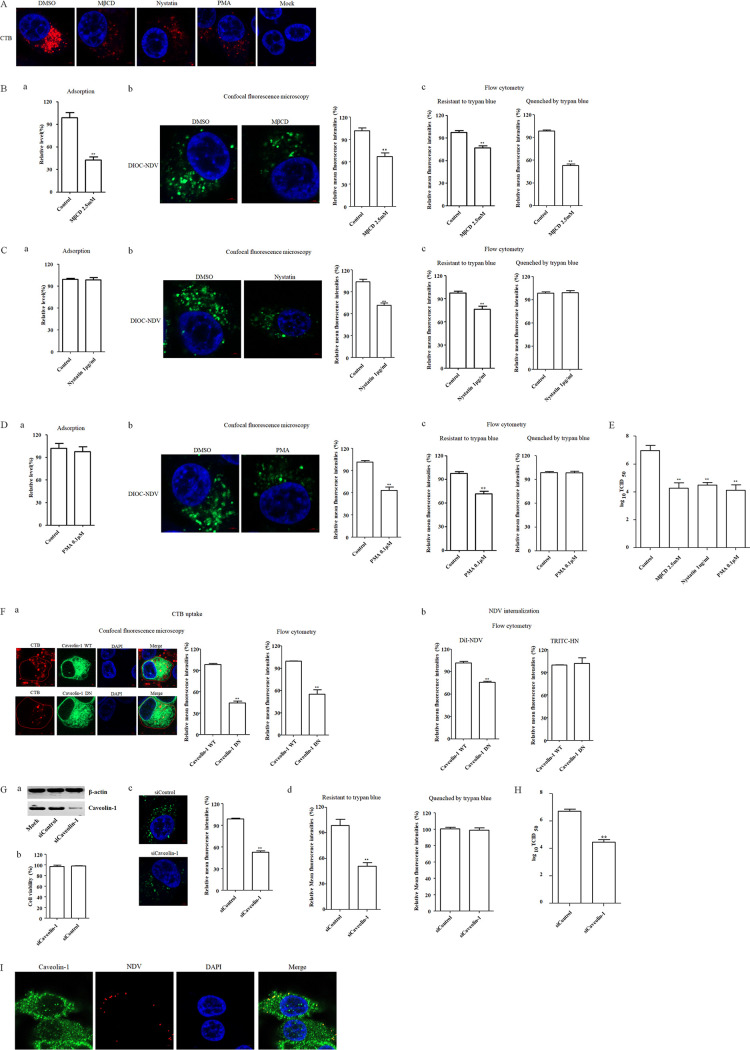
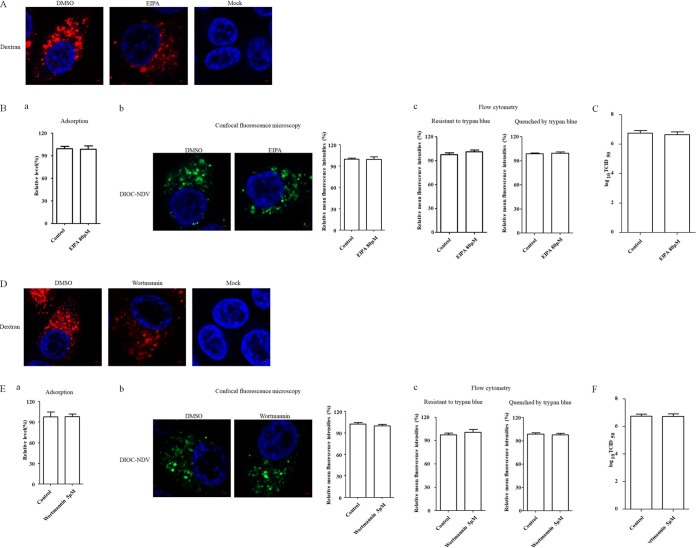
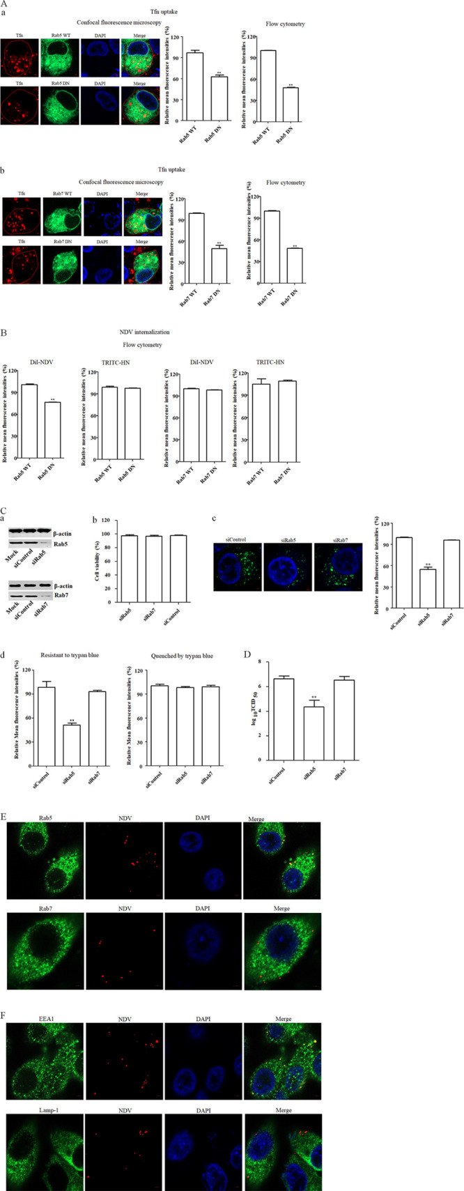
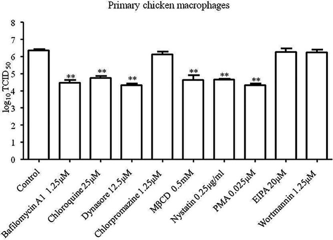
The cellular entry pathways and the mechanisms of Newcastle disease virus (NDV) entry into cells are poorly characterized. In this study, we demonstrated that chicken interferon-induced transmembrane protein 1 (chIFITM1), which is located in the early endosomes, could limit the replication of NDV in chicken macrophage cell line HD11, suggesting the endocytic entry of NDV into chicken macrophages. Then, we presented a systematic study about the entry mechanism of NDV into chicken macrophages. First, we demonstrated that a low-pH condition and dynamin were required during NDV entry. However, NDV entry into chicken macrophages was independent of clathrin-mediated endocytosis. We also found that NDV entry was dependent on membrane cholesterol. The NDV entry and replication were significantly reduced by nystatin and phorbol 12-myristate 13-acetate treatment, overexpression of dominant-negative (DN) caveolin-1, or knockdown of caveolin-1, suggesting that NDV entry depends on caveola-mediated endocytosis. However, macropinocytosis did not play a role in NDV entry into chicken macrophages. In addition, we found that Rab5, rather than Rab7, was involved in the entry and traffic of NDV. The colocalization of NDV with Rab5 and early endosome suggested that NDV virion was transported to early endosomes in a Rab5-dependent manner after internalization. Of particular note, the caveola-mediated endocytosis was also utilized by NDV to enter primary chicken macrophages. Moreover, NDV entered different cell types using different pathways. Collectively, our findings demonstrate for the first time that NDV virion enters chicken macrophages via a pH-dependent, dynamin and caveola-mediated endocytosis pathway and that Rab5 is involved in the traffic and location of NDV. IMPORTANCE Although the pathogenesis of Newcastle disease virus (NDV) has been extensively studied, the detailed mechanism of NDV entry into host cells is largely unknown. Macrophages are the first-line defenders of host defense against infection of pathogens. Chicken macrophages are considered one of the main types of target cells during NDV infection. Here, we comprehensively investigated the entry mechanism of NDV in chicken macrophages. This is the first report to demonstrate that NDV enters chicken macrophages via a pH-dependent, dynamin and caveola-mediated endocytosis pathway that requires Rab5. The result is important for our understanding of the entry of NDV in chicken macrophages, which will further advance the knowledge of NDV pathogenesis and provide useful clues for the development of novel preventive or therapeutic strategies against NDV infection. In addition, this information will contribute to our further understanding of pathogenesis with regard to other members of the Avulavirus genus in the Paramyxoviridae family.

Keywords: Newcastle disease virus; Rab5; chicken macrophage; endocytic pathway.


Figures

FIG 1
FIG 1
NDV undergoes endocytosis and chIFITM1 located in the early endosome restricts the NDV infection. (A) NDV can be endocytosed. HD11 cells were inoculated with DiOC-labeled NDV F48E9 at 37°C for 60 min. After incubation, cells were washed with low-pH buffer to remove noninternalized viruses, and then the cells were fixed and treated with trypan blue to quench the green fluorescence of DiOC in the plasma membrane surface. The fluorescence of DiOC was examined by confocal fluorescence microscopy (a). HD11 cells were inoculated with DiOC-labeled NDV F48E9 at 37°C for different time. The cells were treated with low-pH buffer to inactivate and remove noninternalized viruses and then collected and fixed in 4% paraformaldehyde. Each cell sample was divided into two. One sample was subjected directly for the flow cytometric analysis, and the other was treated with trypan blue. The fluorescence of DiOC was examined by flow cytometry. The fluorescence intensity that was resistant (b) and quenched (c) by trypan blue at 60, 90, and 120 min was calculated relative to that of 30 min (×100%). (B) Cellular localization of chIFITM1. HD11 cells were transfected with pCAGGS-HA-chIFITM1 or pCAGGS-HA. The cells were fixed at 24 h after transfection and then subjected to indirect immunofluorescence to detect HA-tagged chIFITM1 and early endosomes marker, EEA1, using anti-HA antibody (red) and anti-EEA1 antibody (green). The nuclei were stained with DAPI (blue). (C) chIFITM1 did not inhibit NDV adsorption. HD11 cells transfected with pCAGGS-HA-chIFITM1 or pCAGGS-HA were incubated with NDV F48E9 for 1 h at 4°C to allow for viral adsorption. Viral RNA was quantified by real-time RT-PCR. (D) chIFITM1 did not inhibit NDV internalization. HD11 cells transfected with pCAGGS-HA-chIFITM1 or pCAGGS-HA were incubated with DiOC-labeled NDV F48E9 for 1 h at 4°C to allow for viral adsorption. Cells were then washed and further incubated for 1 h at 37°C to allow for internalization. After incubation, the cells were washed with low-pH buffer to remove noninternalized viruses and then collected and fixed in 4% paraformaldehyde. Each cell sample was divided into two. One sample was subjected directly for the flow cytometric analysis, and the other was treated with trypan blue. The HA-chIFITM1 was detected by using an HA tag antibody. The green fluorescence of DiOC-labeled NDV that was resistant and quenched by trypan blue in HA-chIFITM1-positive and-negative cells was measured by flow cytometry. (E) chIFITM1 reduced the replication of NDV in HD11 cells. HD11 cells were transfected with pCAGGS-HA-chIFITM1 or pCAGGS-HA. At 48 h posttransfection, cells were infected with F48E9 at an MOI of 0.1. At 18 hpi, viral titers in the culture supernatants of infected cells were determined. The bars represent means ± the SD. Data were analyzed by using the Student t test (*, P < 0.05; **, P < 0.01).
FIG 2
FIG 2
NDV entry into HD11cells requires acidic endosomal pH. (A) Bafilomycin A1 and chloroquine did not affect the adsorption of NDV. HD11 cells were treated with the indicated concentration of inhibitors for 1 h at 37°C. Cells were then incubated with NDV F48E9 in the presence of inhibitors for 1 h at 4°C to allow for viral adsorption. DMSO was included as a negative control. Viral RNA was quantified by real-time RT-PCR. (B) Bafilomycin A1 and chloroquine inhibited the internalization of NDV. DiOC-labeled NDV F48E9 was adsorbed onto HD11 cells and then incubated with indicated concentrations of inhibitors for 1 h at 37°C to permit internalization. After a low-pH buffer wash and quenching the green fluorescence of DiOC in the plasma membrane surface with trypan blue, the green fluorescence of internalized virus was determined by confocal fluorescence microscopy (a). Meanwhile, the cells with the same treatment were subjected to flow cytometry. Each cell sample was divided into two. One sample was subjected directly for the flow cytometric analysis, and the other was treated with trypan blue (b). The fluorescence intensity which resistant and quenched by trypan blue in inhibitor-treated cells was calculated relative to that of control cells (×100%). (C) Bafilomycin A1 and chloroquine reduced the replication of NDV. HD11 cells were infected with NDV F48E9 at an MOI of 0.1 in the presence of the inhibitors at 37°C for 1 h. DMSO was included as a negative control. Cells were then washed with PBS and incubated in medium containing 5% FBS at 37°C. At 18 hpi, viral titers in the culture supernatants of infected cells were determined. The bars represent means ± the SD. Data were analyzed by using the Student t test (*, P < 0.05; **, P < 0.01).
FIG 3
FIG 3
NDV entry into HD11cells depends on dynamin. (A) Dynasore inhibited Tfn uptake. HD11 cells were treated with 50 μM dynasore or DMSO for 1 h at 37°C, followed by incubation with 10 μg/ml Tfn for 1 h at 4°C, and then shifted to 37°C for 1 h; after a wash with low-pH buffer, the cells were fixed and stained with DAPI. (B) Dynasore inhibited the internalization of NDV. HD11 cells were treated with the indicated concentration of inhibitors for 1 h at 37°C. The cells were then incubated with NDV F48E9 in the presence of inhibitors for 1 h at 4°C to allow for viral adsorption. DMSO was included as a negative control. Viral RNA was quantified by real-time RT-PCR (a). Alternatively, DiOC-labeled NDV F48E9 was adsorbed onto HD11 cells and then incubated with indicated concentrations of inhibitors for 1 h at 37°C to permit internalization. After a wash with low-pH buffer and quenching the green fluorescence of DiOC in the plasma membrane surface with trypan blue, the green fluorescence of internalized virus was determined by confocal fluorescence microscopy (b). Meanwhile, the treated cells were subjected to flow cytometry. Each cell sample was divided into two. One sample was subjected directly for the flow cytometric analysis, and another was treated with trypan blue (c). The fluorescence intensity which was resistant and quenched by trypan blue in inhibitor-treated cells was calculated relative to that of control cells (×100%). (C) Dynasore reduced the replication of NDV. HD11 cells were infected with NDV F48E9 at an MOI of 0.1 in the presence of the inhibitors at 37°C for 1 h. DMSO was included as a negative control. The cells were then washed with PBS and incubated in medium containing 5% FBS at 37°C. At 18 hpi, the viral titers in the culture supernatants of infected cells were determined. (D) Effects of dynamin II on Tfn uptake and NDV internalization. HD11 cells transfected with the EGFP-tagged WT or DN dynamin II were incubated with 10 μg/ml Tfn and DiI-labeled or unlabeled NDV F48E9 for 1 h at 4°C and then shifted to 37°C for 1 h. After a wash with low-pH buffer, the red fluorescence of Tfn in EGFP-positive cells was determined by confocal fluorescence microscopy and flow cytometry, respectively (a). The red fluorescence of DiI-labeled NDV in EGFP-positive cells was determined by flow cytometry (b). In addition, the level of HN protein on the cell surface in unlabeled NDV-infected EGFP-positive cells was determined using anti-HN antibody via flow cytometry (b). (E) Dynamin II knockdown inhibited the internalization of NDV. HD11 cells were transfected with siRNA targeting dynamin (siDynamin II) or a control siRNA (siControl). The effect of siRNA knockdown on dynamin expression was determined by Western blotting (a). Cell viability upon siDynamin II and siControl transfection was assessed as described in the text (b). At 48 h posttransfection, the internalization assay was performed with DiOC-labeled NDV F48E9 as described above. The green fluorescence of DiOC that was resistant and quenched by trypan blue was determined by confocal fluorescence microscopy (c) and flow cytometry (d), respectively. (F) Dynamin knockdown reduced the replication of NDV. HD11 cells were transfected with siDynamin II or siControl. At 48 h posttransfection, cells were infected with NDV F48E9 at an MOI of 0.1, and the viral titers in the culture supernatants of infected cells were determined at 18 hpi. The bars represent means ± the SD. Data were analyzed by using the Student t test (*, P < 0.05; **, P < 0.01).
FIG 4
FIG 4
Clathrin-mediated endocytosis is not required for NDV entry. (A) Chlorpromazine inhibited Tfn uptake. HD11 cells were treated with 5 μM chlorpromazine or DMSO for 1 h at 37°C, followed by incubation with 10 μg/ml Tfn for 1 h at 4°C, and then shifted to 37°C for 1 h; after a wash with low-pH buffer, the cells were fixed and stained with DAPI. (B) Chlorpromazine did not inhibit NDV adsorption and internalization. HD11 cells were treated with indicated concentration of inhibitors for 1 h at 37°C. Cells were then incubated with NDV F48E9 in the presence of inhibitors for 1 h at 4°C to allow for viral adsorption. DMSO was included as a negative control. Viral RNA was quantified by real-time RT-PCR (a). Alternatively, DiOC-labeled NDV F48E9 was adsorbed onto HD11 cells and then incubated with the indicated concentrations of inhibitors for 1 h at 37°C. The internalization assay was performed as described above. The green fluorescence of DiOC that was resistant and quenched by trypan blue was determined by confocal fluorescence microscopy (b) and flow cytometry (c), respectively. (C) Chlorpromazine did not affect the replication of NDV. HD11 cells were infected with NDV F48E9 at an MOI of 0.1 in the presence of the inhibitors at 37°C for 1 h. DMSO was included as a negative control. Cells were then washed with PBS and incubated in medium containing 5% FBS at 37°C. At 18 hpi, viral titers in the culture supernatants of infected cells were determined. (D) Effects of EPS15 on Tfn uptake and NDV internalization. HD11 cells transfected with the EGFP-tagged WT or DN EPS15 were incubated with 10 μg/ml Tfn and DiI-labeled or unlabeled NDV F48E9 for 1 h at 4°C and then shifted to 37°C for 1 h. After a wash with low-pH buffer, the red fluorescence of Tfn (a) and DiI-labeled NDV (b) and HN protein on the cell surface (panel b) was determined as described above. (E) Clathrin knockdown showed no effect on the internalization of NDV. HD11 cells were transfected with siRNA targeting clathrin heavy chain (siClathrinHC) or a control siRNA (siControl). The effect of siRNA knockdown on clathrin heavy chain expression was determined by Western blotting (a). Cell viability upon siClathrinHC and siControl transfection was assessed as described in the text (b). At 48 h posttransfection, the internalization assay was performed with DiOC-labeled NDV F48E9 as described above. The green fluorescence of DiOC that was resistant and quenched by trypan blue was determined by confocal fluorescence microscopy (c) and flow cytometry (d), respectively. (F) Clathrin knockdown showed no effect on the replication of NDV. HD11 cells were transfected with siClathrinHC or siControl. At 48 h posttransfection, the cells were infected with NDV F48E9 at an MOI of 0.1, and the viral titers in the culture supernatants of infected cells were determined at 18 hpi. The bars represent means ± the SD. Data were analyzed by using the Student t test (*, P < 0.05; **, P < 0.01).
FIG 5
FIG 5
Caveola-mediated endocytosis is required for NDV entry. (A) MβCD-, nystatin-, and PMA-inhibited CTB uptake. HD11 cells were treated with 2.5 mM MβCD, 1 μg/ml nystatin, 0.1 μM PMA, or DMSO for 1 h at 37°C, followed by incubation with 10 μg/ml CTB for 1 h at 4°C, and then shifted to 37°C for 1 h; a after wash with low-pH buffer, the cells were fixed and stained with DAPI. (B to D) Effects of inhibitors of caveola-mediated endocytosis on the adsorption and internalization of NDV. The effects of MβCD (B), nystatin (C), and PMA (D) on the adsorption and internalization of NDV were determined. HD11 cells were treated with the indicated concentrations of inhibitors for 1 h at 37°C. The cells were then incubated with NDV F48E9 in the presence of inhibitors for 1 h at 4°C to allow for viral adsorption. DMSO was included as a negative control. Viral RNA was quantified by real-time RT-PCR (subpanel a in panels B, C, and D). Alternatively, DiOC-labeled NDV F48E9 was adsorbed onto HD11 cells and then incubated with the indicated concentrations of inhibitors for 1 h at 37°C; the internalization assay was then performed as described above. The green fluorescence of DiOC that was resistant and quenched by trypan blue was determined by confocal fluorescence microscopy (subpanel b in panels B, C, and D) and flow cytometry (subpanel c in panels B, C, and D), respectively. (E) MβCD, nystatin, and PMA reduced the replication of NDV. HD11 cells were infected with NDV F48E9 at an MOI of 0.1 in the presence of the inhibitors at 37°C for 1 h. DMSO was included as a negative control. Next, the cells were washed with PBS and incubated in medium containing 5% FBS at 37°C. At 18 hpi, the viral titers in the culture supernatants of infected cells were determined. (F) Effects of caveolin-1 on CTB uptake and NDV internalization. HD11 cells transfected with plasmids expressing EGFP-tagged WT and DN caveolin-1 were incubated with 10 μg/ml CTB and DiI-labeled or unlabeled NDV F48E9 for 1 h at 4°C and then shifted to 37°C for 1 h. After a wash with low-pH buffer, the red fluorescence of Tfn (a) and DiI-labeled NDV (b) and HN protein (b) on the cell surface was determined as described above. (G) Caveolin-1 knockdown inhibited NDV internalization. HD11 cells were transfected with siRNA targeting caveolin-1 (siCaveolin-1) or siControl. The effect of siRNA knockdown on caveolin-1 expression was determined by Western blotting (a). Cell viability upon siCaveolin-1 and siControl transfection was assessed as described in the text (b). At 48 h posttransfection, an internalization assay was performed with DiOC-labeled NDV F48E9, as described above. The green fluorescence of DiOC that was resistant and quenched by trypan blue was determined by confocal fluorescence microscopy (c) and flow cytometry (d), respectively. (H) Caveolin-1 knockdown reduced the replication of NDV. HD11 cells were transfected with siCaveolin-1 or siControl. At 48 h posttransfection, the cells were infected with NDV F48E9 at an MOI of 0.1, and the viral titers in the culture supernatants of infected cells were determined at 18 hpi. (I) The colocalization of NDV with caveolin-1. HD11 cells were incubated with NDV F48E9 at an MOI of 10 at 4°C for 1 h and then shifted to 37°C for 1 h. The cells were then fixed and reacted with anti-caveolin-1 antibody and NDV HN antibody and visualized by confocal microscopy. The bars represent means ± the SD. Data were analyzed by using the Student t test (*, P < 0.05; **, P < 0.01).
FIG 6
FIG 6
Macropinocytosis is not required for NDV entry. (A and D) EIPA and wortmannin inhibited dextran uptake. HD11 cells were treated with 80 μM EIPA, 5 μM wortmannin, or DMSO for 1 h at 37°C, followed by incubation with 10 μg/ml dextran for 1 h at 4°C, and then shifted to 37°C for 1 h; after a wash with low-pH buffer, the cells were fixed and stained with DAPI. (B and E) EIPA and wortmannin did not inhibit NDV adsorption and internalization. HD11 cells were treated with indicated concentration of inhibitors for 1 h at 37°C. The cells were then incubated with NDV F48E9 in the presence of inhibitors for 1 h at 4°C to allow viral adsorption. DMSO was included as a negative control. Viral RNA was quantified by real-time RT-PCR (subpanel a in panels B and E). Alternatively, DiOC-labeled NDV F48E9 was adsorbed onto HD11 cells and then incubated with indicated concentrations of inhibitors for 1 h at 37°C. AN internalization assay was performed as described above. The green fluorescence of DiOC that was resistant and quenched by trypan blue was determined by confocal fluorescence microscopy (subpanel b in panels B and E) and flow cytometry (subpanel c in panels B and E), respectively. (C and F) EIPA and wortmannin did not affect the replication of NDV. HD11 cells were infected with NDV F48E9 at an MOI of 0.1 in the presence of the inhibitors at 37°C for 1 h. DMSO was included as a negative control. The cells were washed with PBS and incubated in medium containing 5% FBS at 37°C. At 18 hpi, the viral titers in the culture supernatants of infected cells were determined. The bars represent means ± the SD. Data were analyzed by using the Student t test (*, P < 0.05; **, P < 0.01).
FIG 7
FIG 7
Effects of Rabs on NDV infection. (A and B) Effects of Rab5 and Rab7 on Tfn uptake (A) and NDV internalization (B). HD11 cells transfected with plasmids expressing EGFP-tagged WT or DN Rab5 and Rab7 were incubated with 10 μg/ml Tfn, and DiI-labeled or unlabeled NDV F48E9 for 1 h at 4°C and then shifted to 37°C for 1 h. After a wash with low-pH buffer, the red fluorescence of Tfn (subpanels a and b in panel A), DiI-labeled NDV (B), and HN protein (B) on the cell surface was determined as described above. (C) Rab5 knockdown reduced the internalization of NDV. HD11 cells were transfected with siRNA targeting Rab 5 (siRab5), Rab7 (siRab7), or siControl. The effect of siRNA knockdown of Rab5 and Rab7 expression was determined by Western blotting (a). Cell viability upon siRab5, siRab7, and siControl transfection was assessed, as described in the text (b). At 48 h posttransfection, an internalization assay was performed with DiOC-labeled NDV F48E9 as described above. The green fluorescence of DiOC that was resistant and quenched by trypan blue was determined by confocal fluorescence microscopy (c) and flow cytometry (d), respectively. (D) Rab 5 knockdown reduced the replication of NDV. HD11 cells were transfected with siRab5, siRab7, or siControl. At 48 h posttransfection, the cells were infected with NDV F48E9 at an MOI of 0.1, and the viral titers in the culture supernatants of infected cells were determined at 18 hpi. (E) Colocalization of NDV with Rab5. HD11 cells were incubated with NDV F48E9 at an MOI of 10 at 4°C for 1 h and then shifted to 37°C for 1 h. Cells were then fixed and reacted with anti-Rab5 antibody, anti-Rab7 antibody, and NDV HN antibody, respectively, and visualized by confocal microscopy. (F) Colocalization of NDV with the early endosome marker EEA1. HD11 cells were incubated with NDV F48E9 at an MOI of 10 at 4°C for 1 h and then shifted to 37°C for 1 h. The cells were fixed and reacted with anti-EEA1 antibody, anti-Lamp-1 antibody, and NDV HN antibody, respectively, and visualized by confocal microscopy. The bars represent means ± the SD. Data were analyzed by using the Student t test (*, P < 0.05; **, P < 0.01).
FIG 8
FIG 8
NDV utilizes caveola-mediated endocytosis to enter primary chicken macrophages. Primary chicken macrophages cultured in 24-well plates were treated with the highest concentrations of bafilomycin A1, chloroquine, dynasore, chlorpromazine, MβCD, nystatin, PMA, EIPA, and wortmannin without causing obvious cytotoxic effects for 1 h at 37°C. Cells treated with DMSO were included as a control. The cells were infected with F48E9 at an MOI of 0.1 in the presence of the inhibitors at 37°C for 1 h. After incubation, the cells were washed with PBS and incubated in medium containing 5% FBS at 37°C. At 24 hpi, the viral titers in the culture supernatants of infected cells were determined. The bars represent means ± the SD. Data were analyzed by using the Student t test (*, P < 0.05; **, P < 0.01).
FIG 9
FIG 9
Effects of inhibitors on the replication of NDV in DF-1 (A) and LMH (B) cells. DF-1 and LMH cells cultured in 12-well plates were treated with the highest concentrations of bafilomycin A1, chloroquine, dynasore, chlorpromazine, MβCD, nystatin, PMA, EIPA, and wortmannin without causing obvious cytotoxic effects for 1 h at 37°C. Cells treated with DMSO were included as a control. The cells were infected with F48E9 at an MOI of 0.1 in the presence of the inhibitors at 37°C for 1 h. After incubation, the cells were washed with PBS and incubated again in medium containing 5% FBS at 37°C. At 18 hpi, the viral titers in the culture supernatants of infected DF-1 and LMH cells were determined. The bars represent means ± the SD. Data were analyzed by using the Student t test (*, P < 0.05; **, P < 0.01).

Similar articles

Cited by

References

    1. Alexander DJ. 2009. Ecology and epidemiology of Newcastle disease, p 19–26. In Capua I, Alexander DJ (ed), Avian influenza and Newcastle disease. Springer, New York, NY.
    1. Ganar K, Das M, Sinha S, Kumar S. 2014. Newcastle disease virus: current status and our understanding. Virus Res 184:71–81. 10.1016/j.virusres.2014.02.016. - DOI - PMC - PubMed
    1. Alexander DJ, Aldous EW, Fuller CM. 2012. The long view: a selective review of 40 years of Newcastle disease research. Avian Pathol 41:329–335. 10.1080/03079457.2012.697991. - DOI - PubMed
    1. Cox RM, Plemper RK. 2017. Structure and organization of paramyxovirus particles. Curr Opin Virol 24:105–114. 10.1016/j.coviro.2017.05.004. - DOI - PMC - PubMed
    1. El Najjar F, Schmitt AP, Dutch RE. 2014. Paramyxovirus glycoprotein incorporation, assembly and budding: a three-way dance for infectious particle production. Viruses 6:3019–3054. 10.3390/v6083019. - DOI - PMC - PubMed

Publication types

LinkOut - more resources