. 2021 Mar 24;6(1):133.
doi: 10.1038/s41392-021-00542-2.

FGF1ΔHBS prevents diabetic cardiomyopathy by maintaining mitochondrial homeostasis and reducing oxidative stress via AMPK/Nur77 suppression

Affiliations

FGF1ΔHBS prevents diabetic cardiomyopathy by maintaining mitochondrial homeostasis and reducing oxidative stress via AMPK/Nur77 suppression

Dezhong Wang et al. Signal Transduct Target Ther. .

Abstract

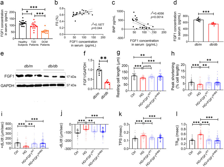
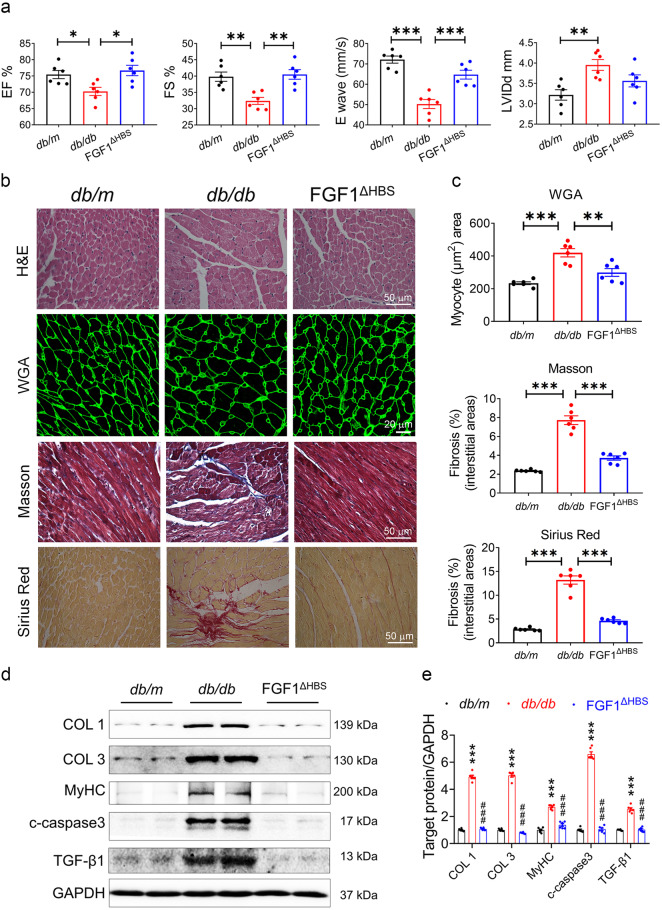
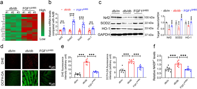
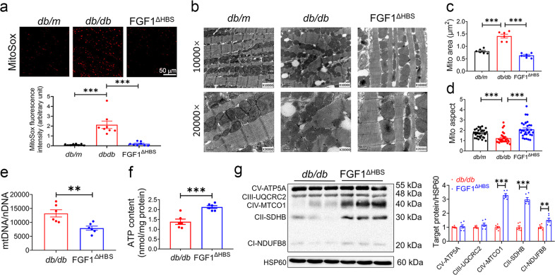
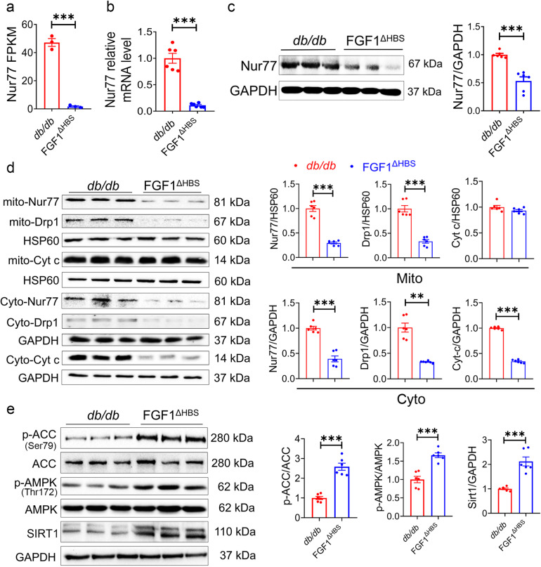
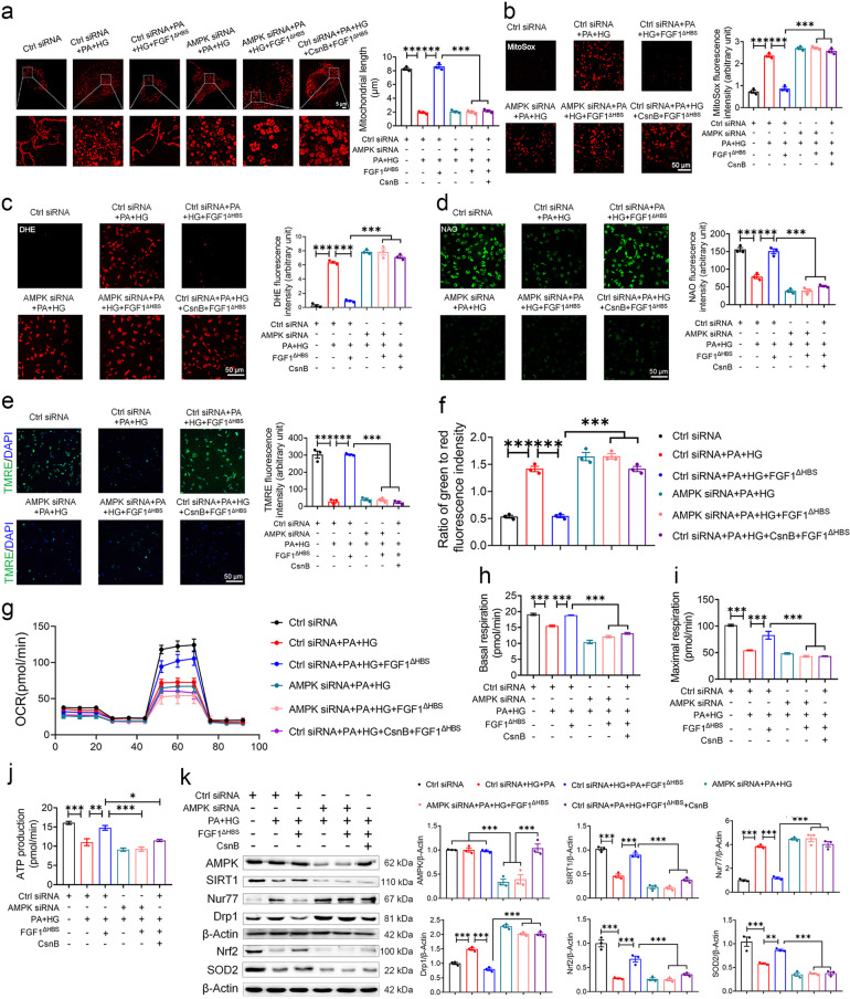
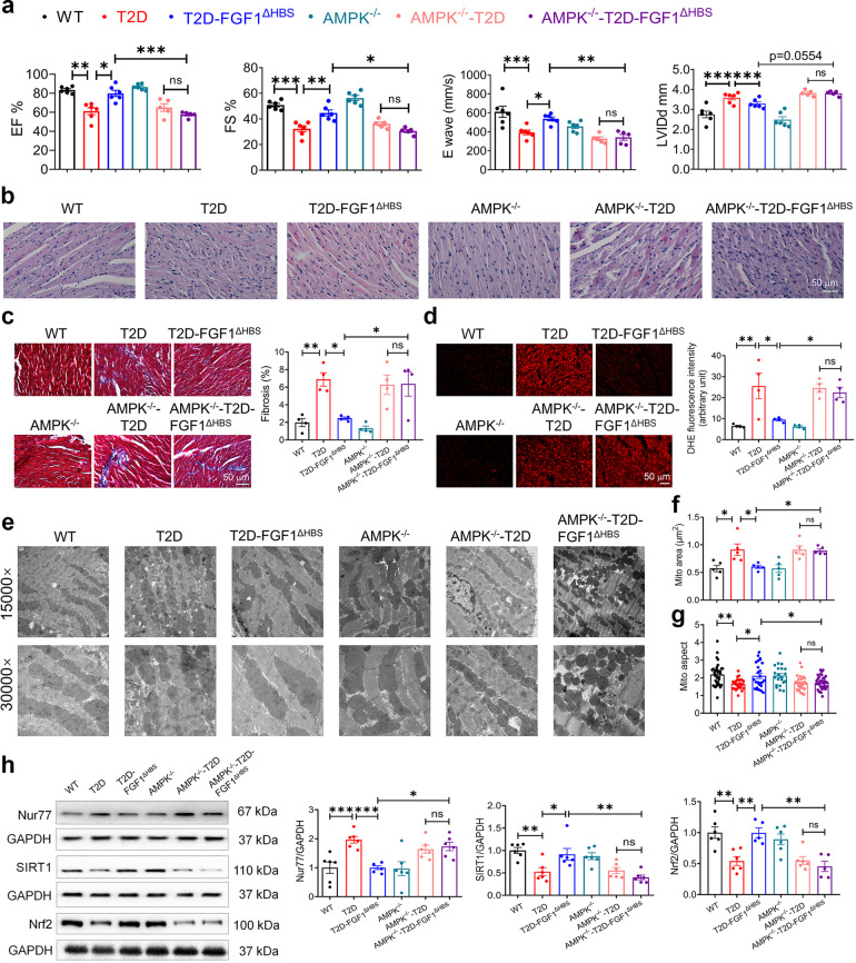
As a classically known mitogen, fibroblast growth factor 1 (FGF1) has been found to exert other pleiotropic functions such as metabolic regulation and myocardial protection. Here, we show that serum levels of FGF1 were decreased and positively correlated with fraction shortening in diabetic cardiomyopathy (DCM) patients, indicating that FGF1 is a potential therapeutic target for DCM. We found that treatment with a FGF1 variant (FGF1∆HBS) with reduced proliferative potency prevented diabetes-induced cardiac injury and remodeling and restored cardiac function. RNA-Seq results obtained from the cardiac tissues of db/db mice showed significant increase in the expression levels of anti-oxidative genes and decrease of Nur77 by FGF1∆HBS treatment. Both in vivo and in vitro studies indicate that FGF1∆HBS exerted these beneficial effects by markedly reducing mitochondrial fragmentation, reactive oxygen species (ROS) generation and cytochrome c leakage and enhancing mitochondrial respiration rate and β-oxidation in a 5' AMP-activated protein kinase (AMPK)/Nur77-dependent manner, all of which were not observed in the AMPK null mice. The favorable metabolic activity and reduced proliferative properties of FGF1∆HBS testify to its promising potential for use in the treatment of DCM and other metabolic disorders.


Conflict of interest statement

The authors declare no competing interests.

Figures

Fig. 1
Fig. 1
Decreased endogenous FGF1 levels in diabetic individuals and comparably protective function of FGF1WT and FGF1ΔHBS in high-glucose-treated cardiomyocytes. a Serum levels of FGF1 in healthy subjects (n = 17), T2D patients with (n = 10) and without (n = 12) DCM. b Correlation between serum FGF1 levels and fractional shortening (FS) in T2D patients. c Correlation between serum FGF1 and BNP levels in T2D patients. d Serum FGF1 levels in db/m (Ctrl) and db/db (T2D) mice determined by ELISA. n = 6. e Representative western blot analysis of FGF1 in cardiac tissues from db/m and db/db mice. GAPDH was a loading control. f Densitometric quantification of western blots as shown in e. n = 6. gl Contractile properties of primary cardiomyocytes from adult C57Bl/6J mice were treated with FGF1WT or FGF1ΔHBS (500 ng/mL for 1 h) and exposed to high glucose (HG, 35 mM) for 5 h. n = 62–65. g Resting cell length. h Peak shortening normalized to resting cell length. i Maximal velocity of shortening (+dL/dt). j Maximal velocity of re-lengthening (−dL/dt). k Time to peak shortening. l Time to 90% re-lengthening. Data were mean ± SEM; *P < 0.05, **P < 0.01, ***P < 0.001
Fig. 2
Fig. 2
FGF1ΔHBS prevents cardiac dysfunction and remodeling in db/db mice. ae db/db mice were treated with FGF1ΔHBS (0.5 mg/kg body weight) or vehicle every other day for 16 weeks, and littermate db/m mice served as controls. a Echocardiographic parameters. n = 6. b Representative images of hematoxylin-eosin (H&E), WGA, Masson’s trichrome, Sirius red staining in cardiac tissues. c Quantification of myocyte area and cardiac fibrosis area in WGA, Masson’s trichrome and Sirius Red staining. n = 6. d Western blot analysis of collagen I (COL 1), collagen III (COL 3), myosin heavy chain (MyHC), cleaved caspase 3 (c-caspase 3) and transforming growth factor β1 (TGF-β1) in cardiac tissues; GAPDH was a loading control. e Densitometric quantification of western blots shown in d. n = 6. Data were mean ± SEM; *P < 0.05, **P < 0.01, ***P < 0.001 in a, c; ***P < 0.001 vs. db/m, ###P < 0.001 vs. db/db in e
Fig. 3
Fig. 3
FGF1ΔHBS preserves anti-oxidant capacity in the hearts of db/db mice. a Hierarchical clustering of FGF1ΔHBS-upregulated genes related to anti-oxidative stress based on the RNA sequencing analysis in the hearts from from db/db and db/db + FGF1ΔHBS mice. n = 3. b The mRNA levels of Nrf2 and its target genes Gsta3, Sod-2 and Ho-1 in the cardiac tissues. n = 6. c Western blot analysis (left panel) and densitometric quantification (right panel) of Nrf2, SOD2 and HO-1 in the cardiac tissues. GAPDH was a loading control. n = 6. d Representative images of DHE and DCFH-DA staining in the cardiac tissues. e Quantification of the corresponding fluorescence intensity in d. n = 6. f Relative concentration of NADP+ normalized to the db/m group. n = 6. Data were mean ± SEM; **P < 0.01 ***P < 0.001 in (b, e, f); ***P < 0.001 vs. db/m, ###P < 0.001 vs db/db in c
Fig. 4
Fig. 4
FGF1ΔHBS prevents mitochondrial ROS production and dysfunction in the hearts of db/db mice. a Representative images (upper panel) and quantification (lower panel) of fluorescence intensity of MitoSox in cardiac tissues from db/db and db/db + FGF1ΔHBS mice. n = 6. b Representative transmission electron micrographs of cardiac tissues in each group. c Mitochondrial area in cardiac tissues of each group. n = 6. d Mitochondrial aspect ratio (long/short axis) in the cardiac tissues. n = 30–31. e mtDNA copy number per nuclear genome in the cardiac tissues. n = 6. f ATP content in the cardiac tissues. n = 6. g Western blot analysis (left panel) and densitometric quantification (right panel) of mitochondrial respiratory complex in the cardiac tissues. HSP60 was a loading control. n = 6. Data were mean ± SEM; **P < 0.01, ***P < 0.001
Fig. 5
Fig. 5
FGF1ΔHBS suppresses Nur77 expression and activates AMPK in the hearts of db/db mice. a FPKM values of Nur77 gene in the RNA-Seq analysis. n = 3. b Nur77 mRNA levels confirmed by qPCR. n = 6. c Western blot analysis (left panel) and densitometric quantification (right panel) of Nur77 in the cardiac tissues. GAPDH was a loading control. n = 6. d Western blot analysis (left panel) and densitometric quantification (right panel) of Drp1, Nur77 and Cyt C in cardiac tissues of each group. HSP60 and GAPDH were loading controls. n = 6. e Western blot analysis (left panel) and densitometric quantification (right panel) of p-ACC, ACC, p-AMPK, AMPKα2 and SIRT1 in the cardiac tissues. GAPDH was a loading control. n = 6. Data were mean ± SEM; **P < 0.01, ***P < 0.001
Fig. 6
Fig. 6
FGF1∆HBS attenuates mitochondrial dysfunction in primary cardiomyocytes via AMPK-Nur77 pathway. ak Primary cardiomyocytes were transfected with control or AMPK siRNA and starved for 12 h, and then cardiomyocytes were treated with palmitate (500 μM) and high glucose (35 mM) with or without CsnB (10 μg/mL) in FBS free medium for 1 h, followed by incubation with FGF1∆HBS (500 ng/mL) for additional 48 h. Mannitol + control siRNA group was an osmotic control. a Representative images of MitoTracker staining (left panel) and mitochondrial length (right panel) of primary cardiomyocytes. bd Representative images of MitoSox (b), DHE (c), and 10-N-nonyl acridine orange (NAO) (d) staining and corresponding quantitative analysis of fluorescence intensity. e Representative images of TMRE staining (left panel) and quantitative analysis (right panel) of fluorescence intensity. f Mitochondrial membrane potential was evaluated by the ratio of JC-10 fluorescence intensities at 529 nm (green) and 590 nm (red). gj Mitochondrial respiratory function was assessed by OCR assay. k Western blot analysis (left panel) and densitometric quantification (right panel) of AMPKα2, SIRT1, Nur77, Drp1, Nrf2 and SOD2 in the cardiomyocytes. n = 3 independent experiments for each group. Data were mean ± SEM; *P < 0.05, **P < 0.01, ***P < 0.001
Fig. 7
Fig. 7
AMPK mediates the protective effect of FGF1ΔHBS against DCM in vivo. ah At the age of 8 weeks, male wild type (WT) or AMPKα2 knockout mice were fed with a high-fat diet for 8 weeks and intraperitoneal injected with STZ, and then treated with FGF1∆HBS (0.5 mg/kg body weight) or saline every other day for 8 weeks. a Echocardiographic assessment for each group. n = 5–6. b H&E staining of the cardiac tissues. c Masson’s trichrome staining (left panel) and quantitative analysis (right panel) of cardiac fibrosis area. n = 4. d Representative images (left panel) and quantification (right panel) of fluorescence intensity of DHE in cardiac tissues. n = 4. e Representative transmission electron micrographs of the cardiac tissues. f Mitochondrial area of the cardiac tissues. n = 5. g Mitochondrial aspect ratio (long/short axis) of the cardiac tissues. n = 21–37. h Western blot analysis (left panel) and densitometric quantification (right panel) of Nur77, SIRT1 and Nrf2 in the cardiac tissues. n = 5–6. Data were mean ± SEM; *P < 0.05, **P < 0.01, ***P < 0.001; ns, not significant

Similar articles

Cited by

References

    1. Tan, Y. et al. Mechanisms of diabetic cardiomyopathy and potential therapeutic strategies: preclinical and clinical evidence. Nat. Rev. Cardiol. 17, 585–607 (2020). - PMC - PubMed
    1. Jia G, Hill MA, Sowers JR. Diabetic cardiomyopathy: an update of mechanisms contributing to this clinical entity. Circ. Res. 2018;122:624–638. doi: 10.1161/CIRCRESAHA.117.311586. - DOI - PMC - PubMed
    1. Kenny HC, Abel ED. Heart failure in type 2 diabetes mellitus. Circ. Res. 2019;124:121–141. doi: 10.1161/CIRCRESAHA.118.311371. - DOI - PMC - PubMed
    1. Kan M, et al. Heparin-binding growth factor type 1 (acidic fibroblast growth factor): a potential biphasic autocrine and paracrine regulator of hepatocyte regeneration. Proc. Natl Acad. Sci. USA. 1989;86:7432–7436. doi: 10.1073/pnas.86.19.7432. - DOI - PMC - PubMed
    1. Nabel EG, et al. Recombinant fibroblast growth factor-1 promotes intimal hyperplasia and angiogenesis in arteries in vivo. Nature. 1993;362:844–846. doi: 10.1038/362844a0. - DOI - PubMed

Publication types

MeSH terms

Substances