. 2021 Mar 24;12(4):312.
doi: 10.1038/s41419-021-03598-8.

HIF1α/HIF2α-Sox2/Klf4 promotes the malignant progression of glioblastoma via the EGFR-PI3K/AKT signalling pathway with positive feedback under hypoxia

Affiliations

HIF1α/HIF2α-Sox2/Klf4 promotes the malignant progression of glioblastoma via the EGFR-PI3K/AKT signalling pathway with positive feedback under hypoxia

Pan Wang et al. Cell Death Dis. .

Abstract

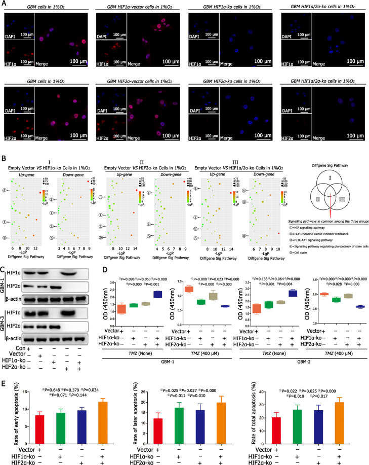
Previous studies have suggested that hypoxic responses are regulated by hypoxia-inducible factors (HIFs), which in turn promote the malignant progression of glioblastoma (GBM) by inhibiting apoptosis and increasing proliferation; these events lead to a poor prognosis of GBM patients. However, there are still no HIF-targeted therapies for the treatment of GBM. We have conducted series of experiments and discovered that GBM cells exhibit features indicative of malignant progression and are present in a hypoxic environment. Knocking out HIF1α or HIF2α alone resulted in no significant change in cell proliferation and cell cycle progression in response to acute hypoxia, but cells showed inhibition of stemness expression and chemosensitization to temozolomide (TMZ) treatment. However, simultaneously knocking out HIF1α and HIF2α inhibited cell cycle arrest and promoted proliferation with decreased stemness, making GBM cells more sensitive to chemotherapy, which could improve patient prognosis. Thus, HIF1α and HIF2α regulate each other with negative feedback. In addition, HIF1α and HIF2α are upstream regulators of epidermal growth factor (EGF), which controls the malignant development of GBM through the EGFR-PI3K/AKT-mTOR-HIF1α signalling pathway. In brief, the HIF1α/HIF2α-EGF/EGFR-PI3K/AKT-mTOR-HIF1α signalling axis contributes to the growth of GBM through a positive feedback mechanism. Finally, HIF1α and HIF2α regulate Sox2 and Klf4, contributing to stemness expression and inducing cell cycle arrest, thus increasing malignancy in GBM. In summary, HIF1α and HIF2α regulate glioblastoma malignant progression through the EGFR-PI3K/AKT pathway via a positive feedback mechanism under the effects of Sox2 and Klf4, which provides a new tumour development model and strategy for glioblastoma treatment.


Conflict of interest statement

The authors declare no competing interests.

Figures

Fig. 1
Fig. 1. HIF1α and HIF2α were highly expressed in GBM under hypoxic conditions.
A The CGGA database showed that both HIF1α and HIF2α were highly expressed in GBM. B Primary GBM tissues showed high levels of HIF1α and HIF2α expression using immunohistochemistry detection. C GBM cells exposed to 21% O2 and 1% O2 for 72 h showed that there was a much higher expression of HIF1α and HIF2α in cells cultured in 1% O2 than in cells cultured in 21% O2. D, E GBM cells cultured in 21% O2 and 1% O2 demonstrated that both HIF1α and HIF2α were highly expressed in 1% O2, while there was almost no HIF1α and HIF2α expression under 21% O2 conditions. P values were determined by the independent samples t test.
Fig. 2
Fig. 2. HIF1α and HIF2α regulated cell proliferation and apoptosis.
A Immunofluorescence confirmed the successful knockout (KO) of HIF1α and HIF2α in HIF1α-KO, HIF2α-KO and HIF1α/HIF2α-KO cells. B We cultured empty vector cells, HIF1α-KO cells, HIF2α-KO cells, HIF1α/HIF2α-KO cells in 1% O2 for 24 h, KEGG pathway analysis revealed five common and significant signalling pathways, including the HIF signalling pathway, EGFR pathway, PI3K–AKT signalling pathway and signalling pathways regulating the pluripotency of stem cells and cell cycle using KEGG. C There were no differences in HIF1α and HIF2α between the control and empty vector groups after culturing both cells in 1% O2 for 72 h. However, the expression of HIF1α increased significantly after knocking out HIF2α; and HIF2α expression increased significantly after knocking out HIF1α. D After individually knocking out HIF1α or HIF2α, there were no differences in cell proliferation between the HIF1α-KO or HIF2α-KO cells and the empty vector cells. However, after simultaneously knocking out HIF1α and HIF2α, the cell proliferation rate increased significantly compared with the cell proliferation rates in other groups, including empty vector cells, HIF1α-KO cells or HIF2α-KO cells. Then, TMZ (400 μM) was added to the culture medium for another 72 h, and the cell proliferation became slower in HIF1α-KO or HIF2α-KO cells than in the empty vector cells; however, the slowest proliferation rate was found in the HIF1α/HIF2α-KO cells. E Cell apoptosis detection showed no difference in early apoptosis, but late and total apoptosis rates increased in HIF1α-KO or HIF2α-KO cells. However, after simultaneously knocking out HIF1α and HIF2α, there was a significant increase in early, late and total apoptosis rates compared with those in other groups. P values were determined by one-way ANOVA.
Fig. 3
Fig. 3. HIF1α and HIF2α upregulated EGF in hypoxia.
A EGF was highly expressed in GBM, according to the CGGA database. B GBM cells were cultured in 21% O2 and 1% O2 for 12 h, and EGF expression was ~3.8–4.6-fold higher in cells cultured in 1% O2 than control cells cultured in 21% O2. C ELISA demonstrated that there were higher levels of EGF in the 1% O2 group than in the control group. D Immunofluorescence showed that there was no EGF expression under normoxia; however, EGF levels increased significantly under hypoxic conditions. E Both HIF1α and HIF2α had a positive relationship with EGF. F After knocking out HIF1α or HIF2α, EGF levels decreased significantly, and EGF expression was lowest after simultaneously knocking out HIF1α and HIF2α. G The addition of EGF had lower early, later and total apoptosis rates in HIF1α-KO or HIF2α-KO cells. P values were determined by the independent samples t test.
Fig. 4
Fig. 4. HIF1α/HIF2α-EGF regulated GBM malignancy via the EGFR–PI3K/AKT pathway in hypoxia.
A There was a higher expression of EGFR, PI3K, PDK1, AKT and mTOR in cells cultured in 1% O2 than cells cultured in control conditions. B, C The cells cultured in 1% O2 for 72 h highly expressed EGFR, PI3K, PDK1, AKT and mTOR, but there was much less expression of EGFR, PI3K, PDK1, AKT and mTOR under normoxic conditions. D Both HIF1α and HIF2α had a positive correlation with EGFR, PI3K, PDK1, AKT and mTOR. E A significant decrease in the expression of EGFR, PI3K, PDK1, AKT and mTOR in HIF1α-KO or HIF2α-KO cells. The addition of EGF into the culture medium of HIF1α-KO or HIF2α-KO cells showed an immediate recovery of the expression of EGFR, PI3K, PDK1, AKT, mTOR and HIF1α. F Adding the EGFR inhibitor (AG1478), PI3K inhibitor (Ly294002) and mTOR inhibitor (rapamycin) into the culture medium of GBM cells in 1% O2 showed that there were no significant differences for early apoptosis; however, there were higher late and total apoptosis rates. P values were determined by the independent samples t test and one-way ANOVA.
Fig. 5
Fig. 5. Sox2, Klf4, CD133 and CD15 were expressed in GBM under hypoxia.
A, B Assessment of samples from the CGGA database showed that Sox2 and Klf4 were expressed in GBM. C GBM cells exposed to 21% O2 and 1% O2 for 72 h showed that Sox2, Klf4, CD133 and CD15 levels increased significantly in 1% O2 when compared with control cells in 21% O2. D, E There was a lower expression of Sox2, Klf4, CD133 and CD15 under normoxia, while these proteins increased significantly under hypoxic conditions. P values were determined by the independent samples t test.
Fig. 6
Fig. 6. HIF1α and HIF2α regulated GBM cell stemness expression through Sox2 and Klf4 in hypoxia.
A Both HIF1α and HIF2α had a positive relationship with Sox2 and Klf4. B There was a lower expression of Sox2 and Klf4 after knocking out HIF1α or HIF2α, and the levels were lowest after simultaneously knocking out HIF1α and HIF2α. C Compared with the control conditions, after knocking out Sox2 and Klf4, CD133 and CD15 expression decreased significantly. D The number of cells in the G1 phase decreased while the number of cells in the G2/M + S phase increased after knocking out Sox2 or Klf4. E After knocking out Sox2 or Klf4, the early, late and total apoptosis rates increased significantly compared with those in the control and empty vector cells. F HIF1α and HIF2α mutually regulate each other with negative feedback. HIF1α and HIF2α act as upstream gene regulators of EGF, and EGF regulates GBM malignancy through the EGFR–PI3K/AKT–mTOR–HIF1α signalling pathway. In addition, HIF1α and HIF2α upregulate Sox2 and Klf4 expression; high expression of Sox2 and Klf4 contributes to an increase in stemness expression and the transformation of cells in the G2/M + S phase to the G1 phase, thus leading to cell survival and therapy resistance. *P < 0.05 and #P > 0.05 were determined by one-way ANOVA.

References

    1. Keith B, Johnson RS, Simon MC. HIF1α and HIF2α: sibling rivalry in hypoxic tumour growth and progression. Nat. Rev. Cancer. 2011;12:9–22. doi: 10.1038/nrc3183. - DOI - PMC - PubMed
    1. Johansson E, et al. CD44 interacts with HIF-2α to modulate the hypoxic phenotype of perinecrotic and perivascular glioma cells. Cell Rep. 2017;20:1641–1653. doi: 10.1016/j.celrep.2017.07.049. - DOI - PubMed
    1. Womeldorff M, Gillespie D, Jensen RL. Hypoxia-inducible factor-1 and associated upstream and downstream proteins in the pathophysiology and management of glioblastoma. Neurosurg. Focus. 2014;37:E8. doi: 10.3171/2014.9.FOCUS14496. - DOI - PubMed
    1. Ivliev AE, Hoen PA, Sergeeva MG. Coexpression network analysis identifies transcriptional modules related to proastrocytic differentiation and sprouty signaling in glioma. Cancer Res. 2010;70:10060–10070. doi: 10.1158/0008-5472.CAN-10-2465. - DOI - PubMed
    1. Velpula KK, Bhasin A, Asuthkar S, Tsung AJ. Combined targeting of PDK1 and EGFR triggers regression of glioblastoma by reversing the Warburg effect. Cancer Res. 2013;73:7277–7289. doi: 10.1158/0008-5472.CAN-13-1868. - DOI - PubMed

Publication types

Substances