. 2021 Mar 24;11(1):6743.
doi: 10.1038/s41598-021-86217-y.

Protein cleavage influences surface protein presentation in Mycoplasma pneumoniae

Affiliations

Protein cleavage influences surface protein presentation in Mycoplasma pneumoniae

Iain J Berry et al. Sci Rep. .

Abstract

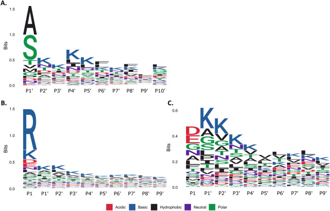
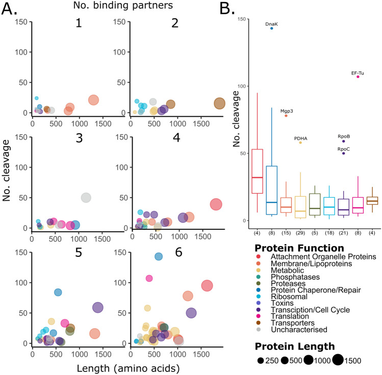
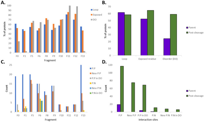
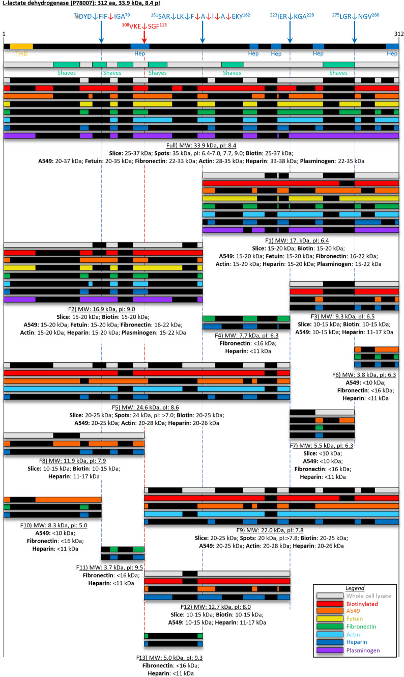
Mycoplasma pneumoniae is a significant cause of pneumonia and post infection sequelae affecting organ sites distant to the respiratory tract are common. It is also a model organism where extensive 'omics' studies have been conducted to gain insight into how minimal genome self-replicating organisms function. An N-terminome study undertaken here identified 4898 unique N-terminal peptides that mapped to 391 (56%) predicted M. pneumoniae proteins. True N-terminal sequences beginning with the initiating methionine (iMet) residue from the predicted Open Reading Frame (ORF) were identified for 163 proteins. Notably, almost half (317; 46%) of the ORFS derived from M. pneumoniae strain M129 are post-translationally modified, presumably by proteolytic processing, because dimethyl labelled neo-N-termini were characterised that mapped beyond the predicted N-terminus. An analysis of the N-terminome describes endoproteolytic processing events predominately targeting tryptic-like sites, though cleavages at negatively charged residues in P1' (D and E) with lysine or serine/alanine in P2' and P3' positions also occurred frequently. Surfaceome studies identified 160 proteins (23% of the proteome) to be exposed on the extracellular surface of M. pneumoniae. The two orthogonal methodologies used to characterise the surfaceome each identified the same 116 proteins, a 72% (116/160) overlap. Apart from lipoproteins, transporters, and adhesins, 93/160 (58%) of the surface proteins lack signal peptides and have well characterised, canonical functions in the cell. Of the 160 surface proteins identified, 134 were also targets of endo-proteolytic processing. These processing events are likely to have profound implications for how the host immune system recognises and responds to M. pneumoniae.


Conflict of interest statement

The authors declare no competing interests.

Figures

Figure 1
Figure 1
Proteolytic sequence logos of the N-terminome of Mycoplasma pneumoniae at (A) the protein N-terminus after initiating methionine removal (N-end rule), (B) internal neo-N-term endoproteolytic sites and (C) N-term endoproteolytic sites excluding tryptic-like sites.
Figure 2
Figure 2
Interaction map of all proteins identified on the surface of Mycoplasma pneumoniae, grouped by gene ontology. Proteins with known interactions provided by STRING, are indicated by connecting lines, representing possible protein networks on the cellular surface. Proteins are coloured by their predicted cellular location: green (cytoplasm), brown (membrane), grey (unknown), red (extracellular). Proteins with signal icons either contained a signal peptide (black signal icon) or were found to be non-classically secreted (blue signal icon). Proteins indicated with a red circle were not found to be processed in our N-terminome, all other proteins were found with endo-proteolytic processing sites.
Figure 3
Figure 3
Endoproteolytic processing and putative binding interactions of M. pneumoniae surface proteins. (A) Multi-panel bubble chart displaying the relationship between protein length and number of cleavage, split by the number of binding partners (1 to 6 ECM molecules). (B) Boxplot showing number of cleavages observed in each functional group. The number of proteins assigned to each group is shown in brackets.
Figure 4
Figure 4
Cleavage map of Uncharacterized lipoprotein MPN052 (P75062). Full length protein designated as “Full” and cleavage fragments are designated F1–F13 as indicated under each set of bars. Cleavage sites identified from N-term dimethyl labelling are indicated by the blue arrows (and blue broken lines) with exact sites shown in the sequence above. Semi-tryptic peptides were also characterised for cleavage sites indicated by the red arrows (and red broken lines). Two hypothetical cleavage sites (black arrows) that span regions 205–219 and 511–571 are shown as the grey boxes, the exact cleavage site is unknown and thus a range is shown. A signal peptide predicted by SignalP is shown. Bioinformatic tools to predict transmembrane domains (TmD, golden boxes), heparin binding domains (Hep, blue boxes), and disordered regions (Disorder, purple boxes) are shown. The peptides shown as black boxes in coloured bars were identified by mass spectrometry from: 1D- and 2D-SDS PAGE of M. pneumoniae whole cell lysates (grey bars), and surface biotinylation (red bars). Peptides are also shown from affinity chromatography of: A549 surface complexes (orange bars), fetuin (yellow bars), fibronectin (green bars), actin (light blue bars), heparin (dark blue bars), and plasminogen (purple bars).
Figure 5
Figure 5
Predicted structural changes to uncharacterized lipoprotein MPN052 (P75062) post-cleavage. (A) The percentage of each fragment (F0 = full length) taken up by loop structures, exposed residues, disordered regions (DO). (B) Relative change in loop, exposed residues and DO pre- and post-cleavage. (C) The number of protein:protein (P:P) and protein:nucleotide (P:N) interaction sites per fragment. (D) Relative change in P:P and P:N sites pre and post-cleavage.
Figure 6
Figure 6
Cleavage map of L-lactate dehydrogenase. Full length protein designated as “Full” and cleavage fragments designated F1–F13, as indicated under each set of bars. Cleavage sites identified from N-term dimethyl labelling are indicated by the blue arrows (and blue broken lines) with exact sites shown in the sequence above. Semi-tryptic peptides were also characterised for cleavage sites indicated by the red arrows (and red broken lines). Bioinformatic tools to predict a transmembrane domain (TmD, golden boxes), heparin binding domains (Hep, blue boxes). The peptides shown as black boxes in coloured bars were identified by mass spectrometry from: 1D- and 2D-SDS PAGE of M. pnuemoniae whole cell lysates (grey bars), and surface biotinylation (red bars). Peptides are also shown from affinity chromatography of: A549 surface complexes (orange bars), fetuin (yellow bars), fibronectin (green bars), actin (light blue bars), heparin (dark blue bars), and plasminogen (purple bars).

Similar articles

Cited by

References

    1. Othman N, Isaacs D, Daley AJ, Kesson AM. Mycoplasma pneumoniae infection in a clinical setting. Pediatr. Int. 2008;50:662–666. doi: 10.1111/j.1442-200X.2008.02644.x. - DOI - PubMed
    1. Atkinson TP, Balish MF, Waites KB. Epidemiology, clinical manifestations, pathogenesis and laboratory detection of Mycoplasma pneumoniae infections. FEMS Microbiol. Rev. 2008;32:956–973. doi: 10.1111/j.1574-6976.2008.00129.x. - DOI - PubMed
    1. Narita M. Pathogenesis of extrapulmonary manifestations of Mycoplasma pneumoniae infection with special reference to pneumonia. J. Infect. Chemother. 2010;16:162–169. doi: 10.1007/s10156-010-0044-X. - DOI - PubMed
    1. Sanchez B, Bressollier P, Urdaci MC. Exported proteins in probiotic bacteria: Adhesion to intestinal surfaces, host immunomodulation and molecular cross-talking with the host. FEMS Immunol. Med. Microbiol. 2008;54:1–17. doi: 10.1111/j.1574-695X.2008.00454.x. - DOI - PubMed
    1. Dandekar T, et al. Re-annotating the Mycoplasma pneumoniae genome sequence: Adding value, function and reading frames. Nucleic Acids Res. 2000;28:3278–3288. doi: 10.1093/nar/28.17.3278. - DOI - PMC - PubMed

Publication types