

PDGFB RNA in situ hybridization for the diagnosis of dermatofibrosarcoma protuberans
- PMID: 33762682
- PMCID: PMC8298273
- DOI: 10.1038/s41379-021-00800-2
PDGFB RNA in situ hybridization for the diagnosis of dermatofibrosarcoma protuberans
Abstract
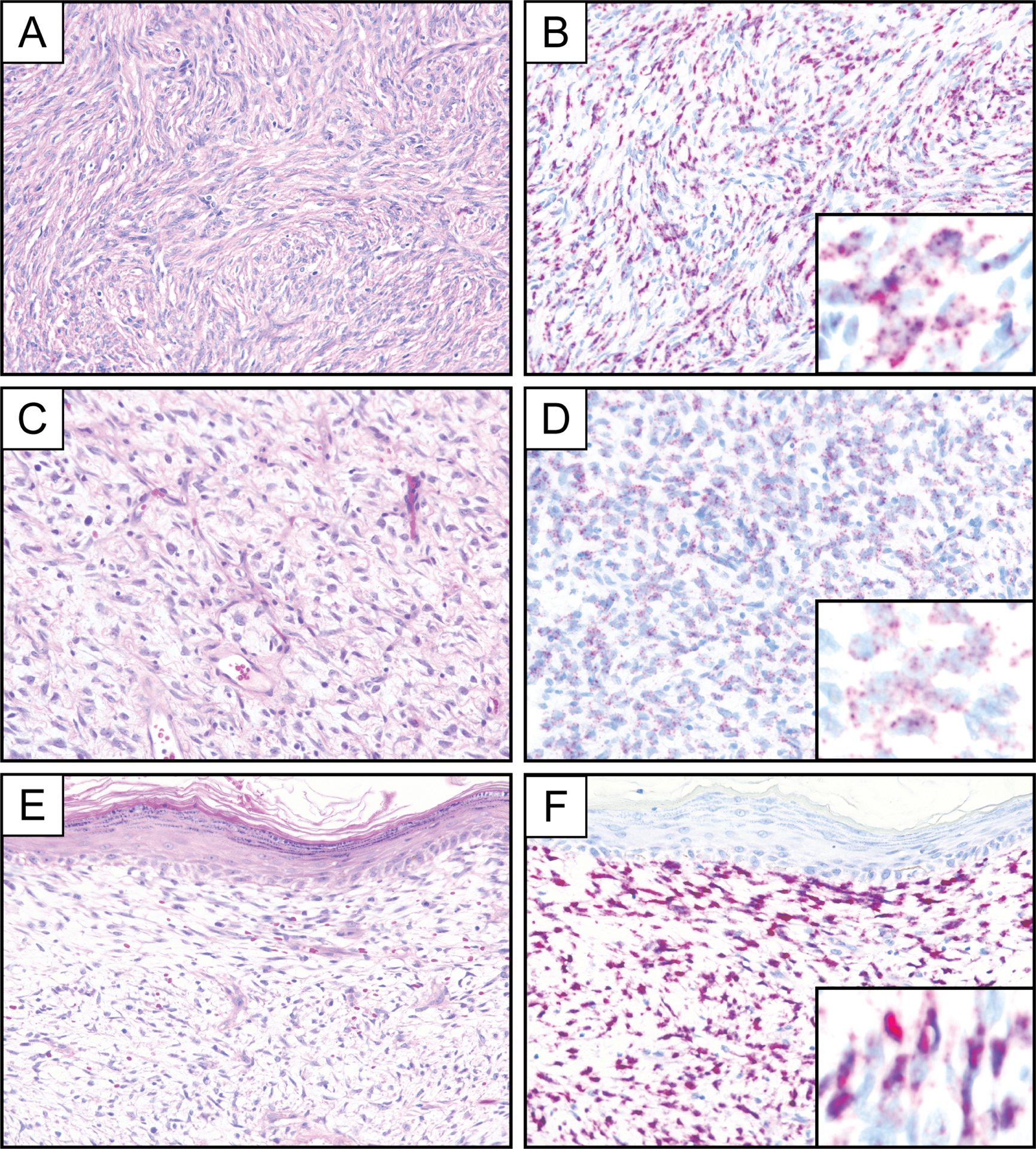
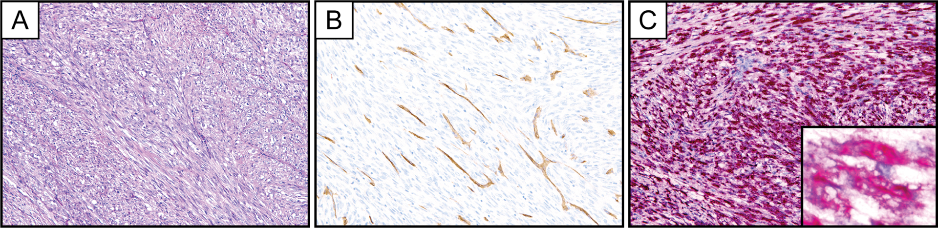
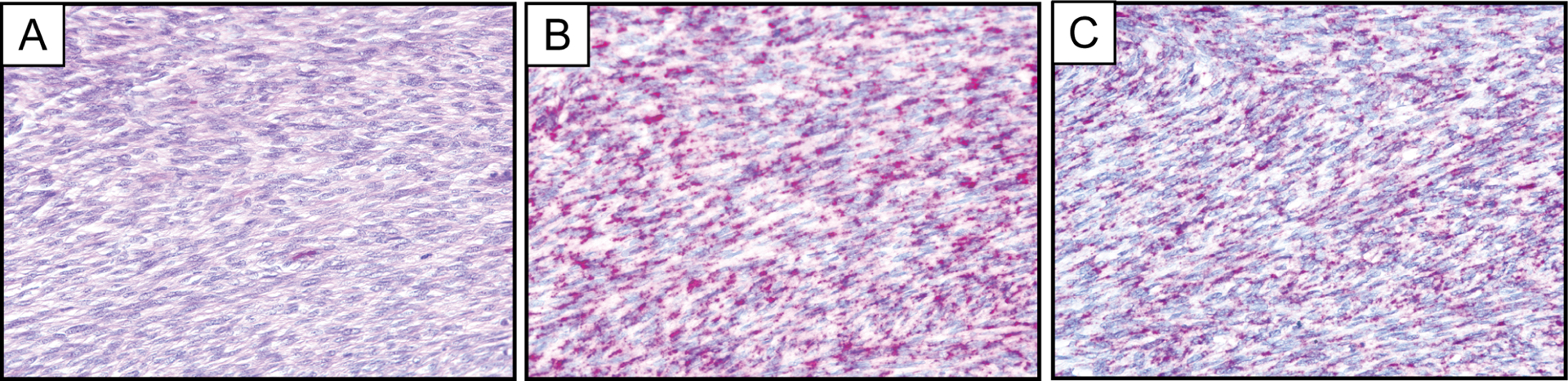
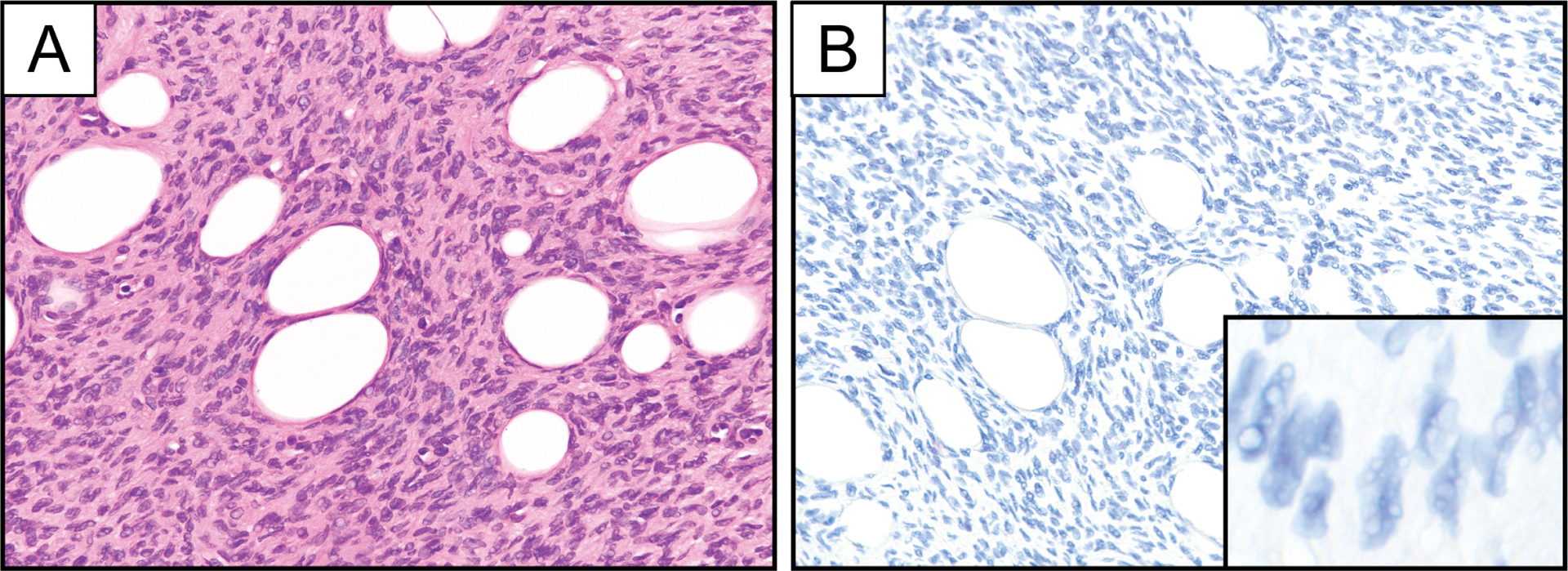
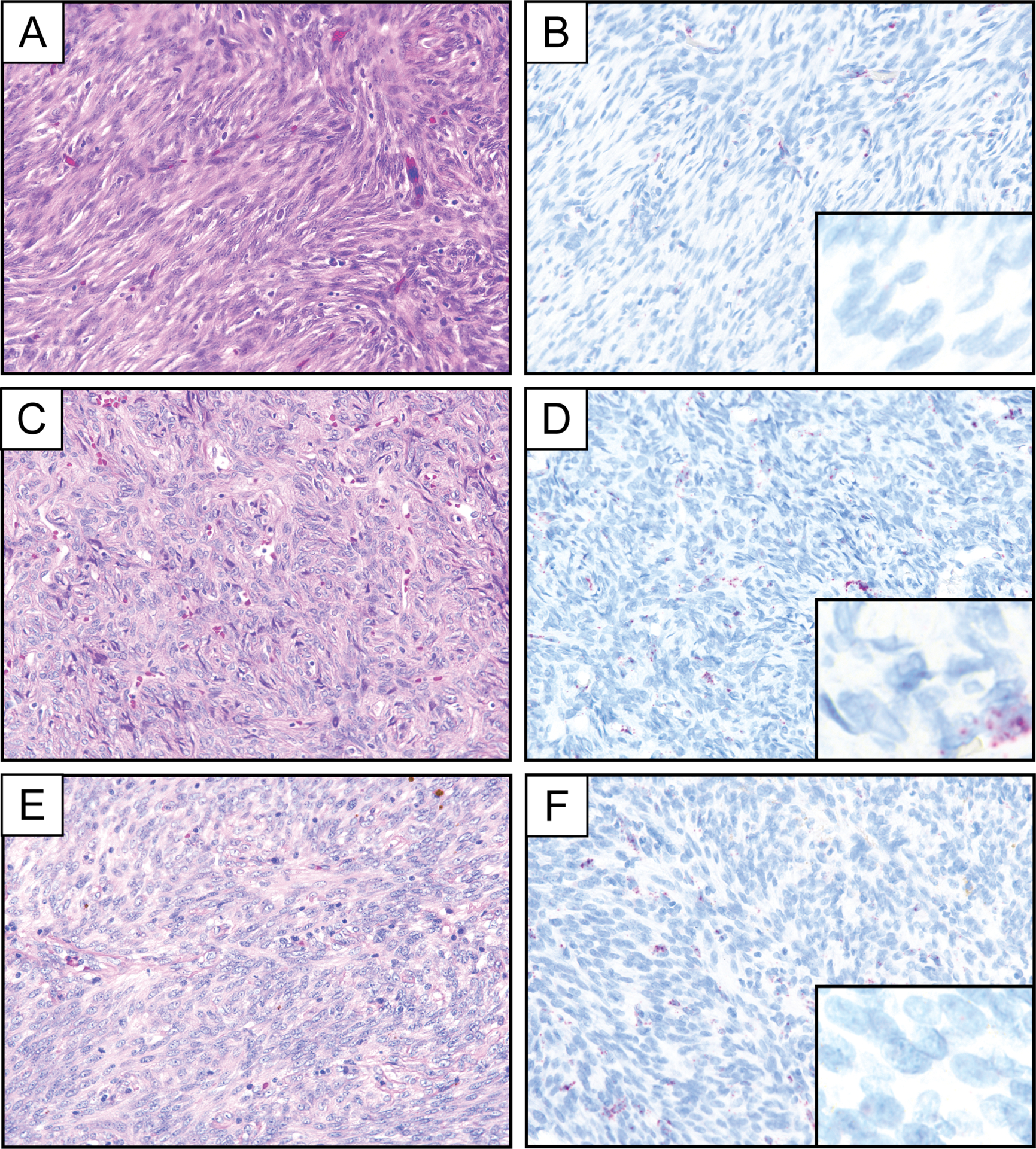
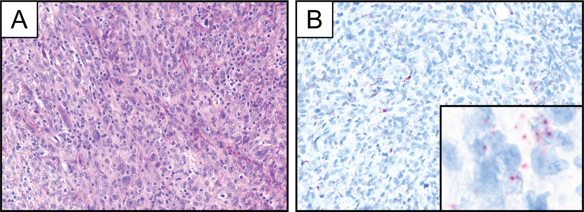
Dermatofibrosarcoma protuberans (DFSP) is a spindle cell neoplasm of the skin and superficial soft tissue with a tendency for locally aggressive behavior; metastatic potential coincides with fibrosarcomatous transformation. The vast majority of DFSPs harbor the t(17;22) translocation resulting in a COL1A1-PDGFB fusion that drives autocrine growth stimulation via PDGFB overexpression. Here, we examined the utility of PDGFB RNA chromogenic in situ hybridization (CISH) for the diagnosis of DFSP. A total of 337 tumors represented in whole tissue sections and tissue microarrays, including 37 cases of DFSP and 300 histologically similar spindle cell tumors, were subjected to PDGFB RNA CISH using commercially available probes. PDGFB overexpression was observed by light microscopy in 24 of 26 conventional DFSPs (92%) and 11 of 11 fibrosarcomatous DFSPs (100%). One of two DFSPs negative for PDGFB by RNA CISH was found to harbor an uncommon alternative rearrangement involving PDGFD. All examined cases of histologic mimics were negative for PDGFB overexpression; limited PDGFB expression, not reaching an empirical threshold of greater than 5 puncta or one aggregate of chromogen in more than 25% of cells, was observed in 7 of 300 mimics (2.3%), including desmoplastic melanoma, malignant peripheral nerve sheath tumor, angiosarcoma, and pleomorphic dermal sarcoma. Vascular PDGFB expression was seen in several tumor types. We conclude that PDGFB RNA CISH, with careful interpretation and the use of appropriate thresholds, may serve as a surrogate marker of PDGFB rearrangement and a useful ancillary tool for the diagnosis of DFSP.
© 2021. The Author(s), under exclusive licence to United States & Canadian Academy of Pathology.
Conflict of interest statement
DECLARATION OF CONFLICTING INTERESTS
The authors declare no potential conflicts of interest with respect to the research, authorship, and/or publication of this article.
Figures
References
-
- WHO Classification of Tumours Editorial Board. Soft Tissue and Bone Tumours. (Lyon: International Agency for Research on Cancer, 2020).
-
- Reimann JDR, Fletcher CDM. Myxoid dermatofibrosarcoma protuberans: a rare variant analyzed in a series of 23 cases. Am J Surg Pathol 31, 1371–1377 (2007). - PubMed
-
- Dupree WB, Langloss JM, Weiss SW. Pigmented dermatofibrosarcoma protuberans (Bednar tumor). A pathologic, ultrastructural, and immunohistochemical study. Am J Surg Pathol 9, 630–639 (1985). - PubMed
-
- Calonje E, Fletcher CD. Myoid differentiation in dermatofibrosarcoma protuberans and its fibrosarcomatous variant: clinicopathologic analysis of 5 cases. J Cutan Pathol 23, 30–36 (1996). - PubMed
-
- Mentzel T, Beham A, Katenkamp D, Dei Tos AP, Fletcher CDM. Fibrosarcomatous (‘High-Grade’) Dermatofibrosarcoma Protuberans: Clinicopathologic and Immunohistochemical Study of a Series of 41 Cases With Emphasis on Prognostic Significance. Am J Surg Pathol 22, 576–587 (1998). - PubMed
Publication types
MeSH terms
Substances
Grants and funding
LinkOut - more resources
Full Text Sources
Other Literature Sources
Medical
Miscellaneous
