. 2021 Apr;592(7855):596-600.
doi: 10.1038/s41586-021-03369-7. Epub 2021 Mar 24.

Presymptomatic training mitigates functional deficits in a mouse model of Rett syndrome

Affiliations

Presymptomatic training mitigates functional deficits in a mouse model of Rett syndrome

Nathan P Achilly et al. Nature. 2021 Apr.

Abstract

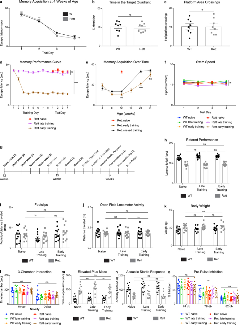
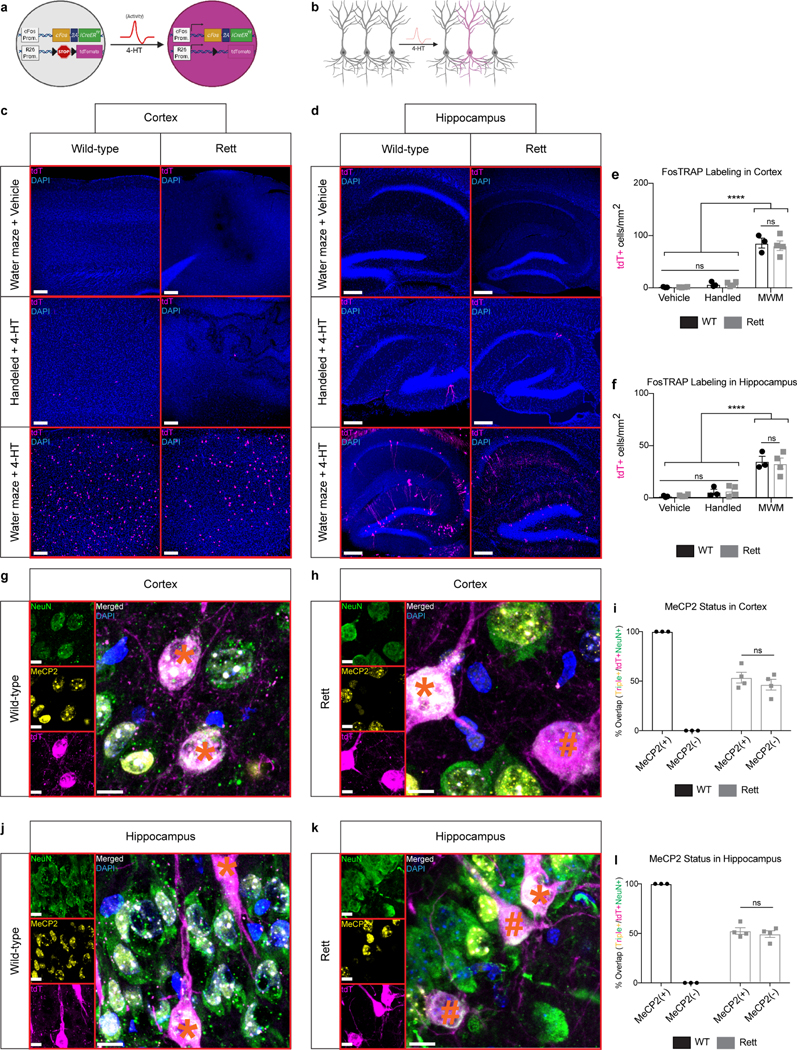
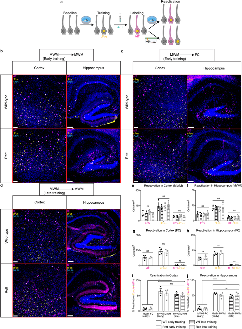
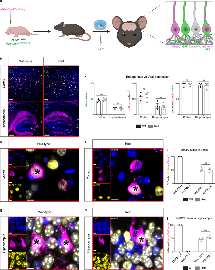
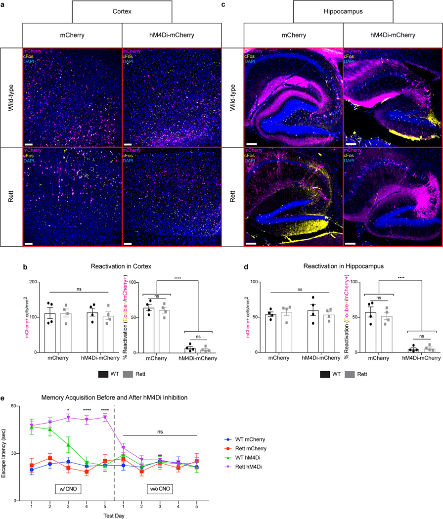
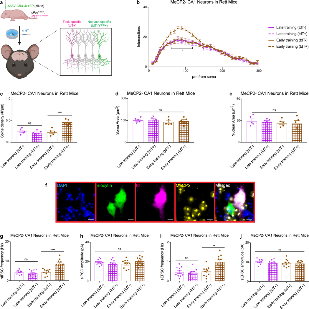
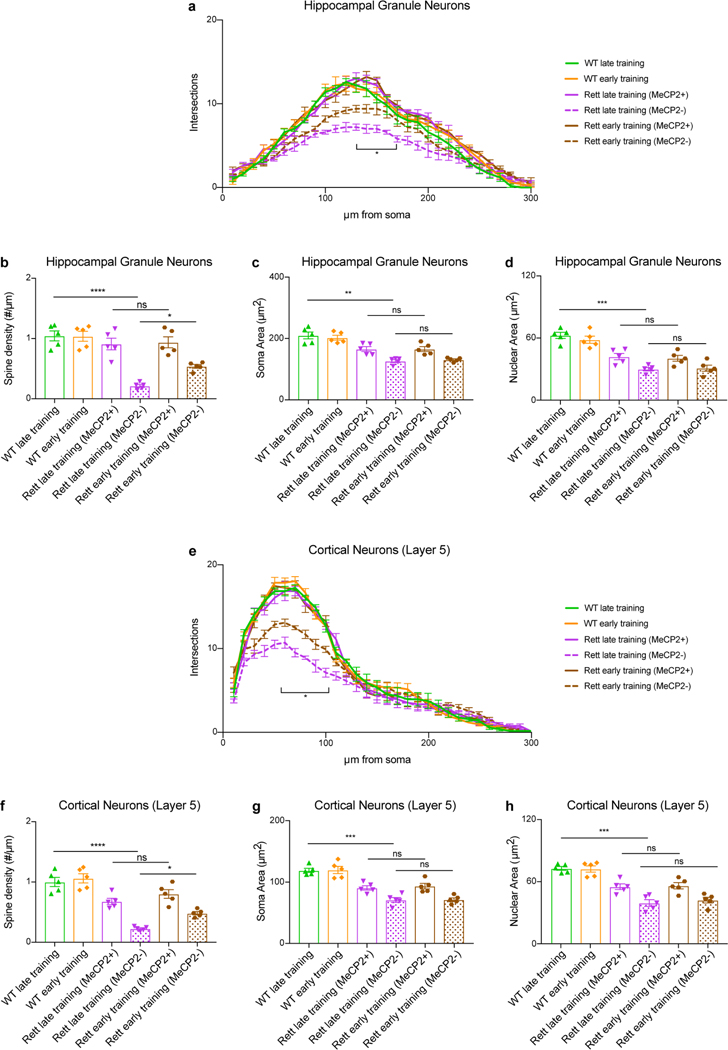
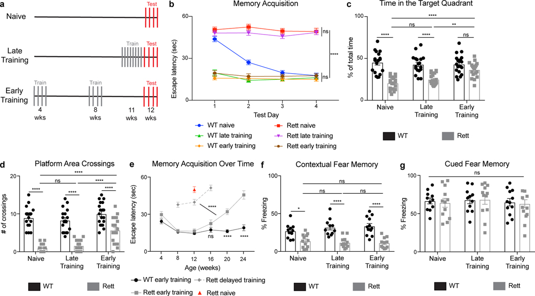
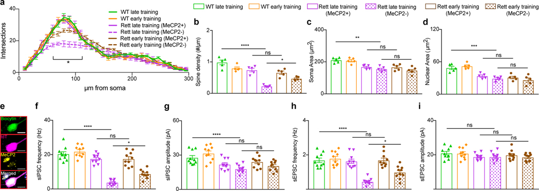
Mutations in the X-linked gene MECP2 cause Rett syndrome, a progressive neurological disorder in which children develop normally for the first one or two years of life before experiencing profound motor and cognitive decline1-3. At present there are no effective treatments for Rett syndrome, but we hypothesized that using the period of normal development to strengthen motor and memory skills might confer some benefit. Here we find, using a mouse model of Rett syndrome, that intensive training beginning in the presymptomatic period dramatically improves the performance of specific motor and memory tasks, and significantly delays the onset of symptoms. These benefits are not observed when the training begins after symptom onset. Markers of neuronal activity and chemogenetic manipulation reveal that task-specific neurons that are repeatedly activated during training develop more dendritic arbors and have better neurophysiological responses than those in untrained animals, thereby enhancing their functionality and delaying symptom onset. These results provide a rationale for genetic screening of newborns for Rett syndrome, as presymptomatic intervention might mitigate symptoms or delay their onset. Similar strategies should be studied for other childhood neurological disorders.


Figures

Extended Data Fig. 1 |
Extended Data Fig. 1 |. Presymptomatic motor training on the rotarod does not improve other behavioral deficits in Rett mice.
a, Motor performance on the rotarod. WT (n = 12) and Rett (n = 10) mice were tested over 4 days beginning at 8 weeks of age. b, Timeline for additional behavioral assays in rotarod-trained mice. c, Performance curves of rotarod-trained Rett naive (n = 19), Rett late-trained (n = 18), and Rett early-trained (n = 19) mice across training and test days. d-o, After rotarod testing at 24 weeks of age, WT naive (n = 12), Rett naive (n = 12), WT late-trained (n = 12), Rett late-trained (n = 11), WT early-trained (n = 11), Rett early-trained (n = 12) mice were tested on a variety of behavioral assays. Motor function was tested in the footslip (d) and open field assays (e). Spatial memory was tested in the Morris water maze (f-h). Social behavior was tested in the 3-chamber assay (i). Anxiety was tested in the elevated plus maze (j). Sensorimotor gating was tested in the acoustic startle (k) and pre-pulse inhibition assays (l). Contextual (m) and cued (n) memory were assessed in the fear conditioning assays. Body weight was measured (o). The sample size (n) corresponds to the number of biologically independent mice. Data are represented as mean ± s.e.m. Statistical significance was determined using a two-way ANOVA with Tukey’s multiple comparisons test; ns (p>0.05), **** (p<0.0001).
Extended Data Fig. 2 |
Extended Data Fig. 2 |. Presymptomatic memory training in the Morris water maze does not improve other behavioral deficits in Rett mice.
a-c, Spatial memory performance in the Morris water maze. WT (n = 8) and Rett (n = 8) mice were tested over 4 days beginning at 4 weeks of age (a). A probe trial on day 5 revealed the time spent in the target quadrant (b) and the number of platform area crossings (c). d, Performance curves of Morris water maze-trained Rett naive (n = 19), Rett late-trained (n = 19), and Rett early-trained (n = 18) mice across training and test days. e, Spatial memory in the Morris water maze over time in early-trained Rett mice (n = 7) and Rett mice (n = 8) that missed a training session. f, Swim speed in the Morris water maze of WT naive (n = 18), WT late-trained (n = 18), WT early-trained (n = 19), Rett naive (n = 19), Rett late-trained (n = 19), and Rett early-trained (n = 18) mice. g, Timeline for additional behavioral assays in Morris water maze-trained mice. h-o, After Morris water maze testing at 12 weeks of age, WT naive (n = 11), Rett naive (n = 12), WT late-trained (n = 11), Rett late-trained (n = 12), WT early-trained (n = 12), Rett early-trained (n = 11) mice were tested on a variety of behavioral assays. Motor function was tested in rotarod (h), footslip (i), and open field assays (j). Body weight was measured (k). Social behavior was tested in the 3-chamber assay (l). Anxiety was tested in the elevated plus maze (m). Sensorimotor gating was tested in the acoustic startle (n) and pre-pulse inhibition (o) assays. The sample size (n) corresponds to the number of biologically independent mice. Data are represented as mean ± s.e.m. Statistical significance was determined using a two-tailed, unpaired student’s t-test (b-c) or two-way ANOVA with Tukey’s multiple comparisons test (a, d-o); ns (p>0.05), **** (p<0.0001).
Extended Data Fig. 3 |
Extended Data Fig. 3 |. FosTRAP labels task-specific neurons, including MeCP2+ and MeCP2 neurons in Rett mice.
a, Schematic of FosTRAP. In the presence of 4-hydroxytamoxifen (4-HT), neural activity activates cFos expression and subsequent expression of a Cre-dependent reporter, tdTomato. b, FosTRAP labels a subset of neurons following 4-HT administration. c-f, FosTRAP labeling during the Morris water maze labels neurons in multiple brain regions. Labeled neurons in the cortex (c) and hippocampus (d) from 13-week-old mice that were trained in the Morris water maze and injected with vehicle or 4-HT as well as mice that were handled and injected with 4-HT. Quantification in the cortex (e) and hippocampus (f) of WT (n = 3) and Rett (n = 4) mice. Scale bar, 200 μm. g-l, FosTRAP labeling in MeCP2+ and MeCP2 neurons. FosTRAP labels NeuN+/MeCP2+ neurons in the cortex (g) and hippocampus (j) of WT mice. FosTRAP labels NeuN+/MeCP2+ and NeuN+/MeCP2 in the cortex (h) and hippocampus (k) of Rett mice. Quantification in the cortex (i) and hippocampus (l) of WT (n = 3) and Rett (n = 4) mice. (*) denotes a MeCP2+ neuron. (#) denotes a MeCP2 neuron. Scale bar, 20 μm. The sample size (n) corresponds to the number of biologically independent mice. 3–4 sections were analyzed per mouse. Data are represented as mean ± s.e.m. Statistical significance was determined using a two-way ANOVA with Tukey’s multiple comparisons test; ns (p>0.05), **** (p<0.0001).
Extended Data Fig. 4 |
Extended Data Fig. 4 |. Morris water maze-associated neurons are reactivated during the Morris water maze but not during fear conditioning.
a, FosTRAP labeling of Morris water maze-associated neurons, with subsequent reactivation of task-specific neurons during retesting in the Morris water maze or fear conditioning assay. b-j, cFos reactivation in Morris water maze (MWM)-associated neurons of early-trained mice retested in the Morris water maze (b), early-trained mice tested in a novel fear conditioning (FC) assay (c), and late-trained mice retested in the Morris water maze (d) at 13 weeks of age. The number of tdTomato+ (tdT+), cFos+, and tdT+cFos+ neurons were quantified in the cortex (e) and hippocampus (f) of early-trained WT (n = 3) and Rett (n = 4) mice and late-trained WT (n = 3) and Rett (n = 3) mice after retesting in the Morris water maze. The number of tdTomato+ (tdT+), cFos+, and tdT+cFos+ neurons were quantified in the cortex (g) and hippocampus (h) of early-trained WT (n = 4) and Rett (n = 3) mice after fear conditioning. The reactivation percentage, defined as the percentage of tdT+ neurons that were also cFos+, were quantified in the cortex (i) and hippocampus (j) of Morris water maze-trained mice after retesting in the Morris water maze or the fear conditioning assay. Scale bar, 200 μm. The sample size (n) corresponds to the number of biologically independent mice. 3–4 sections were analyzed per mouse. Data are represented as mean ± s.e.m. Statistical significance was determined using a two-way ANOVA with Tukey’s multiple comparisons test; ns (p>0.05), ** (p<0.01), **** (p<0.0001).
Extended Data Fig. 5 |
Extended Data Fig. 5 |. Viral delivery of DREADD-containing AAVs labels task-specific neurons, including MeCP2+ and MeCP2 neurons in Rett mice.
a, P0 AAV delivery of Cre-dependent mCherry into cFosCreER mice that also contain a Cre-dependent GFP in the Rosa26 locus. Expression of mCherry in GFP+ neurons demonstrates that the AAVs are reactivated in task-specific neurons. b-c, Overlap between injected AAV (mCherry) and endogenous reporter (GFP) in early-trained mice at 13 weeks of age. Quantification of GFP+ cells, mCherry+ cells, and overlap in the cortex and hippocampus of WT (n = 3) and Rett (n = 4) mice (c). Scale bar, 200 μm. d-i, FosTRAP labeling in MeCP2+ and MeCP2 neurons from AAV-injected mice trained in the Morris water maze. FosTRAP labels MeCP2+ neurons in the cortex (d) and hippocampus (g) of WT mice. FosTRAP labels MeCP2+ and MeCP2 neurons in the cortex (e) and hippocampus (h) of Rett mice. Quantification in the cortex (f) and hippocampus (i) of WT (n = 3) and Rett (n = 4) mice. (*) denotes a MeCP2+ neuron. (#) denotes a MeCP2 neuron. Scale bar, 20 μm. The sample size (n) corresponds to the number of biologically independent mice. 3–4 sections were analyzed per mouse. Data are represented as mean ± s.e.m. Statistical significance was determined using a two-way ANOVA with Tukey’s multiple comparisons test; ns (p>0.05).
Extended Data Fig. 6 |
Extended Data Fig. 6 |. CNO prevents reactivation of hM4Di-expressing neurons.
a-d, cFos reactivation in task-specific neurons expressing mCherry or hM4Di-mCherry. Overlap between cFos and mCherry in the cortex (a) and hippocampus (c) of mice injected with CNO during testing in the Morris water maze at 13 weeks of age. Quantification of mCherry+ cells and reactivation from cortex (b) and hippocampus (d) in WT (n = 4) and Rett (n = 4) mice. Scale bar, 200 μm. e, The effect of removing CNO on Morris water maze performance in WT mCherry (n = 6), Rett mCherry (n = 5), WT hM4Di (n = 5), and Rett hM4Di (n = 6) mice at 13 weeks of age. The sample size (n) corresponds to the number of biologically independent mice. 3–4 sections were analyzed per mouse. Data are represented as mean ± s.e.m. Statistical significance was determined using a two-way ANOVA with Tukey’s multiple comparisons test; ns (p>0.05), * (p<0.05), **** (p<0.0001).
Extended Data Fig. 7 |
Extended Data Fig. 7 |. CNO reactivates hM3Dq-expressing neurons.
a-d, cFos reactivation in task-specific neurons expressing mCherry or hM3Dq-mCherry. Overlap between cFos and mCherry in the cortex (a) and hippocampus (c) of mice injected with CNO in the homecage at 13 weeks of age. Quantification of mCherry+ cells and reactivation from cortex (b) and hippocampus (d) in WT (n = 4) and Rett (n = 4) mice. Scale bar, 200 μm. The sample size (n) corresponds to the number of biologically independent mice. 3–4 sections were analyzed per mouse. Data are represented as mean ± s.e.m. Statistical significance was determined using a two-way ANOVA with Tukey’s multiple comparisons test; ns (p>0.05), **** (p<0.0001).
Extended Data Fig. 8 |
Extended Data Fig. 8 |. Repeated activation of a random subset of neurons does not alter the performance of early-trained mice.
a, AAV injection paradigm to express DREADDs in a random subset of neurons and manipulate their activity with CNO. High-titer AAV is delivered to cFosCreER mice, which express Cre in task-specific neurons following 4-hydroxytamoxifen administration. Low-titer AAV is delivered to Camk2aCreER mice, which express Cre ubiquitously in excitatory neurons following 4-hydroxytamoxifen administration. cFosCreER and Camk2aCreER mice both express the same number of labeled neurons, but those in cFosCreER mice are task-specific. b-d, Overlap between injected AAV (mCherry) and endogenous reporter (GFP) in early-trained mice at 13 weeks of age. Quantification of mCherry+ cells in the cortex (c) and hippocampus (d) of WT (n = 3) and Rett (n = 4) mice expressing cFosCreER and WT (n = 4) and Rett (n = 4) expressing Camk2aCreER. Scale bar, 200 μm. e-f, cFos reactivation in Camk2aCreER mice expressing mCherry. Quantification of reactivation in the cortex and hippocampus of WT (n = 4) and Rett (n = 3) mice after Morris water maze training at 13 weeks of age (f). g-i, Morris water maze performance in early-trained WT and Rett mice expressing Camk2aCreER and hM4Di-mCherry or mCherry. WT mCherry (n = 7), Rett mCherry (n = 8), WT hM4Di (n = 8), and Rett hM4Di (n = 6) mice were tested at 13 weeks of age and injected with CNO during testing (g). A probe trial on day 6 measured the time spent in target quadrant (h) and the number of platform area crossings (i). j-k, Morris water maze performance in early-trained WT and Rett mice expressing Camk2aCreER and hM3Dq-mCherry or mCherry. A probe trial on WT mCherry (n = 7), Rett mCherry (n = 8), WT hM3Dq (n = 7), and Rett hM3Dq (n = 7) measured the time spent in the target quadrant (j) and the number of platform area crossings (k). The sample size (n) corresponds to the number of biologically independent mice. Data are represented as mean ± s.e.m. Statistical significance was determined using a two-way ANOVA with Tukey’s multiple comparisons test; ns (p>0.05).
Extended Data Fig. 9 |
Extended Data Fig. 9 |. Morphological and electrophysiological benefits are evident in task-specific neurons after presymptomatic training.
a, P0 AAV delivery of YFP to assess the morphology of neurons that are not task-specific (tdT-). b-e, Morphological analysis of MeCP2 hippocampal CA1 neurons that are task-specific (tdT+) and not task-specific (tdT-/YFP+) in trained Rett mice at 13 weeks of age. Sholl analysis (b), spine density (c), soma area (d), and nuclear area (e) were measured in MeCP2 neurons of late-trained (n = 5) and early-trained (n = 5) Rett mice. f-j, Electrophysiological recordings of MeCP2 CA1 neurons in late- and early-trained Rett mice at 13 weeks of age. f, Representative image of neurons that are task-specific (magenta) and not task-specific (no magenta), both of which were injected with biocytin (green) during recording and immunostained to determine the MeCP2 status (yellow). Frequency (g) and amplitude (h) of sIPSCs and frequency (i) and amplitude (j) of sEPSCs were measured in MeCP2 neurons from late-trained (n = 10) and early-trained (n = 10) Rett mice. The sample size (n) corresponds to the number of biologically independent mice. For b-c, 10–15 neurons were analyzed per mouse. For d-e, 50–100 neurons were analyzed per mouse. For g-j, 1–3 neurons were analyzed per mouse. Data are represented as mean ± s.e.m. Statistical significance was determined using a one-way (b) or two-way (c-e, g-j) ANOVA with Tukey’s multiple comparisons test; ns (p>0.05), ** (p<0.01), **** (p<0.0001).
Extended Data Fig. 10 |
Extended Data Fig. 10 |. Presymptomatic training improves dendritic morphology of task-specific hippocampal granule and cortical neurons in Rett mice.
a-d, Morphological analysis of task-specific hippocampal granule neurons in late- and early-trained WT and Rett mice at 13 weeks of age. Sholl analysis (a), spine density (b), soma area (c), and nuclear area (d) were measured in neurons of WT late-trained (n = 5), WT early-trained (n = 5), Rett late-trained MeCP2 (n = 5), Rett early-trained MeCP2+ (n = 5), and Rett early-trained MeCP2 (n = 5) neurons. e-h, Morphological analysis of layer 5 cortical task-specific neurons in late- and early-trained WT and Rett mice. Sholl analysis (e), spine density (f), soma area (g), and nuclear area (h) were measured in neurons of WT late-trained (n = 5), WT early-trained (n = 5), Rett late-trained MeCP2+ (n = 5), Rett late-trained MeCP2 (n = 5), Rett early-trained MeCP2+ (n = 5), and Rett early-trained MeCP2 (n = 5) neurons. The sample size (n) corresponds to the number of biologically independent mice. For a-b and e-g, 10–15 neurons were analyzed per mouse. For c-d and g-h, 50–100 neurons were analyzed per mouse. Data are represented as mean ± s.e.m. Statistical significance was determined using a one-way (b-d, f-h) and two-way (a, e) ANOVA with Tukey’s multiple comparisons test; ns (p>0.05), * (p<0.05), ** (p<0.01), *** (p<0.001), **** (p<0.0001).
Fig. 1 |
Fig. 1 |. Presymptomatic training improves motor performance on the rotarod in Rett mice.
a, Training regimen for naive, late-trained, and early-trained mice; each line represents 4 trials/day. b, Average motor performance on the rotarod. WT naive (n = 19), WT late-trained (n = 19), WT early-trained (n = 18), Rett naive (n = 19), Rett late-trained (n = 18), and Rett early-trained (n = 19) mice were tested across 4 days at 24 weeks of age. c, A subset of these mice underwent additional behavioral tests (see Extended Data Fig. 1) and the rest of the early-trained mice were followed for rotarod performance until 32 weeks of age. Early training delayed the onset of motor symptoms in Rett mice (n = 7) until after 22 weeks, with some benefit still apparent at 32 weeks; comparisons are WT (n = 7) at 32 weeks of age and naive Rett mice (n = 12) at 12 weeks of age. In another group of Rett mice (n = 8), we delayed training initiation until 16 weeks, following the same early-training protocol, but this yielded no benefit. The sample size (n) corresponds to the number of biologically independent mice. Data are represented as mean ± s.e.m. Statistical significance was determined using a two-way ANOVA with Tukey’s multiple comparisons test; ns (p>0.05), ** (p<0.01), *** (p<0.001), **** (p<0.0001).
Fig. 2 |
Fig. 2 |. Presymptomatic training improves spatial memory in the Morris water maze task in Rett mice.
a, Training regimen for naive, late-trained, and early-trained mice; each line represents 8 trials/day. Early training started at 4 weeks of age; late training started at 11 weeks of age. Late- and early-trained mice received the same number of training trials. b-d, Spatial memory performance in the water maze. (b) WT naive (n = 18), WT late-trained (n = 18), WT early-trained (n = 19), Rett naive (n = 19), Rett late-trained (n = 19), and Rett early-trained (n = 18) mice were tested over 4 consecutive days at 12 weeks of age. A probe trial on day 5 measured the time spent in the target quadrant (c) and number of platform area crossings (d). e, Early-trained WT (n = 7) and Rett mice (n = 7) mice were tested until 24 weeks of age and then compared to naive Rett mice (n = 12) at 12 weeks of age. In a subset of Rett mice (n = 8), training was delayed until 8 weeks of age. f-g, Conditioned fear memory in water maze-trained mice. Contextual (f) and cued memory (g) were assessed in WT naive (n = 11), WT late-trained (n = 11), WT early-trained (n = 12), Rett naive (n = 12), Rett late-trained (n = 12), and Rett early-trained (n= 11) mice. The sample size (n) corresponds to the number of biologically independent mice. Data are represented as mean ± s.e.m. Statistical significance was determined using a two-way ANOVA with Tukey’s multiple comparisons test; ns (p>0.05), ** (p<0.01), **** (p<0.0001).
Fig. 3 |
Fig. 3 |. Repeated activation of task-specific neurons mediates the beneficial effects of presymptomatic training in Rett mice.
a, Training timeline for mice expressing hM4Di-mCherry or mCherry at 4, 8, and 12 weeks of age. b-d, Morris water maze performance in early-trained mice. (b) WT mCherry + CNO (n = 7), Rett mCherry + CNO (n = 6), WT hM4Di + Vehicle (n = 6), Rett hM4Di + Vehicle (n = 5), WT hM4Di + CNO (n = 7), and Rett hM4Di + CNO (n = 7) mice were tested at 13 weeks of age. A probe trial on day 6 measured the time spent in the target quadrant (c) and the number of platform area crossings (d). e, Training timeline for mice expressing hM3Dq-mCherry or mCherry at 4, 8, and 12 weeks of age. f-g, Morris water maze performance in early-trained mice. A probe trial was performed on WT mCherry + CNO (n = 7), Rett mCherry + CNO (n = 8), WT hM3Dq + Vehicle (n = 5), Rett hM3Dq + Vehicle (n = 5), WT hM3Dq + CNO (n = 7), and Rett hM3Dq + CNO (n = 10) mice at 13 weeks of age and measured the time spent in the target quadrant (f) and the number of platform area crossings (g). The sample size (n) corresponds to the number of biologically independent mice. Data are represented as mean ± s.e.m. Statistical significance was determined using a two-way ANOVA with Tukey’s multiple comparisons test; ns (p>0.05), * (p<0.05), ** (p<0.01), **** (p<0.0001).
Fig. 4 |
Fig. 4 |. Presymptomatic training improves morphological and electrophysiological defects in neurons of Rett mice.
a-d, Morphological analysis of hippocampal CA1 task-specific neurons in late- and early-trained mice. Sholl analysis (a), spine density (b), soma area (c), and nuclear area (d) were measured in neurons of WT late-trained (n = 5), WT early-trained (n = 5), Rett late-trained (n = 5), and Rett early-trained (n = 5) mice at 13 weeks of age. e-i, Electrophysiological recordings from task-specific hippocampal CA1 neurons in late- and early-trained mice. e, Representative image of a task-specific neuron (magenta) injected with biocytin (green) during recording and immunostained to determine the MeCP2 status (yellow). Frequency (f) and amplitude (g) of sIPSCs and frequency (h) and amplitude (i) of sEPSCs were measured in WT late-trained (n = 10), WT early-trained (n = 10), Rett late-trained (n = 10), Rett early-trained (n = 10) mice at 13 weeks of age. The sample size (n) corresponds to the number of biologically independent mice. Data are represented as mean ± s.e.m. Statistical significance was determined using a one-way (b-d, f-i) or two-way (a) ANOVA with Tukey’s multiple comparisons test; ns (p>0.05), * (p<0.05), ** (p<0.01), *** (p<0.001), **** (p<0.0001).

References

    1. Amir RE et al. Rett syndrome is caused by mutations in X-linked MECP2, encoding methyl-CpG-binding protein 2. Nat Genet 23, 185–188 (1999). - PubMed
    1. Hagburg B, Aicardi J, Dias K, Ramos O. A progressive syndrome of autism, dementia, ataxia, and loss of purposeful hand use in girls: Rett’s syndrome: report of 35 cases. Ann Neurol 14, 471–479 (1983). - PubMed
    1. Sandweiss AJ, Brandt VL, Zoghbi HY Advances in understanding of Rett syndrome and MECP2 duplication syndrome: prospects for future therapies. Lancet Neurol 19, 689–698 (2020). - PubMed
    1. Laurvick CL et al. Rett syndrome in Australia: a review of the epidemiology. J Pediatr 148, 347–353 (2006). - PubMed
    1. Neul JL et al. Developmental delay in Rett syndrome: data from the natural history study. J Neurodevelop Disord 6, 20 (2014). - PMC - PubMed

Publication types