Meta-Analysis
. 2021 Aug;28(8):2517-2535.
doi: 10.1038/s41418-021-00767-2. Epub 2021 Mar 24.

The DJ1-Nrf2-STING axis mediates the neuroprotective effects of Withaferin A in Parkinson's disease

Affiliations
Meta-Analysis

The DJ1-Nrf2-STING axis mediates the neuroprotective effects of Withaferin A in Parkinson's disease

Miao Zhao et al. Cell Death Differ. 2021 Aug.

Abstract

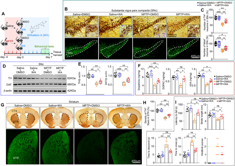
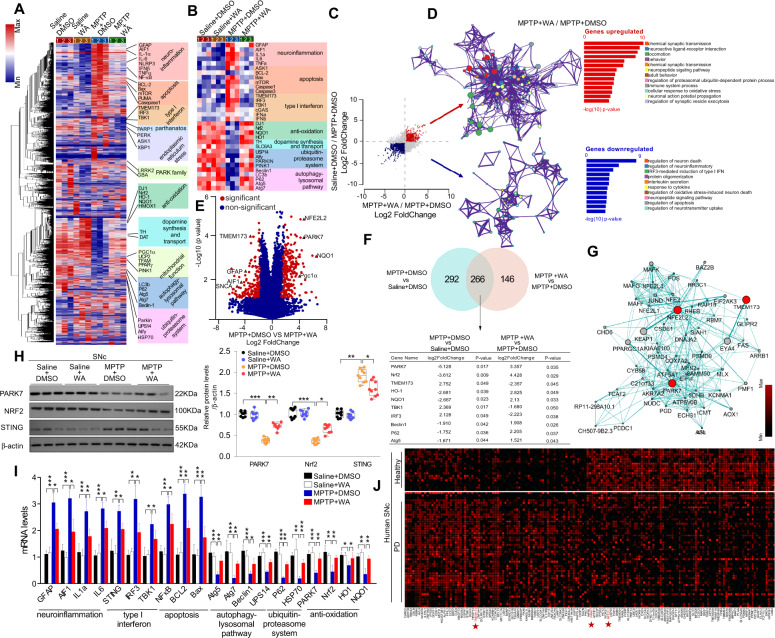
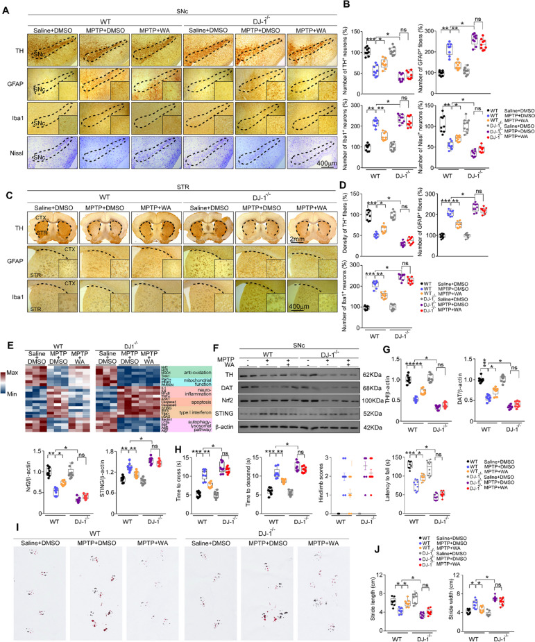
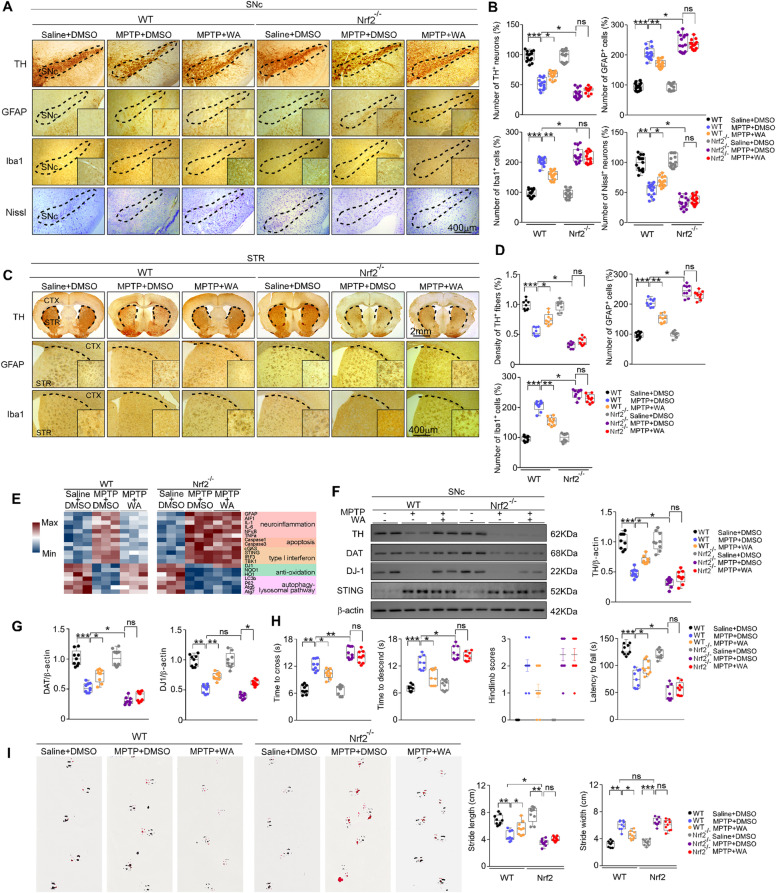
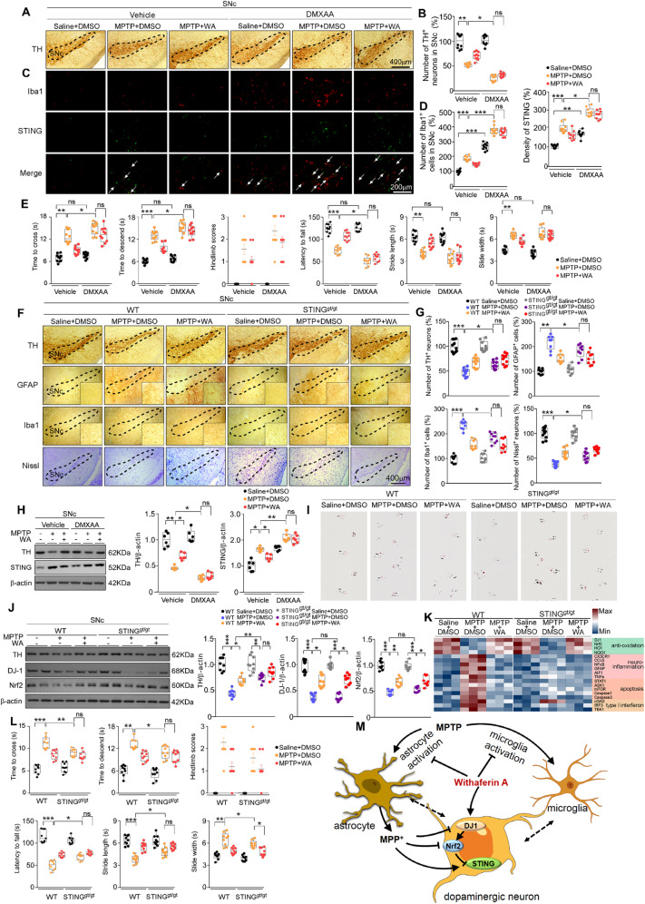
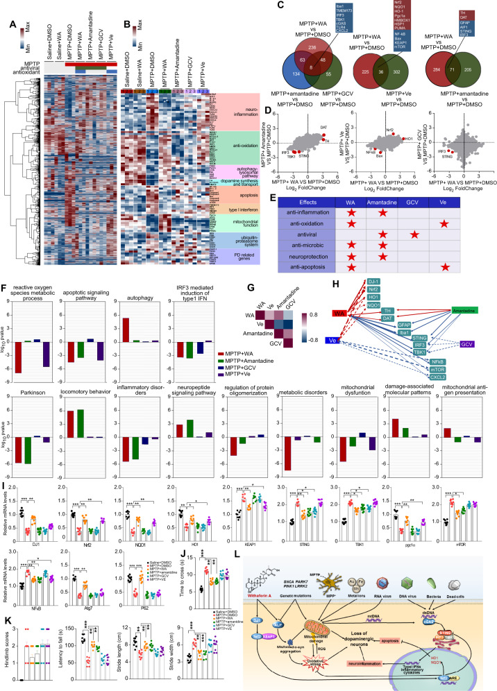
The pathogenesis of Parkinson's disease (PD) remains unclear, and there is no disease-modifying agent for PD. Withaferin A (WA), a naturally occurring compound, has emerged as a neuroprotective agent. However, the mechanisms by which WA is neuroprotective in PD are unknown. Here we show that WA protected against loss of dopaminergic neurons, neuroinflammation, and motor deficits in MPTP-induced PD mouse models. Whole-genome deep sequencing analysis combined with Meta-analysis of human PD studies reveal that DJ1, Nrf2, and STING in substantia nigra pars compacta (SNc) are linked to anti-PD effect of WA. We found that WA activated DJ1 and Nrf2, and suppressed STING within SNc; and overexpression of STING in SNc dampened the effect of WA. Using genetically modified mice (DJ1-KO, Nrf2-KO, STINGgt/gt and STING-KO) and immunolabeling technique, we identified that WA targeted DJ1-Nrf2-STING pathway in dopaminergic neurons; and we demonstrate that STING might be an important factor in PD pathogenesis. In addition, WA alleviated accumulation of phosphorylated α-synuclein (p-α-syn) and insoluble α-syn within SNc in adeno-associated virus (AAV)-mediated human α-syn overexpression PD model. Our comparative analysis on whole-genome transcriptome profiles suggests that STING might be a key target of WA and amantadine in PD treatment. This study highlights a multifaceted role for WA in neuroprotection, and suggests that WA can be a potential candidate for treatment of PD.


Conflict of interest statement

The authors declare no competing interests.

Figures

Fig. 1
Fig. 1. Neuroprotective effects of Withaferin A in MPTP-induced PD mouse model.
A Diagram of the experimental design. MPTP (20 mg/kg) or vehicle (saline) was injected (i.p.) for 5 consecutive days starting on day -4 after acclimation (3 days), then mice intraperitoneally (i.p.) received WA (20 μg/kg) or vehicle (DMSO) per day for 7 days, tissues were harvested for molecular analyses at day 8 after the last behavior test. B Representative photomicrographs of TH and Nissl staining in SNc. Scale bars, 400 μm for low-magnification images and 40 μm for high-magnification images, respectively. C Unbiased stereological counts of TH-positive and Nissl-positive neurons in SNc. Data are mean ± s.e.m.; n = 30 biologically independent animals; *P = 0.0425 **P = 0.007, ***P < 0.001 by one-way ANOVA with Bonferroni’s post hoc test. D Representative immunoblots of TH, DAT, and β-actin in SNc (cropped blot images are shown, see Supplementary Fig. 16 for full immunoblots). E Quantification of TH and DAT protein levels in SNc. Data are mean ± s.e.m.; n = 9 biologically independent animals. *P < 0.05, **P < 0.01, and ***P < 0.001. F HPLC assessment of dopamine concentrations in striatum of vehicle (saline) or MPTP injected mice treated with vehicle or WA. Data are mean ± s.e.m.; n = 6 biologically independent animals; *P < 0.05, **P < 0.01, and ***P < 0.001 by one-way ANOVA with Bonferroni’s post hoc test. G Representative photomicrographs of TH staining in striatum, scale bar, 2 mm (upper panel) and 400 μm (down panel). H Quantification of TH-positive striatal fiber density. Data are mean ± s.e.m.; n = 9 biologically independent animals. I Time to traverse beam apparatus, time to descend pole, Hind-limb clasping reflex score, fall latency from an accelerating rotarod and gait analysis. Data are mean ± s.e.m.; n = 9 biologically independent animals. The one-way ANOVAs were used for statistical analysis followed by Bonferroni’s post hoc test. *P < 0.05, **P < 0.01, and ***P < 0.001. ns, not significant.
Fig. 2
Fig. 2. Gene expression analysis of SNc in MPTP-induced PD mice treated with Withaferin A.
A Hierarchical clustered heatmap of gene expression profiles for WA or vehicle treatment in SNc of MPTP-induced PD mice. B Heatmap of DEGs of MPTP-received mice treated with WA or vehicle. C Scatter plot highlights the DEGs of WA treatment compared with vehicle in MPTP-recieved mice, upregulated genes are colored in red, downregulated genes are colored in blue. (P < 0.05 with unpaired two-tailed Student’s t tests). D Gene Ontology enrichment was based on DEGs that have a P value smaller than 0.05. Enrichment analysis for Gene Ontology terms among the genes of a gene–trait correlation module was performed using Metascape. E Volcano plot displays DEGs of WA treatment compared with vehicle in MPTP-received mice. Significantly altered genes are colored in red, insignificantly altered genes are colored in blue. F Venn diagram of overlapping significantly changed genes (±1.2 fold, P < 0.05). The top ten overlapping genes are presented. G Protein–protein interaction network identified among DJ1, Nrf2 and STING using GeneMANIA (direct interaction database). H Representative immunoblots of DJ1, Nrf2, STING and β-actin, and quantification of DJ1, Nrf2, STING protein levels in SNc (cropped blot images are shown, see Supplementary Fig. 16 for full immunoblots). Data are mean±s.e.m.; n = 9 biologically independent animals; *P < 0.05, **P < 0.01, and ***P < 0.001. I Relative mRNA expression of the indicated genes in SNc. Data are mean ± s.e.m.; n = 8 biologically independent animals. The one-way ANOVAs were used for statistical analysis followed by Bonferroni’s post hoc test. *P < 0.05, **P < 0.01, and ***P < 0.001. J Heatmap of the top 100 significant genes of SNc of human PD samples analyzed by Meta-analysis (FDR < 0.05). Expression values of each gene are standardized within each dataset. Hierarchical clustering was used to cluster samples and genes.
Fig. 3
Fig. 3. Neuroprotective effects of Withaferin A in PD are DJ1 dependent.
A Representative TH, GFAP, Iba1 and Nissl staining of SNc in WT and DJ1-KO mice, scale bar, 400 μm. B Unbiased stereological counts of TH+, GFAP+, Iba1+, and Nissl+ cells in SNc of WT and DJ1-KO mice. Data are mean ± s.e.m.; n = 9 biologically independent animals; *P < 0.05, **P < 0.01, and ***P < 0.001 by two-way ANOVAs followed by Tukey’s multiple comparisons test. C Representative photomicrographs of TH, GFAP, Iba1 staining in STR of WT and DJ1-KO mice, scale bar, 2 mm. D Stereological counts of TH, GFAP, Iba1 positive cells in STR of WT and DJ1-KO mice. Data are mean ± s.e.m.; n = 9 biologically independent animals; *P < 0.05, **P < 0.01, and ***P < 0.001 by two-way ANOVAs followed by Tukey’s multiple comparisons test. E Relative mRNA expression in SNc of WT and DJ1-KO mice. Data are mean ± s.e.m.; n = 8 biologically independent animals. F, G Representative immunoblots of TH, DAT, Nrf2 and STING in SNc (cropped blot images are shown, see Supplementary Fig. 16 for full immunoblots), quantification of TH, DAT, Nrf2, and STING levels. Data are mean ± s.e.m.; n = 9 biologically independent animals; *P < 0.05, **P < 0.01, and ***P < 0.001 by two-way ANOVAs followed by Tukey’s multiple comparisons test. H Time to traverse beam apparatus, time to descend pole, hind-limb clasping reflex score, fall latency from an accelerating rotarod and (I and J) gait analysis. Data are mean ± s.e.m.; n = 9 biologically independent animals. Two-way ANOVA followed by Tukey’s post hoc test. *P < 0.05, **P < 0.01, and ***P < 0.001. ns, not significant.
Fig. 4
Fig. 4. Neuroprotective actions of Withaferin A in PD are mediated by DJ1-Nrf2 axis.
A Representative TH, GFAP, Iba1 and Nissl staining of SNc in WT and Nrf2-KO mice, scale bar, 400 μm. B Unbiased stereological counts of TH+, GFAP+, Iba1+ and Nissl+ cells in SNc of WT and Nrf2-KO mice. Data are mean ± s.e.m.; n = 15 biologically independent animals; *P < 0.05, **P < 0.01, and ***P < 0.001 by two-way ANOVAs followed by Tukey’s multiple comparisons test. C Representative photomicrographs of TH, GFAP and Iba1 staining in STR of WT and Nrf2-KO mice, scale bar, 2 mm. D Stereological counts in STR of WT and Nrf2-KO mice. Data are mean ± s.e.m.; n = 9 biologically independent animals; *P < 0.05, **P < 0.01, and ***P < 0.001 by two-way ANOVAs followed by Tukey’s multiple comparisons test. E Relative mRNA expression in SNc of WT and Nrf2-KO mice. Data are mean ± s.e.m.; n = 8 biologically independent animals. F Representative immunoblots of TH, DAT, DJ1 and STING in SNc (cropped blot images are shown, see Supplementary Fig. 16 for full immunoblots). G Quantification of TH, DAT, DJ1 and STING levels. Data are mean ± s.e.m.; n = 9 biologically independent animals. H Time to traverse beam apparatus, time to descend pole, Hind-limb clasping reflex score, fall latency from an accelerating rotarod. I Gait analysis. Data are mean ± s.e.m.; n = 9 biologically independent animals. Two-way ANOVA followed by Tukey’s post hoc test. *P < 0.05, **P < 0.01, and ***P < 0.001. ns, not significant.
Fig. 5
Fig. 5. Withaferin A suppresses STING to protect dopaminergic neurons from MPTP neurotoxicity.
A Representative TH staining of SNc dopaminergic neurons in DMXAA or vehicle-treated mice, scale bar, 400 μm. B Unbiased stereological counts of TH+ neurons in SNc of DMXAA or vehicle-treated mice. Data are mean ± s.e.m.; n = 10 biologically independent animals; *P < 0.05 and **P < 0.01 by two-way ANOVAs followed by Tukey’s multiple comparisons test. C Representative Iba1 and STING immunostaining in SNc of DMXAA or vehicle-treated mice, scale bar, 200 μm. D Quantification of Iba1 and STING levels. Data are mean ± s.e.m.; n = 10 biologically independent animals; *P < 0.05, **P < 0.01, and ***P < 0.001 by two-way ANOVAs followed by Tukey’s multiple comparisons test. E Time to traverse beam apparatus, time to descend pole, hind-limb clasping reflex score, fall latency from an accelerating rotarod and gait analysis. Data are mean ± s.e.m.; n = 9 biologically independent animals; *P < 0.05, **P < 0.01, and ***P < 0.001 by two-way ANOVAs followed by Tukey’s multiple comparisons test. F Representative TH, GFAP, Iba1 and Nissl staining of SNc in WT and STINGgt/gt mice, scale bar, 400 μm. G Unbiased stereological counts of TH+, GFAP+, Iba1+ and Nissl+ cells in SNc of WT and STINGgt/gt mice. Data are mean ± s.e.m.; n = 9 biologically independent animals; *P < 0.05, **P < 0.01, and ***P < 0.001 by two-way ANOVAs followed by Tukey’s multiple comparisons test. H Representative immunoblots and quantification of TH and STING in SNc of DMXAA treated mice (cropped blot images are shown, see Supplementary Fig. 16 for full immunoblots). I Representative gait test images measured in WT and STINGgt/gt mice; n = 9 biologically independent animals. J Representative immunoblots and quantification of TH, DJ1, Nrf2 in SNc of WT and STINGgt/gt mice (cropped blot images are shown, see Supplementary Fig. 16 for full immunoblots). Data are mean ± s.e.m.; n = 9 biologically independent animals; *P < 0.05, **P < 0.01, and ***P < 0.001 by two-way ANOVAs followed by Tukey’s multiple comparisons test. K Relative mRNA expression in SNc of WT and STINGgt/gt mice. Data are mean ± s.e.m.; n = 6 biologically independent animals. L Time to traverse beam apparatus, time to descend pole, hind-limb clasping reflex score, fall latency from an accelerating rotarod. Data are mean ± s.e.m.; n = 10 biologically independent animals; *P < 0.05, **P < 0.01, and ***P < 0.001 by two-way ANOVAs followed by Tukey’s multiple comparisons test. M Schematic summary of the mechanism underlying the neuroprotective actions of WA in PD.
Fig. 6
Fig. 6. Withaferin A has multiple neuroprotective effects; Withaferin A and amantadine may treat PD by suppressing STING.
A Hierarchical clustered heatmap of gene expression profiles for WA, amantadine, Ve, ganciclovir treatment in SNc of MPTP-induced PD mice. B The heatmap of DEGs of MPTP-received mice treated with WA, amantadine, Ve, ganciclovir. C Venn diagram of overlapping significantly changed genes (±1.2 fold, P < 0.05). D Scatter plot highlights the DEGs of amantadine, Ve and GCV treatment compared with vehicle in MPTP-recieved mice, significantly altered genes are colored in red. (P < 0.05 with Benjamini–Hochberg multiple testing correction). E The pharmacological actions of WA, amantadine, Ve and GCV from published literatures. F Gene Ontology enrichment was based on DEGs that have a P value smaller than 0.05. G Spearman correlation between aggregated response of WA, amantadine, Ve and GCV gene signatures to different interventions. H Network of interventions based on similarity of their gene expression profiles. The width of edge is defined by statistical significance of Spearman correlation between interventions. Only significant connections are shown. I Relative mRNA levels in SNc of WA, amantadine, Ve and GCV treated mice. Data are mean ± s.e.m.; n = 9 biologically independent animals; *P < 0.05, **P < 0.01, and ***P < 0.001 by one-way ANOVA with Bonferroni’s post hoc test. J, K Time to traverse beam apparatus, time to descend pole, hind-limb clasping reflex score, fall latency from an accelerating rotarod and gait analysis. Data are mean ± s.e.m.; n = 9 biologically independent animals. The one-way ANOVAs were used for statistical analysis followed by Bonferroni’s post hoc test. *P < 0.05, **P < 0.01, and ***P < 0.001. L Schematic summary of the mechanism underlying the neuroprotective actions of WA in PD. The neuroprotective role of WA against PD is dependent on relief of oxidative stress, mitigation of the STING-mediated neuroinflammation, enhancement of mitochondrial function, and reduction of apoptosis, through activation of DJ1-Nrf2-STING axis in dopaminergic neurons of SNc.

Similar articles

Cited by

References

    1. Burbulla LF, Jeon S, Zheng J, Song P, Silverman RB, Krainc D. A modulator of wild-type glucocerebrosidase improves pathogenic phenotypes in dopaminergic neuronal models of Parkinson’s disease. Sci Transl Med. 2019;11:eaau6870. - PMC - PubMed
    1. Johnson ME, Stecher B, Labrie V, Brundin L, Brundin P. Triggers, facilitators, and aggravators: redefining Parkinson’s disease pathogenesis. Trends Neurosci. 2019;42:4–13. doi: 10.1016/j.tins.2018.09.007. - DOI - PMC - PubMed
    1. Mattson MP. Infectious agents and age-related neurodegenerative disorders. Ageing Res Rev. 2004;3:105–120. doi: 10.1016/j.arr.2003.08.005. - DOI - PMC - PubMed
    1. Laperle AH, Sances S, Yucer N, Dardov VJ, Garcia VJ, Ho R, et al. iPSC modeling of young-onset Parkinson’s disease reveals a molecular signature of disease and novel therapeutic candidates. Nat Med. 2020;26:289–299. doi: 10.1038/s41591-019-0739-1. - DOI - PubMed
    1. Koprich JB, Kalia LV, Brotchie JM. Animal models of alpha-synucleinopathy for Parkinson disease drug development. Nat Rev Neurosci. 2017;18:515–529. doi: 10.1038/nrn.2017.75. - DOI - PubMed

Publication types