Review
. 2020 Oct 26;45(1):1-16.
doi: 10.1080/01658107.2020.1810286. eCollection 2021.

Diagnosing Optic Disc Drusen in the Modern Imaging Era: A Practical Approach

Collaborators, Affiliations
Review

Diagnosing Optic Disc Drusen in the Modern Imaging Era: A Practical Approach

F Costello et al. Neuroophthalmology. .

Abstract

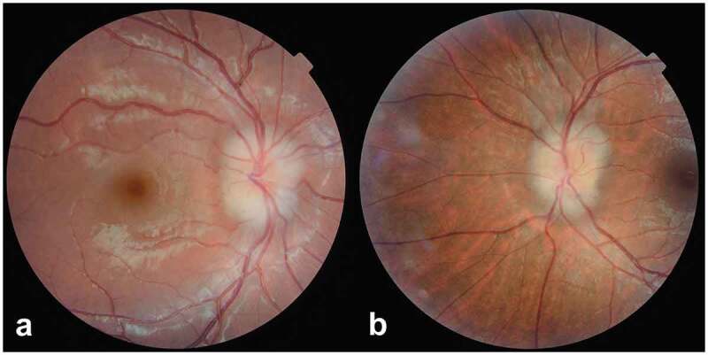
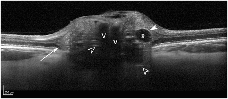
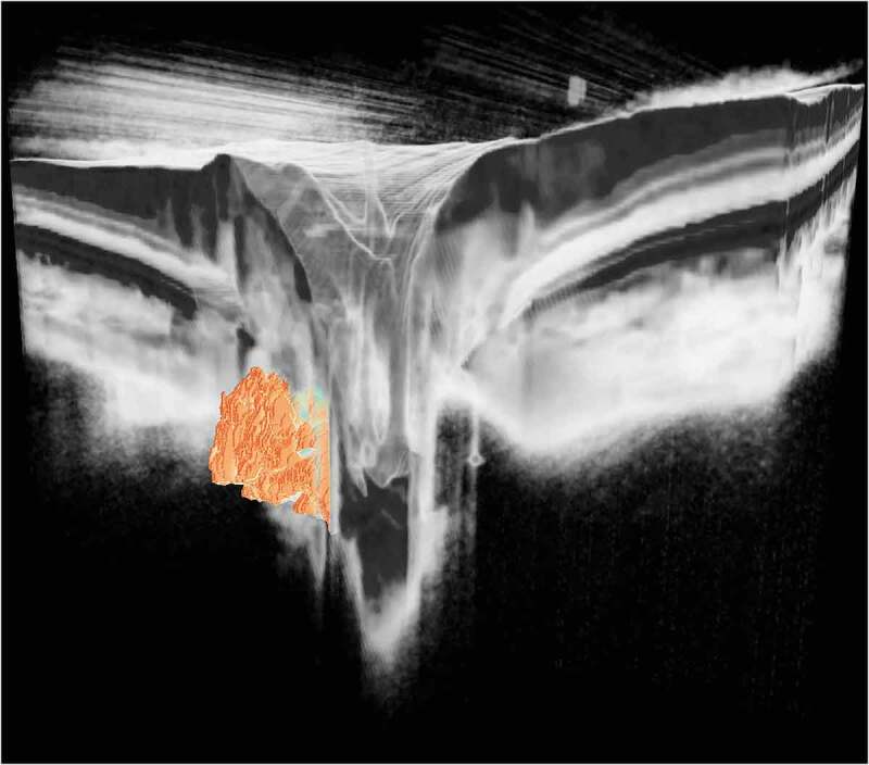
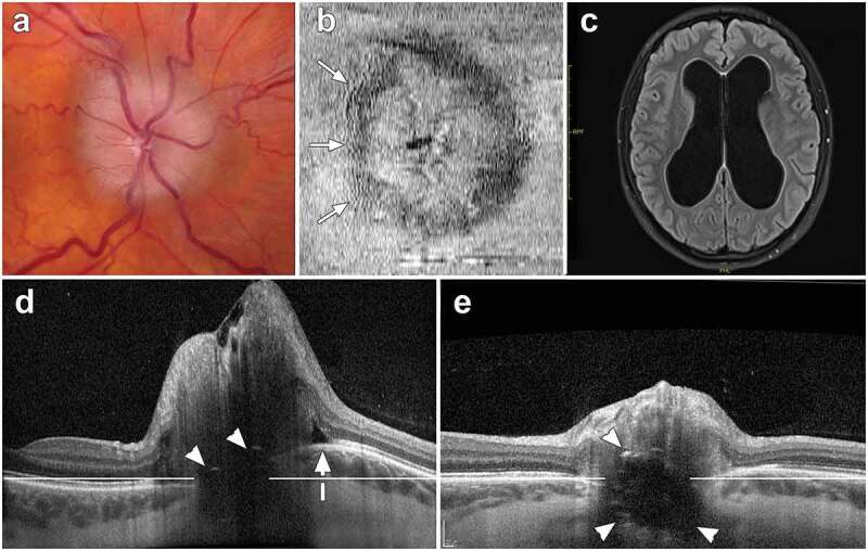
Optic disc drusen (ODD) are a well-recognised cause of an elevated optic disc appearance. When visible with ophthalmoscopy and fundus photography, ODD are readily identified. Yet, in more subtle cases of ODD, ancillary testing may be needed to render the diagnosis. Facilitating the diagnosis of ODD has clinical relevance, because affected individuals may otherwise undergo unnecessary costly and invasive investigations to rule out raised intracranial pressure and other causes of optic disc oedema. In this review, the role of established and emerging optical coherence tomography (OCT) techniques in the diagnosis and management of ODD cases is reviewed. A practical approach is taken to explain how to optimise use of commercially available OCT technology in the clinical setting. Optical coherence tomography provides many advantages over other imaging modalities in the diagnosis of ODD, including the ability to correlate retinal measures of neuroaxonal structure with drusen characteristics. Earlier spectral domain OCT techniques, however, were hindered by poor penetrance. In the modern imaging era, enhanced depth imaging OCT and swept source OCT enable higher resolution of ODD and other optic nerve head structures that might otherwise be mistaken for drusen. Ongoing studies featuring OCT angiography indicate that this technique may provide complementary information about microvascular supply that correlate with structural measures of optic nerve injury. Advances in OCT will continue to improve diagnostic accuracy and inform clinical understanding regarding structure-function correlations germane to the longitudinal follow up of ODD patients.

Keywords: Optic disc drusen (ODD); enhanced depth imaging OCT (EDI-OCT); optical coherence tomography (OCT); optical coherence tomography angiography (OCTA); papilloedema; pseudo-papilloedema; swept source OCT (SS-OCT).


Figures

Figure 1.
Figure 1.
Colour fundus photographs showing the comparable appearance of optic disc elevation in (a) an 8-year-old boy with buried optic disc drusen (right eye) and, (b) a 23-year-old woman with idiopathic intracranial hypertension and associated papilloedema (left eye)
Figure 2.
Figure 2.
Spectral domain optical coherence tomography (OCT) images of a 25-year-old woman with large optic disc drusen (ODD) (white asterisk). Enhanced depth imaging OCT in the lower image clearly shows the advantage of this technique, by depicting the posterior delineation of the ODD and lack of shadowing of the drusen, as compared with the top image
Figure 3.
Figure 3.
Optic disc drusen morphology in a 22-year-old woman, shown with enhanced depth imaging optical coherence tomography. The drusen are seen as signal-poor structures (asterisk) with a partial hyper-reflective margin (solid white arrowhead). Prelaminar hyper-reflective lines (open white arrowheads) are thought to represent drusen precursors. Peripapillary hyper-reflective ovoid mass-like structures (longer white arrow) circumscribe the disc and likely represent herniated nerve fibres due to crowded optic nerve head conditions. Blood vessel shadows are denoted with v’s
Figure 4.
Figure 4.
Enhanced depth imaging optical coherence tomography from five different patients with optic disc drusen (ODD). Upper panel: a line connecting the Bruch’s membrane openings on either side of the optic disc can be used to categorise ODD by location, namely (a) superficial and (b) deep depending on where the bulk of the drusen material is located. Lower panel: The scale bar in the lower left corner can be used to categorise ODD by size including large (c), referring to drusen measuring greater than 200 μm in at least one direction (arrow), and small (d), referring to drusen that are smaller than 200 μm in all directions (arrow). Conglomerates of ODD (e) are made up of multiple smaller drusen (arrow) that cannot be clearly differentiated from one another
Figure 5.
Figure 5.
Enhanced-depth imaging optical coherence tomography imaging shows peripapillary hyper-reflective ovoid mass-like structures (PHOMS) in a 23-year-old woman with idiopathic intracranial hypertension (upper panel), and in a 13-year-old boy with optic disc drusen (lower panel). The two main characteristics of the PHOMS are: the upward deflection of retinal layers on top of the drusen creating the ‘ski slope sign’ (arrowheads), and the visible hyper-reflective Bruch’s membrane (longer white arrows) underneath the PHOMS. The PHOMS (encircled in white) are annotated to the right
Figure 6.
Figure 6.
Comparing spectral domain (SD) optical coherence tomography (OCT) (a) and swept source (SS) OCT (b) images from a volume scan of an eye with deep ODD. The SD-OCT scan was performed with enhanced-depth imaging (EDI), and included frame averaging techniques. Both OCT scans depict comparable images of optic disc drusen, showing a hypo-reflective core (white asterisk) and hyper-reflective margins. The associated prelaminar hyper-reflective lines (white arrow) are slightly more visible with the SD-OCT EDI scan (a)
Figure 7.
Figure 7.
Three-dimensional visualisation of deeply localised optic disc drusen derived from a swept source optical coherence tomography optic disc volume scan. The large, solid drusen are buried within the optic nerve head tissue (which are faded in this artistic rendering, to allow better visibility). (Image processing and visualisation by Peter Maloca, Institute of Molecular and Clinical Ophthalmology Basel, Switzerland)
Figure 8.
Figure 8.
Images from an 18-year-old female with papilloedema, obstructive hydrocephalus and optic disc drusen (ODD). (a) Fundus photograph of an elevated optic nerve head, showing mild vessel tortuosity and obscuration of the entire disc margin. (b) Infrared en face optical coherence tomography (OCT) depicting peripapillary wrinkles on the temporal side of the optic disc (arrows). (c) Magnetic resonance imaging demonstrating ventricular enlargement caused by obstructive hydrocephalus. (d) Transverse axial OCT (30°, vertically scaled 3x, enhanced-depth imaging) at baseline. This image demonstrates ODD with signal-poor region and two small hyper-reflective caps (arrowheads). Bruch’s membrane layer is anteriorly deformed towards the vitreous (white arrow) relative to a horizontal reference line. The mean peripapillary retinal nerve fibre layer thickness measures 316 μm. [e] Follow-up transverse axial OCT performed after a ventriculostomy procedure shows a significant decrease in the disc elevation (mean peripapillary RNFL thickness measures 87 μm). The ODD are more visible. Moreover, the shape of Bruch’s membrane layer is flatter, moving posteriorly away from the vitreous towards the reference line

References

    1. Hamann S, Malmqvist L, Costello F.. Optic disc drusen: understanding an old problem from a new perspective. Acta Ophthalmol. 2018;96(7):673–684. doi:10.1111/aos.13748. - DOI - PubMed
    1. Lorentzen SE. Drusen of the optic disk. A clinical and genetic study. Acta Ophthalmol (Copenh). 1966;Suppl 90:91–180. - PubMed
    1. Fraser JA, Bursztyn LLCD. Optical coherence tomography in optic disc drusen. Annal Eye Sci. 2020;5:5. doi:10.21037/aes.2019.12.03. - DOI
    1. Chang MY, Pineles SL. Optic disk drusen in children. Surv Ophthalmol. 2016;61(6):745–758. doi:10.1016/j.survophthal.2016.03.007. - DOI - PMC - PubMed
    1. Duncan JE, Freedman SF, El-Dairi MA. The incidence of neovascular membranes and visual field defects from optic nerve head drusen in children. J Aapos. 2016;20(1):44–48. doi:10.1016/j.jaapos.2015.10.013. - DOI - PubMed

LinkOut - more resources