Case Reports
. 2020 Jul 20;45(1):36-40.
doi: 10.1080/01658107.2020.1773507. eCollection 2021.

Morning Glory Disc Anomaly with Contractile Peripapillary Staphyloma in an 18-Month-Old Girl

Affiliations
Case Reports

Morning Glory Disc Anomaly with Contractile Peripapillary Staphyloma in an 18-Month-Old Girl

Kalina Trifonova et al. Neuroophthalmology. .

Abstract

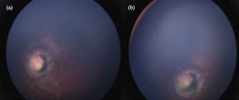
We present an 18-month-old girl with strabismus and a variable esotropia of the left eye. Fixation of the affected eye was intermittent with a relative afferent pupillary defect. A fundus photography of the left eye displayed a combination of features of both morning glory disc anomaly and peripapillary staphyloma. A B-scan ultrasonography examination of the left eye showed a conical excavation of the posterior pole. Cycloplegic refraction measurements showed a large amount of anisometropia. Correction with glasses and part-time occlusion was prescribed and a strict follow-up routine was advised. No other systemic associations with the disease have been discovered so far in our patient. We support the theory that morning glory disc anomaly and peripapillary staphyloma may represent two different morphologies in the spectrum of the same disease.

Keywords: Morning glory disc anomaly; amblyopia; contractile staphyloma; esotropia.


Figures

Figure 1.
Figure 1.
RetCam fundal photograph of the left eye demonstrating an enlarged papilla with an annulus of chorioretinal pigmentary disposition, blood vessels abandoning the papilla in a radial fashion to continue in a straight course, gliosis in the centre and deep peripapillary excavation with surrounding mottled pigment epithelium
Figure 2.
Figure 2.
RetCam fundal photograph of the right eye demonstrating a normal fundus
Figure 3.
Figure 3.
Changes in the visible part of the left optic disc. (a) Relaxed phase. (b) Contractile phase
Figure 4.
Figure 4.
Ultrasonography of the left eye demonstrating a conical excavation of the posterior pole (arrowed)

Similar articles

References

    1. Brodsky M. Congenital optic disk anomalies. Surv Ophthalmol. 1994;39(2):89–112. doi:10.1016/0039-6257(94)90155-4. - DOI - PubMed
    1. Miller NR, Walsh FB, Hoyt WF, eds. ‎Walsh and Hoyt’s Clinical Neuro-ophthalmology. Lippincott Williams & Wilkins; Vol. 1. 2005:59.
    1. Quah BL, Hamilton J, Blaser S, Héon E, Tehrani NN.. Morning glory disc anomaly, midline cranial defects and abnormal carotid circulation: an association worth looking for. Pediatr Radiol. 2005;35(5):525–528. doi:10.1007/s00247-004-1345-y. - DOI - PubMed
    1. Cavazos-Adame H, Olvera-Barrios A, Martinez-Lopez-Portillo A, Mohamed-Hamsho J. Morning glory disc anomaly, a report of a successfully treated case of functional amblyopia. J Clin Diagn Res. October 9, 2015;10:ND01–ND03. - PMC - PubMed
    1. BJ L, EI T. Update on the morning glory disc anomaly. Ophthalmic Genet. 2008. June;29(2):47–52. doi:10.1080/13816810801901876. - DOI - PubMed

Publication types

LinkOut - more resources