. 2020 Nov-Dec;22(6):21-29.
doi: 10.1109/MCSE.2020.3015511. Epub 2020 Aug 11.

Revealing the mechanism of SARS-CoV-2 spike protein binding with ACE2

Affiliations

Revealing the mechanism of SARS-CoV-2 spike protein binding with ACE2

Yixin Xie et al. Comput Sci Eng. 2020 Nov-Dec.

Abstract

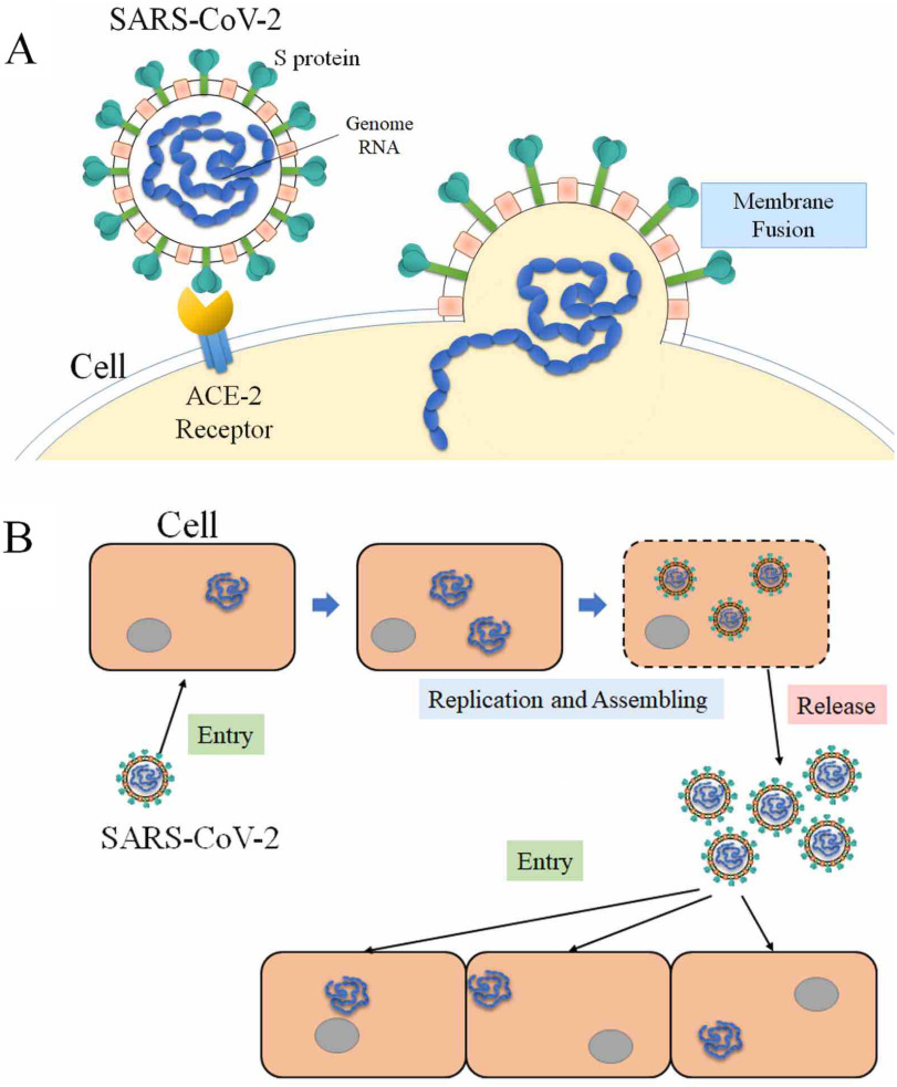
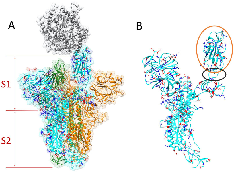
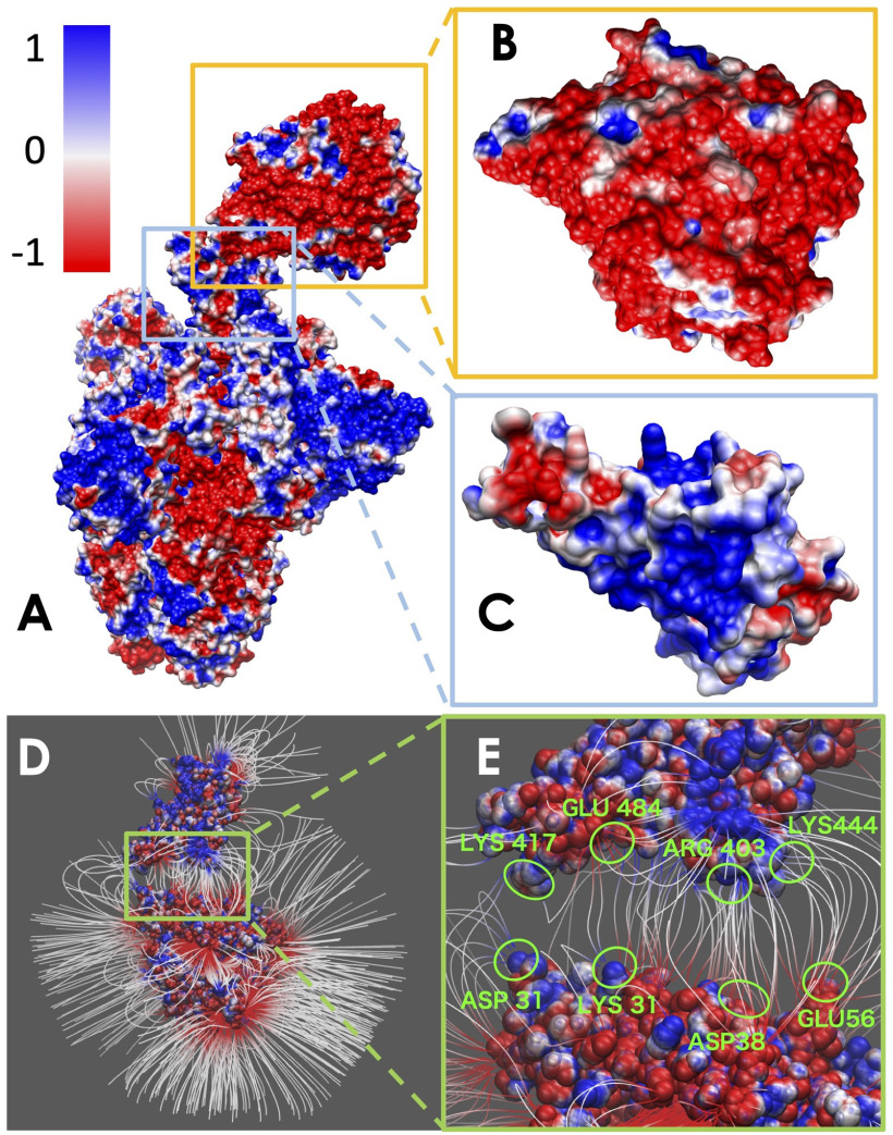
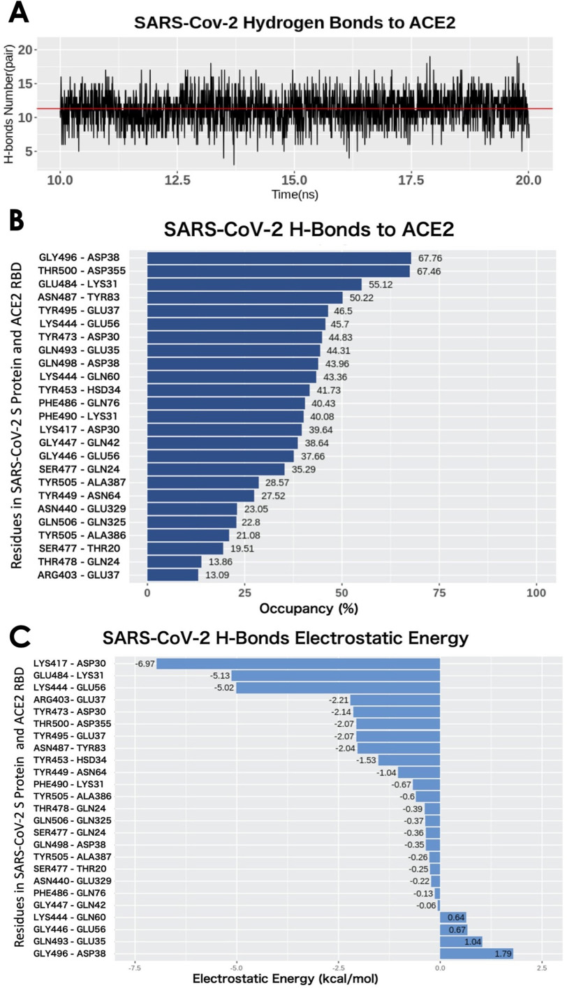
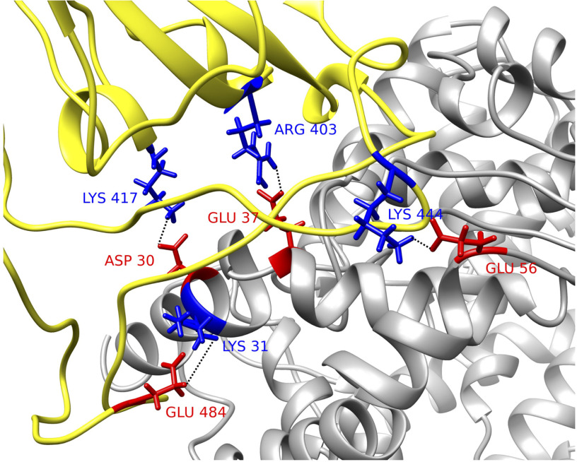
A large population in the world has been infected by COVID-19. Understanding the mechanisms of Severe Acute Respiratory Syndrome CoronaVirus 2 (SARS-CoV-2) is important for management and treatment of the COVID-19. When it comes to the infection process, one of the most important proteins in SARS-CoV-2 is the spike (S) protein, which is able to bind to human Angiotensin-Converting Enzyme 2 (ACE2) and initializes the entry of the host cell. In this study, we implemented multi-scale computational approaches to study the electrostatic features of the interfaces of the SARS-CoV-2 S protein Receptor Binding Domain (RBD) and ACE2. The simulations and analyses were performed on high-performance computing resources in Texas Advanced Computing Center (TACC). Our study identified key residues on the SARS-CoV-2, which can be used as targets for future drug design. The results shed lights on future drug design and therapeutic targets for COVID-19.

Keywords: ACE2; COVID-19; Computational biophysics; SARS-CoV-2; coronavirus; protein-protein interactions; spike protein.


Figures

Figure 1.
Figure 1.
Process of SARS-CoV-2 infecting host cells. (A) SARS-CoV-2 S protein binds to ACE2 of host cell, and enters the host cell after binding. (B) SARS-CoV-2 utilizes the host cell as a factory to reproduce more SARS-CoV-2 and release them to infect more cells.
Figure 2.
Figure 2.
Structure of S protein homotrimer and ACE2. Positive and negative residues on a monomer of S protein are colored in blue and red, respectively. (A) ACE2 is colored in gray. The S protein trimer contains three identical monomers, colored in cyan, green, and orange. One monomer (cyan)'s RBD flips out to bind with the ACE2. The S protein is composed by S1 and S2 subunits. (B) Single S protein monomer structure. The orange circle shows the RBD and the black circle marks the flexible hinge connecting RBD and other part of the S protein.
Figure 3.
Figure 3.
Electrostatic surface and electric field lines of SARS-CoV-2 S protein and ACE2 binding domain. (A) Electrostatic surface of S protein and ACE2. (B) Binding interface of ACE2. (C) Binding interface of S protein RBD. (D) Electric field lines of S protein RBD and ACE2 binding domain. (E) A closeup of electric field lines at the binding interfaces. The residues forming salt bridges are highlighted.
Figure 4.
Figure 4.
H-bonds between SARS-CoV-2 RBD and ACE2 binding domain. (A) Numbers of H-bonds at different time stamps from 10 to 20 ns, the average value is marked as red line with the value of 11.30. (B) Occupancy of H-bonds ordered by decreasing values of occupancy, and the residues are labeled in y-axis with the SARS-Cov-2 in the left and ACE2 in the right. (C) List of hydrogen bonds that are ranked by their electrostatic energy values.
Figure 5.
Figure 5.
Structural demonstration of key residues in salt bridges. SARS-CoV-2 S protein RBD (yellow) with human ACE2 binding domain (gray). Key residues are marked with its amino acid types, sequence numbers, and charges, where blue stands for positively charged amino acid and red stands for the negative ones. Four pairs of salt bridges are found by VMD. All the structures in this figure are rendered by Chimera.

References

    1. World Health Organization, Coronavirus Disease 2019 (COVID-19): Situation Report, 85, 2020.
    1. Fehr A. R. and Perlman S., “Coronaviruses: An overview of their replication and pathogenesis,” in Coronaviruses. Berlin, Germany: Springer, 2015, pp. 1–23. - PMC - PubMed
    1. Li G., et al. , “Coronavirus infections and immune responses,” J. Med. Virol., vol. 92, no. 4, pp. 424–432, 2020. - PMC - PubMed
    1. Li L., Wang L., and Alexov E., “On the energy components governing molecular recognition in the framework of continuum approaches,” Frontiers Mol. Biosci., vol. 2, 2015, Art. no. 5. - PMC - PubMed
    1. Li L., Alper J., and Alexov E., “Cytoplasmic dynein binding, run length, and velocity are guided by long-range electrostatic interactions,” Sci. Rep., vol. 6, 2016, Art. no. 31523. - PMC - PubMed

LinkOut - more resources