Review
. 2021 Mar 8:11:612452.
doi: 10.3389/fimmu.2020.612452. eCollection 2020.

Cross Kingdom Immunity: The Role of Immune Receptors and Downstream Signaling in Animal and Plant Cell Death

Affiliations
Review

Cross Kingdom Immunity: The Role of Immune Receptors and Downstream Signaling in Animal and Plant Cell Death

Thibault Roudaire et al. Front Immunol. .

Abstract

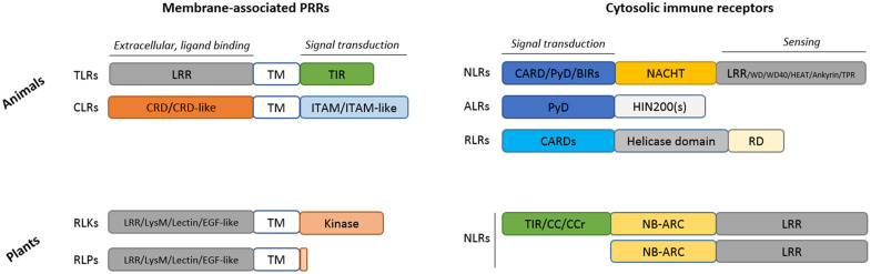
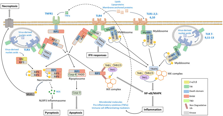
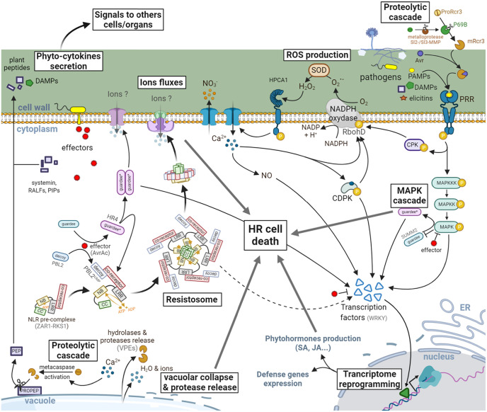
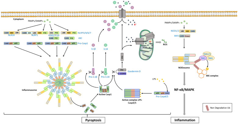
Both plants and animals are endowed with sophisticated innate immune systems to combat microbial attack. In these multicellular eukaryotes, innate immunity implies the presence of cell surface receptors and intracellular receptors able to detect danger signal referred as damage-associated molecular patterns (DAMPs) and pathogen-associated molecular patterns (PAMPs). Membrane-associated pattern recognition receptors (PRRs), such as Toll-like receptors (TLRs), C-type lectin receptors (CLRs), receptor-like kinases (RLKs), and receptor-like proteins (RLPs) are employed by these organisms for sensing different invasion patterns before triggering antimicrobial defenses that can be associated with a form of regulated cell death. Intracellularly, animals nucleotide-binding and oligomerization domain (NOD)-like receptors or plants nucleotide-binding domain (NBD)-containing leucine rich repeats (NLRs) immune receptors likely detect effectors injected into the host cell by the pathogen to hijack the immune signaling cascade. Interestingly, during the co-evolution between the hosts and their invaders, key cross-kingdom cell death-signaling macromolecular NLR-complexes have been selected, such as the inflammasome in mammals and the recently discovered resistosome in plants. In both cases, a regulated cell death located at the site of infection constitutes a very effective mean for blocking the pathogen spread and protecting the whole organism from invasion. This review aims to describe the immune mechanisms in animals and plants, mainly focusing on cell death signaling pathways, in order to highlight recent advances that could be used on one side or the other to identify the missing signaling elements between the perception of the invasion pattern by immune receptors, the induction of defenses or the transmission of danger signals to other cells. Although knowledge of plant immunity is less advanced, these organisms have certain advantages allowing easier identification of signaling events, regulators and executors of cell death, which could then be exploited directly for crop protection purposes or by analogy for medical research.

Keywords: NOD-like receptors; Toll-like receptors; damage-associated molecular patterns; hypersensitive response; pathogen-associated molecular patterns; pattern recognition receptors; regulated cell death.


Conflict of interest statement

The authors declare that the research was conducted in the absence of any commercial or financial relationships that could be construed as a potential conflict of interest.

Figures

Figure 1
Figure 1
Structural comparison of the main immune receptors found in animals and plants. ALR, AIM2 (absent in melanoma 2)-like receptors; BIR, Baculovirus Inhibitor of apoptosis protein Repeat; CARD, caspase recruitment domain; CC, coiled coil domain; CCr, CC-RPW8; CLR; C-type lectin receptors; CRD, Carbohydrate-Recognition Domain; EGF-like, Epidermal Growth Factor like; HIN200(s), Hematopoietic Interferon-inducible Nuclear protein with a 200 amino acid repeat; ITAM, Immunoreceptor Tyrosine-based Activation Motif; LRR, leucine-rich repeat; LysM, Lysin Motif; NACHT, NAIP (neuronal apoptosis inhibitory protein), CIITA (MHC class II transcription activator), HET-E (incompatibility locus protein from Podospora anserina) or TP1 (telomerase-associated protein); NB-ARC, Nucleotide-Binding domain Apaf1, Resistance, CED4; NLR, Nucleotide-binding and oligomerization domain (NOD)-Like Receptor (animals) or Nucleotide-Binding Domain (NBD)-containing LRRs (plants); PYD, Pyrin effector Domain; RD, Regulator Domain; RLK, Receptor-Like Kinase; RLP, Receptor-Like Protein (contain a short cytoplasmic domain devoid of kinase activity); RLR, RIG-I–like receptors; TIR, Toll/Interleukin-1 receptor; TM; Transmembrane; TLRs, Toll-like receptors.
Figure 2
Figure 2
TLR-mediated signaling pathway. The activation of TLR (toll like receptor) 1–2, 4–6, or 10 by lipids, lipoproteins or membrane-anchored proteins and the activation of TLR7-9, 11–13 by virus-derived nucleic acids induce the dimerization of TLRs and the recruitment of Myd88 (myeloid differentiation factor 88) and IRAKs via homotypic interaction domains, forming the Myddosome. IRAKs catalyze phosphorylation cascade leading to the recruitment of TRAF6 [tumor necrosis factor (TNF) receptor associated factor 6]. In turn, TRAF6 promotes the activation by proximity of TAK1 (tumor growth factor-β-activated kinase 1)/TAB1-3 (transforming growth factor-activated kinase1-binding protein 1, 2, and 3) and the IKK (Inhibitor of κB kinase) complexes that result in the activation of MAPK (Mitogen-activated protein kinases), NF-κB (nuclear factor-kappa B)-signaling pathways, and pro-inflammatory response. TLR3 and TLR4 stimulation induces the recruitment of TRIF (TIR-domain-containing adaptor-inducing IFN-β) through a TIR-TIR homotypic interaction. The adaptor TRAM (Trif-related adaptor molecule) serves as a bridge between TLR4 and TRIF. In turn TRIF can recruit TRAF3 that engages IFN (Interferon) response, TRAF6 that leads to MAPK and NF-κB activation, or the kinases RIP (Receptor Interacting Protein) 1 and/or RIP3. When poly-ubiquitinated, RIP1 can recruit TAK1/TAB1-3 and IKK complexes and activates the pro-inflammatory response. In a non-ubiquitinated form, RIP1 can assemble with FADD and caspase-8 to form the ripoptosome that leads to caspase cascade activation and apoptotic cell death or can activate RIP3 in the necrosome. RIP3 catalyzes the activating phosphorylation of MLKL, which oligomerizes and translocates into the plasma membrane to form pores and induces necroptosis. The DAI (DNA-dependent activator of IRFs) can also directly recruit RIP3 via RHIM homotypic interaction to induce MLKL phosphorylation and necroptosis in response to virus-derived nucleic acids. The necrosome has also the ability to induce ROS (Reactive oxygen species) production resulting in NLRP3 (NOD (Nucleotide-binding oligomerization domain)-like receptor protein) inflammasome and pyroptosis (details of pyroptosis available in Figure 4 ). The TLR-mediated production of TNF-α can induce an autocrine stimulation of TNFR1 that can lead to RIP1 engagement and an amplification of the signal.
Figure 3
Figure 3
Signaling events leading to HR in plants. PRRs are activated by the recognition of eliciting molecules resulting from the degradation of plant cells (DAMPs) or released by the pathogens (PAMPs, elicitins, apoplastic avirulence factors: Avr). The signal is then transduced by a cascade of phosphorylation events involving MAPKs, cytoplasmic protein kinases (CPKs), and transcription factors, mainly from the WRKY family. This phosphorylation can also activate the NADPH oxidase RbohD, leading to the production of ROS [O2. transformed into hydrogen peroxide (H2O2) by a superoxide dismutase (SOD)]. An influx of intracellular Ca2+, initiated quickly after perception of H2O2 by HPCA1 leads to the production of nitric oxide (NO), as well as the activation of transcription factors via the calcium dependent protein kinases (CDPKs). This is followed by a reprogramming of the transcriptional activity leading to the expression of defense genes involved in the synthesis of phytohormones (SA, JA, …), the antimicrobial phytoalexins or even the release of hydrolytic enzymes (glucanases, chitinases, …) from the pathogenesis-related proteins family. In the meantime, effectors secreted by pathogens to counter the plant’s defenses can also be directly or indirectly (via the recognition of a modified host-protein) recognized by NLRs. This recognition generally induces a conformational change in the protein (noted here by an asterisk and a color change), allowing the exchange of ADP by an ATP and therefore the activation of the NLR leading in some cases to macromolecular complexes such as the resistosome or to the activation of transcription factors. These larger-scale molecular complexes have been proposed to act via the recruitment of other signaling actors leading to a potentiation of the defenses already in place or by the formation of pores in the plasma membrane. A HR cell death is then observed locally to block the spreading of the pathogen. This will also be associated with the release of DAMPs, phytohormones and phyto-cytokines which will transmit information to neighboring cells and organs to prevent future infections in healthy tissues. Some plant peptides (e.g., PEPs) can be matured by metacaspase-mediated cleavage and released in the apoplast to prime immune responses in neighboring cells, thus enabling the establishment of a local resistance.
Figure 4
Figure 4
NLR-mediated signaling pathway in mammals. The recognition of PAMPs/DAMPs [Pathogen-Associated Molecular Patterns/Damage-Associated Molecular Patterns) by NLRs (NOD (Nucleotide-binding oligomerization domain)-like receptor proteins: NLRCs, NLRPs] promotes the recruitment of procaspase-1 (Pro-Casp1) through homotypic domain interaction, forming a large cytoplasmic complex named inflammasome. In some situation, the adaptor ASC (apoptosis-associated speck-like protein containing a CARD domain) can take part to the complex, bridging the receptor to procaspase-1. The procaspase-1 undergoes auto-activation and processing of its 2 active sub-units (p10 and p20) which assemble into a tetrameric active form. Active caspase-1 catalyzes the cleavage of proIL-18 (proInterleukine-18), proIL-1β (proInterleukine-1β) into mature cytokines, and gasdermin-D releasing the PFD domain that can oligomerizes and anchors into the membrane to form pores. Processing of gasdermin-D can also be ensured by caspase-4 and caspase-5, which are activated after LPS binding. The gasdermin PFD can also form pores into the mitochondrial membrane that induce K+ release and ROS production. In addition, pores created in the plasma membrane by PFD also lead to IL1β and IL18 release and to an ion imbalance that finally results in pyroptosis. The recognition of PAMPs/DAMPs by NOD1/2 promotes the formation of the NODosome composed of the sensor and the protein RIP2 (Receptor Interacting Protein 2) through CARD-dependent homotypic interaction. RIP2 can then recruit TAB1/TAB2/3/TAK1 and IKK complexes that engage MAPK and NF-kB signaling pathways leading to the production of pro-inflammatory cytokines.

Similar articles

Cited by

References

    1. Klessig DF, Choi HW, Dempsey DA. Systemic Acquired Resistance and Salicylic Acid: Past, Present, and Future. Mol Plant Microbe Interact (2018) 31(9):871–88. 10.1094/MPMI-03-18-0067-CR - DOI - PubMed
    1. Amarante-Mendes GP, Adjemian S, Branco LM, Zanetti LC, Weinlich R, Bortoluci KR. Pattern Recognition Receptors and the Host Cell Death Molecular Machinery. Front Immunol (2018) 9:2379. 10.3389/fimmu.2018.02379 - DOI - PMC - PubMed
    1. Galluzzi L, Vitale I, Aaronson SA, Abrams JM, Adam D, Agostinis P, et al. . Molecular mechanisms of cell death: recommendations of the Nomenclature Committee on Cell Death 2018. Cell Death Differ (2018) 25:486–541. 10.1038/s41418-018-0102-y - DOI - PMC - PubMed
    1. Doran AC, Yurdagul A, Jr., Tabas I. Efferocytosis in health and disease. Nat Rev Immunol (2020) 20(4):254–67. 10.1038/s41577-019-0240-6 - DOI - PMC - PubMed
    1. Locato V, De Gara L. Programmed Cell Death in Plants: An Overview. Methods Mol Biol (2018) 1743:1–8. 10.1007/978-1-4939-7668-3_1 - DOI - PubMed

Publication types

LinkOut - more resources