. 2021 Mar 8:9:639279.
doi: 10.3389/fchem.2021.639279. eCollection 2021.

Discovery of New α-Glucosidase Inhibitors: Structure-Based Virtual Screening and Biological Evaluation

Affiliations

Discovery of New α-Glucosidase Inhibitors: Structure-Based Virtual Screening and Biological Evaluation

Shan-Kui Liu et al. Front Chem. .

Abstract

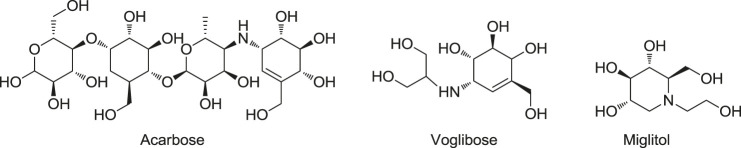
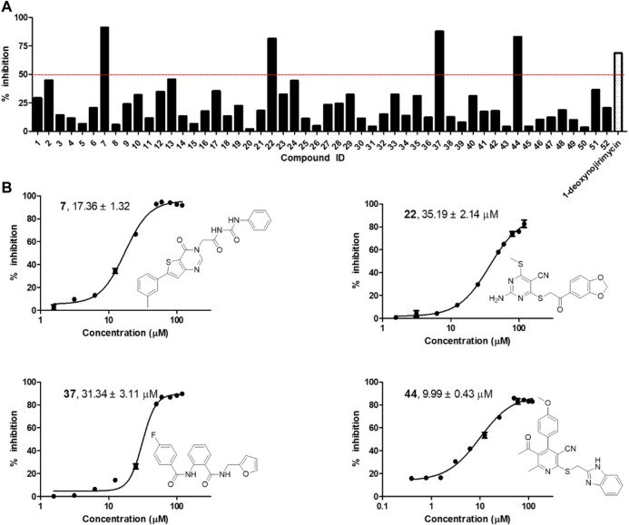
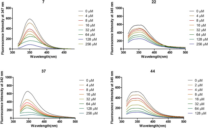
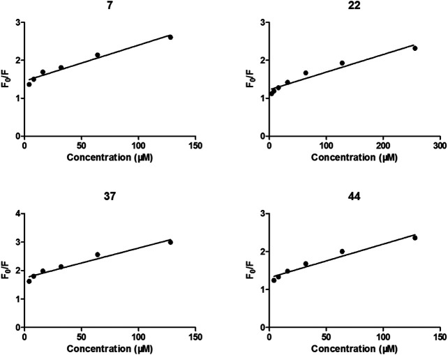
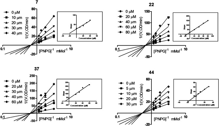
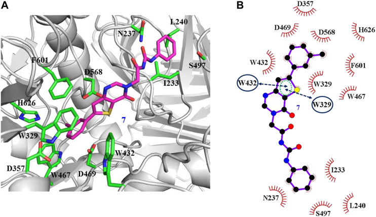
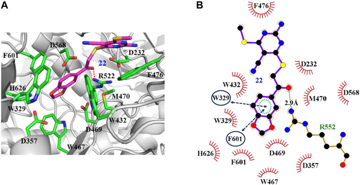
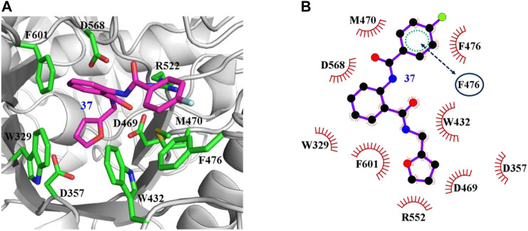
α-Glycosidase inhibitors could inhibit the digestion of carbohydrates into glucose and promote glucose conversion, which have been used for the treatment of type 2 diabetes. In the present study, 52 candidates of α-glycosidase inhibitors were selected from commercial Specs compound library based on molecular docking-based virtual screening. Four different scaffold compounds (7, 22, 37, and 44) were identified as α-glycosidase inhibitors with IC50 values ranging from 9.99 to 35.19 μM. All these four compounds exerted better inhibitory activities than the positive control (1-deoxynojirimycin, IC50 = 52.02 μM). The fluorescence quenching study and kinetic analysis revealed that all these compounds directly bind to α-glycosidase and belonged to the noncompetitive α-glycosidase inhibitors. Then, the binding modes of these four compounds were carefully investigated. Significantly, these four compounds showed nontoxicity (IC50 > 100 μM) toward the human normal hepatocyte cell line (LO2), which indicated the potential of developing into novel candidates for type 2 diabetes treatment.

Keywords: cytotoxicity; molecular docking; type 2 diabetes; virtual screening; α-glycosidase.


Conflict of interest statement

Author JIZ was employed by the company Lunan Pharmaceutical Group Corporation. The remaining authors declare that the research was conducted in the absence of any commercial or financial relationships that could be construed as a potential conflict of interest.

Figures

FIGURE 1
FIGURE 1
Clinically Approved α-glucosidase inhibitors.
FIGURE 2
FIGURE 2
(A) α-glycosidase inhibitory activity of the 52 selected candidates at 100 μM; (B) The chemical structures and IC50 curves of compounds 7, 22, 37, and 44. IC50 data are shown as mean ± SD of three independent experiments.
FIGURE 3
FIGURE 3
Variation of fluorescence emission spectra of α-glycosidase (1 U/ml) in the presence of compounds 7, 22, 37, and 44 with increasing concentration for 30 min at 37°C.
FIGURE 4
FIGURE 4
Stern-Volmer plots for the fluorescence quenching of α-glycosidase by compounds 7, 22, 37, and 44.
FIGURE 5
FIGURE 5
Kinetic assay on α-glycosidase inhibition by compounds 7, 22, 37, and 44, respectively. Lineweaver-Burk reciprocal plots of initial velocity and increasing substrate (PNPG) concentration with secondary plot of slopes vs. the concentration of compounds.
FIGURE 6
FIGURE 6
Docking pose of compound 7 bound to the acarbose binding site in α-glycosidase. (A) The three-dimensional interacting modes between 7 and α-glycosidase. α-Glycosidase, 7 and the interacting residues were shown as cartoon, sticks (carbon atoms colored in magenta), sticks (carbon atom colored in green), respectively. (B) Schematic representation displayed the hydrophobic interactions (shown as starbursts) and Pi-Pi interactions (shown as oval) of 7 with α-Glycosidase.
FIGURE 7
FIGURE 7
Docking pose of compound 22 bound to the acarbose binding site in α-glycosidase. (A) The three-dimensional interacting modes between 22 and α-glycosidase. α-Glycosidase, 22 and the interacting residues were shown as cartoon, sticks (carbon atoms colored in magenta), sticks (carbon atom colored in green), respectively. (B) Schematic representation displayed the hydrophobic interactions (shown as starbursts), Pi-Pi interactions (shown as oval), and H-bond interactions (denoted by dotted green lines) of 22 with α-Glycosidase.
FIGURE 8
FIGURE 8
Docking pose of compound 37 bound to the acarbose binding site in α-glycosidase. (A) The three-dimensional interacting modes between 37 and α-glycosidase. α-Glycosidase, 37 and the interacting residues were shown as cartoon, sticks (carbon atoms colored in magenta), sticks (carbon atom colored in green), respectively. (B) Schematic representation displayed the hydrophobic interactions (shown as starbursts) of 37 with α-Glycosidase.
FIGURE 9
FIGURE 9
Docking pose of compound 44 bound to the acarbose binding site in α-glycosidase. (A) The three-dimensional interacting modes between 44 and α-glycosidase. Compound 44 and the interacting residues were shown as cartoon, sticks (carbon atoms colored in magenta), sticks (carbon atom colored in green), respectively. (B) Schematic representation displayed the hydrophobic interactions (shown as starbursts) and Pi-Pi interactions (shown as oval) of 44 with α-Glycosidase.

References

    1. Abbas G., Al-Harrasi A., Hussain H., Hamaed A., Supuran C. T. (2019). The management of diabetes mellitus-imperative role of natural products against dipeptidyl peptidase-4, α-glucosidase and sodium-dependent glucose Co-transporter 2 (SGLT2). Bioorg. Chem. 86, 305–315. 10.1016/j.bioorg.2019.02.009 - DOI - PubMed
    1. Aguilar-Moncayo M., García-Moreno M. I., Stütz A. E., García, Fernández J. M., Wrodnigg T. M., Ortiz-Mellet C. (2010). Fluorescent-tagged sp2-iminosugars with potent β-glucosidase inhibitory activity. Bioorg. Med. Chem. 18 (21), 7439–7445. 10.1016/j.bmc.2010.09.003 - DOI - PubMed
    1. Chen H., Xiong L., Wang W. J. (2017). Reviews on α-glucosidase inhibitor from plant secondary metabolites. Zhongguo Zhong Yao Za Zhi 42 (15), 2915–2924. 10.19540/j.cnki.cjcmm.20170714.021 - DOI - PubMed
    1. Cohen P., Goedert M. (2004). GSK3 inhibitors: development and therapeutic potential. Nat. Rev. Drug Discov. 3 (6), 479–487. 10.1038/nrd1415 - DOI - PubMed
    1. Dhameja M., Gupta P. (2019). Synthetic heterocyclic candidates as promising α-glucosidase inhibitors: an overview. Eur. J. Med. Chem. 176, 343–377. 10.1016/j.ejmech.2019.04.025 - DOI - PubMed

LinkOut - more resources