. 2021 Mar 8:9:647387.
doi: 10.3389/fcell.2021.647387. eCollection 2021.

LMO7 as an Unrecognized Factor Promoting Pancreatic Cancer Progression and Metastasis

Affiliations

LMO7 as an Unrecognized Factor Promoting Pancreatic Cancer Progression and Metastasis

Xinjian Liu et al. Front Cell Dev Biol. .

Abstract

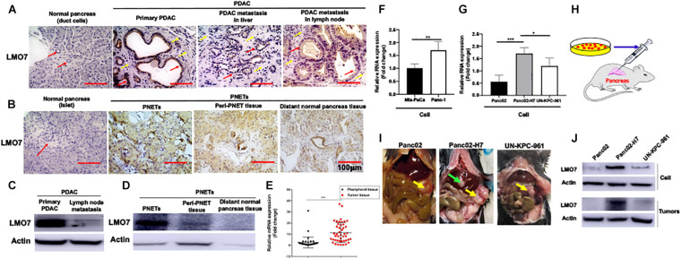
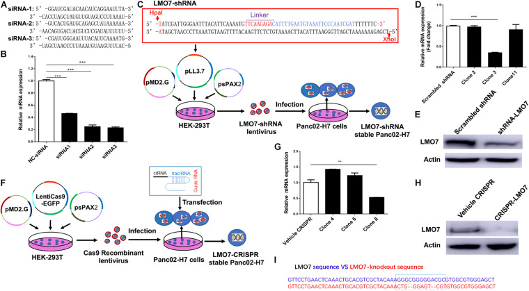
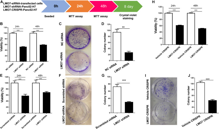
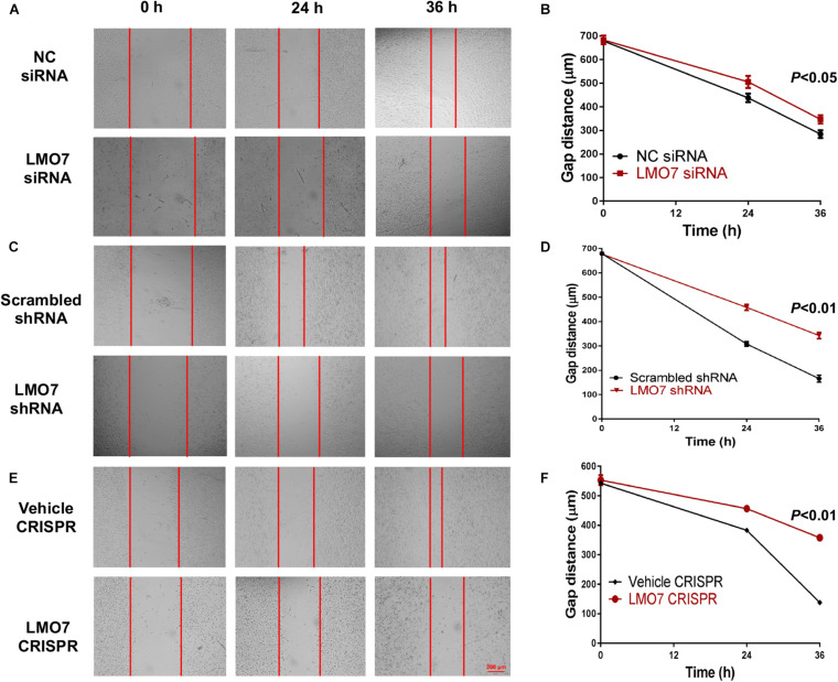
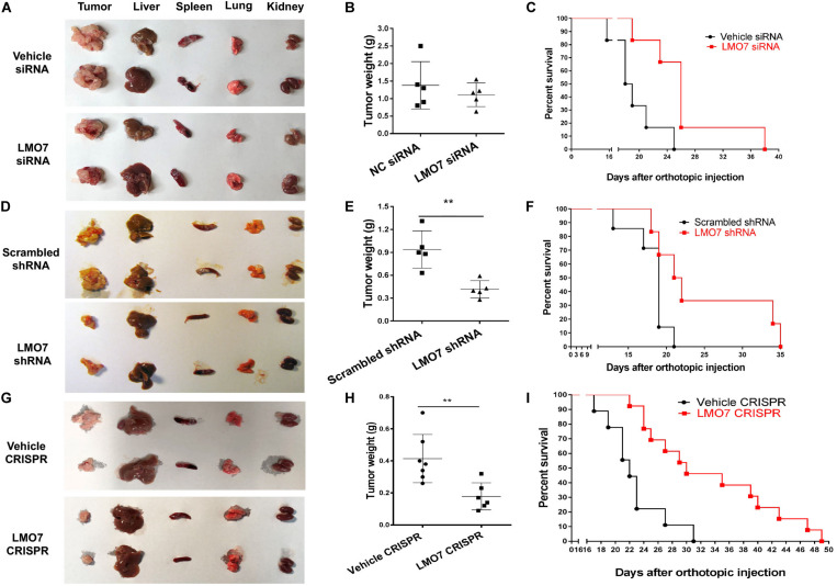
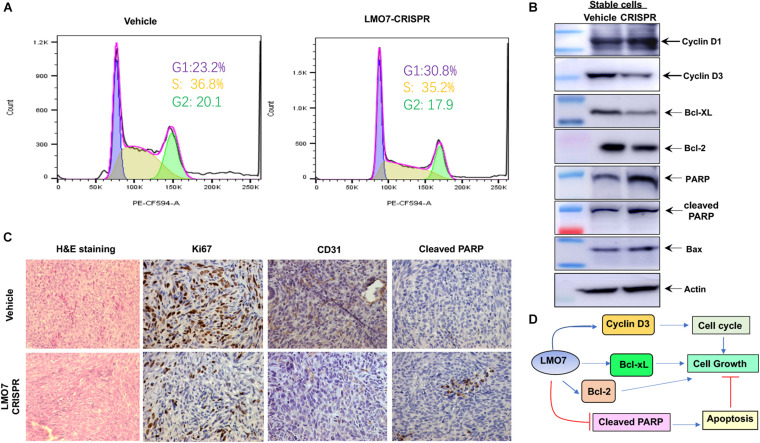
Pancreatic cancer (PC) is one of the most lethal human malignancies without effective treatment. In an effort to discover key genes and molecular pathways underlying PC growth, we have identified LIM domain only 7 (LMO7) as an under-investigated molecule, which highly expresses in primary and metastatic human and mouse PC with the potential of impacting PC tumorigenesis and metastasis. Using genetic methods with siRNA, shRNA, and CRISPR-Cas9, we have successfully generated stable mouse PC cells with LMO7 knockdown or knockout. Using these cells with loss of LMO7 function, we have demonstrated that intrinsic LMO7 defect significantly suppresses PC cell proliferation, anchorage-free colony formation, and mobility in vitro and slows orthotopic PC tumor growth and metastasis in vivo. Mechanistic studies demonstrated that loss of LMO7 function causes PC cell-cycle arrest and apoptosis. These data indicate that LMO7 functions as an independent and unrecognized druggable factor significantly impacting PC growth and metastasis, which could be harnessed for developing a new targeted therapy for PC.

Keywords: CRISPR-Cas9; LIM domain only 7 (LMO7); apoptosis; cell cycle; pancreatic cancer.


Conflict of interest statement

The authors declare that the research was conducted in the absence of any commercial or financial relationships that could be construed as a potential conflict of interest.

Figures

FIGURE 1
FIGURE 1
Increased expression of LMO7 protein and mRNA in human and mouse PC tumors. (A) Detection of LMO7 expression in human primary and metastatic PC tumors. Immunohistochemical staining was used to detect LMO7 in human normal pancreas, primary PDAC, and metastatic PDAC in liver and lymph node. Red arrows point to ductal cells in normal pancreas and PDAC tumors. Weak staining of LMO7 in normal pancreas and strong staining in PDAC tumors were shown. Yellow arrows point to remarkable desmoplasia in primary and metastatic PDAC tumors. (B) Detection of LMO7 expression in human PNETs. Immunohistochemical staining was used to detect LMO7 in normal human pancreas, PNETs, peri-PNET tissue, and distant normal pancreas tissue. Red arrow points to islet in normal pancreas without positive staining of LMO7. On the contrary, a strong staining of LMO7 was detected in primary PNETs; a modest staining of LMO7 in peri-PNET tissue and distant normal pancreas tissue. PNET displayed a typical nested organoid pattern. (C) Western blot detected the expression of LMO7 in primary and metastatic human PDAC tumors. (D) Western blot detected the expression of LMO7 in PNETs and peri-PNET tissue. (E) LMO7 mRNA expression in 45 human PDAC tumors and peritumoral tissues. The paired PDAC tumors and adjacent tissues were harvested from 45 human patients. The significant increase in LMO7 mRNA expression was detected in the tumors compared to peritumoral tissues by qPCR. (F) qPCR detected LMO7 mRNA expression with the level that is higher in human Panc-1 cells than that in Mia-PaCa-2 cells. (G) qPCR detected the LMO7 expression with the level that is higher in mouse Panc02-H7 cells than that in Panc02 cells and UN-KPC-961 cells. (H) Schematic diagram of the establishment of orthotopic murine PC models in wild-type C57BL/6 mice. (I) The representative images show orthotopic murine PC models with or without liver metastasis induced with Panc02, Panc02-H7, or UN-KPC-961 cells. Yellow arrow points to orthotopic PC tumors without liver metastasis. Green arrow points to metastatic tumors in liver. (J) Western blot detects the strong expression of LMO7 protein in Panc02-H7 cells and its derived tumors in comparison to LMO7 expression in Panc02 and UN-KPC-961 cells as well as the derived tumors. *p < 0.05; **p < 0.01; ***p < 0.001.
FIGURE 2
FIGURE 2
Knockdown or knockout of LMO7 in Panc02-H7 cells with siRNA, shRNA, and CRISPR-Cas9 technologies and establishment of relevant stable cells. (A) The sequences of three designed siRNAs for mouse LMO7. (B) qPCR detected the effective knockdown of LMO7 in Panc02-H7 cells with three designed siRNAs. (C) Schematic diagram of the establishment of stable LMO7-knockdown cells with the shRNA approach. (D) qPCR detected the reduced LMO7 mRNA expression in stable LMO7-knockdown cells developed with shRNA. (E) Western blot detected the reduced LMO7 protein expression in stable LMO7-knockdown cells developed with shRNA. (F) Schematic diagram of the establishment of stable LMO7-knockout cells with the CRISPR-Cas9 approach. (G) qPCR detected the reduced LMO7 mRNA expression in the CRISPR-induced stable LMO7-knockout cells. (H) Western blot showed an undetectable LMO7 protein expression in LMO7-CRISPR Panc02-H7 cells. (I) Sanger DNA sequencing shows the depleted nucleotides in the LMO7 gene in the stable knockout cells. **p < 0.01; ***p < 0.001.
FIGURE 3
FIGURE 3
LMO7 defect-caused suppression of cell proliferation and colony formation in Panc02-H7 cells. (A) Schematic diagram for assay of LMO7-mediated cell proliferation and colony formation. (B) MTT detected the suppression of Panc02-H7 cell proliferation post knockdown of LMO7 with siRNAs. (C) Crystal violet staining detected the reduced cell colonies in Panc02-H7 cells post knockdown of LMO7 with siRNAs. (E–G) The reduced cell proliferation and colony formation in stable LMO7-shRNA-Panc02-H7 were detected with the same methods described in (B) to (D). (H–J) The reduced cell proliferation and colony formation in stable LMO7-CRISPR-Panc02-H7 cells were detected with the same methods described in (B) to (D). n = 3, error bars represent mean ± SD. **p < 0.01; ***p < 0.001.
FIGURE 4
FIGURE 4
Silencing LMO7 suppresses Panc02-H7 cell motility. LMO7-siRNA-transfected Panc02-H7 cells, stable LMO7-shRNA-Panc02-H7 cells, or stable LMO7-CRISPR-Panc02-H7 cells were seeded into the 24-well plate with an insert at a dose of 2 × 10⁵ cells/well. On the second day, the inserts were removed and the cell-free gap was measured in the indicated times. Representative images showed the reduced widths of cell-free gaps in siRNA-knockdown Panc02-H7 cells (A,B), stable LMO7-shRNA-Panc02-H7 cells (C,D), or LMO7-CRISPR-Panc02-H7 cells (E,F) in comparison to that in the relevant control cells. n = 3, error bars represent mean ± SD.
FIGURE 5
FIGURE 5
Silencing LMO7 in Panc02-H7 cells slows orthotopic tumor growth and extends the lifespan in recipient mice. LMO7-siRNA-transfected Panc02-H7 cells, stable LMO7-shRNA-Panc02-H7 cells, or stable LMO7-CRISPR-Panc02-H7 cells and corresponding control cells were injected into the head of the pancreas of mice at a dose of 2.5 × 10⁵ cells per mouse. Half of these mice were used to measure tumor size; the rest of them were used to measure the lifespan. 17 days post cell inoculation, some mice were euthanized for isolating tumors and different organs, and the tumors were weighed. Some mice were maintained to daily count survival over time, and the survival curves were made with Kaplan–Meier method. The representative images of tumors and different organs including spleen, liver, lung, and kidney in each mouse were shown (A,D,G); accumulated tumor weights are shown in (B,E,H), and viable mice in each group are shown in (C,F,I). Error bars represent mean ± SD. **p < 0.01.
FIGURE 6
FIGURE 6
LMO7 defect causes cell-cycle arrest and cell apoptosis. (A) Cell-cycle arrest. Flow cytometric assay with PI detected the different frequencies of cells at phases G1, S, and G2 presented in stable LMO7-CRISPR-Panc02-H7 cells and the control cells. (B) Expression of canonical molecules important for cell cycle and apoptosis. Western blotting detected the decreased expression of cyclin D3, Bcl-XL, and Bcl-2 and increased expression of cyclin D1, PARP, cleaved PARP, and Bax in stable LMO7-CRISPR-Panc02-H7 cells in comparison to their control cells. (C) Expression of growth and apoptotic proteins in the tumors. The tumors were harvested and then fixed with formalin to prepare slides. H&E staining showed the tumor structure (left panel); IHC detected the reduced expression of Ki67 and CD31 (middle two panel) and increased expression of cleaved PARP (right panel). (D) A schematic diagram of LMO7-mediated signaling transduction pathways in PC.

References

    1. Amanam I., Chung V. (2018). Targeted therapies for pancreatic cancer. Cancers (Basel) 10:36. 10.3390/cancers10020036 - DOI - PMC - PubMed
    1. Bayard Q., Caruso S., Couchy G., Rebouissou S., Bioulac Sage P., Balabaud C., et al. (2020). Recurrent chromosomal rearrangements of ROS1, FRK and IL6 activating JAK/STAT pathway in inflammatory hepatocellular adenomas. Gut 69 1667–1676. 10.1136/gutjnl-2019-319790 - DOI - PubMed
    1. Chu M., Wang T., Sun A., Chen Y. (2018). Nimesulide inhibits proliferation and induces apoptosis of pancreatic cancer cells by enhancing expression of PTEN. Exp. Ther. Med. 16 370–376. 10.3892/etm.2018.6191 - DOI - PMC - PubMed
    1. di Marco M., Grassi E., Durante S., Vecchiarelli S., Palloni A., Macchini M., et al. (2016). State of the art biological therapies in pancreatic cancer. World J. Gastrointest. Oncol. 8 55–66. 10.4251/wjgo.v8.i1.55 - DOI - PMC - PubMed
    1. Du T. T., Dewey J. B., Wagner E. L., Cui R., Heo J., Park J. J., et al. (2019). LMO7 deficiency reveals the significance of the cuticular plate for hearing function. Nat. Commun. 10:1117. 10.1038/s41467-019-09074-4 - DOI - PMC - PubMed

LinkOut - more resources