Review
. 2021 Mar 8:9:653101.
doi: 10.3389/fcell.2021.653101. eCollection 2021.

Neuron-Oligodendrocyte Interactions in the Structure and Integrity of Axons

Affiliations
Review

Neuron-Oligodendrocyte Interactions in the Structure and Integrity of Axons

Greg J Duncan et al. Front Cell Dev Biol. .

Abstract

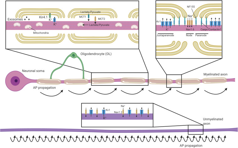
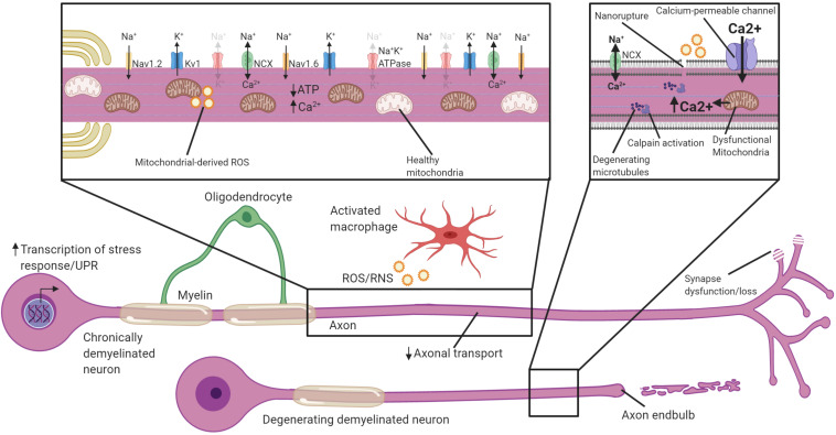
The myelination of axons by oligodendrocytes is a highly complex cell-to-cell interaction. Oligodendrocytes and axons have a reciprocal signaling relationship in which oligodendrocytes receive cues from axons that direct their myelination, and oligodendrocytes subsequently shape axonal structure and conduction. Oligodendrocytes are necessary for the maturation of excitatory domains on the axon including nodes of Ranvier, help buffer potassium, and support neuronal energy metabolism. Disruption of the oligodendrocyte-axon unit in traumatic injuries, Alzheimer's disease and demyelinating diseases such as multiple sclerosis results in axonal dysfunction and can culminate in neurodegeneration. In this review, we discuss the mechanisms by which demyelination and loss of oligodendrocytes compromise axons. We highlight the intra-axonal cascades initiated by demyelination that can result in irreversible axonal damage. Both the restoration of oligodendrocyte myelination or neuroprotective therapies targeting these intra-axonal cascades are likely to have therapeutic potential in disorders in which oligodendrocyte support of axons is disrupted.

Keywords: Wallerian degeneration; axonal degeneration; demyelination; mitochondria; multiple sclerosis; oligodendrocyte; remyelination.


Conflict of interest statement

BE is a co-founder of Autobahn Therapeutics. The remaining authors declare that the research was conducted in the absence of any commercial or financial relationships that could be construed as a potential conflict of interest.

Figures

FIGURE 1
FIGURE 1
Oligodendrocytes regulate axonal structure, conduction and support their survival. Schematic of an axon myelinated by an oligodendrocyte and an unmyelinated axon. Myelin permits saltatory conductance where action potentials are generated at the nodes of Ranvier. The high membrane resistance and low capacitance generated by the myelin sheath promotes rapid current flow along the myelinated section of the axon to the next node, greatly increasing conduction velocities relative to unmyelinated axons. Oligodendrocytes contact axons at the paranode (via NF155-Caspr/Contactin1) and are crucial for the organization, clustering and maintenance of sodium channels (primarily Nav1.6) at the nodes, as well as Kv1 potassium channels at the juxtaparanodes. In contrast, unmyelinated axons have potassium and sodium (primarily Nav1.2 channels) channels dispersed along the axon and are not confined to discrete excitatory clusters. Oligodendrocytes secrete exosomes that can support neuronal health and buffer potassium via the expression of Kir4.1. Oligodendrocytes provide glycolysis byproducts via monocarboxylate transporters (MCTs), which enter the Krebs cycle and are critical to support axonal metabolism.
FIGURE 2
FIGURE 2
Neuronal adaptations to acute demyelination. Schematic of a partially demyelinated neuron early after demyelination. Conduction is reestablished through the demyelinated segment by the increased expression of sodium channels along the axolemma, but it is notably slower. Demyelinated axons require greater Na+ entry to depolarize the axon, necessitating increased activity of the Na+K+ATPase. Mitochondria increase in number and size within demyelinated axons to meet the higher demand for ATP and also uptake Ca2+. If Na+K+ATPase has sufficient ATP, the NCX is rarely activated in the reverse direction (faded arrows). Transcriptional changes occur within the neuron in response to demyelination and could be critical to these adaptions. Faded text and indicates low activity or levels, bolded text or thick arrows indicates increased levels following demyelination.
FIGURE 3
FIGURE 3
Potential mechanisms by which chronically demyelinated axons degenerate. Schematic of a chronically demyelinated intact axon and an additional demyelinated axon undergoing degeneration. Demyelinated axons are exposed to inflammatory mediators including ROS/RNS in MS, have reduced axonal transport, and may have synapse dysfunction/loss. The chronically demyelinated axon is likely to be in an energy crisis in which the lack of oligodendrocyte support coupled with mitochondrial damage and increased energetic demands to sustain AP propagation means there is a shortfall of ATP necessary to drive the Na+K+ATPase. This causes a reversal of the NCX to remove Na+ from the axon, but at the cost of calcium entry. Disruption of the plasma membrane, and calcium entry through calcium-permeable channels including glutamate receptors, ASICs, VGCC and mitochondrial release cause calcium to accumulate in the axon. Intra-axonal calpains are activated when calcium accumulates to high levels and begin the process of degeneration and breakdown critical cytoskeletal structures including microtubules. Activation of cell-stress pathways and the unfolded protein response also are present in many demyelinated neurons that are susceptible to degeneration. Faded text and indicates low activity or levels, bolded text or thick arrows indicates increased activated.

References

    1. Agarwal D., Sandor C., Volpato V., Caffrey T. M., Monzon-Sandoval J., Bowden R., et al. (2020). A single-cell atlas of the human substantia nigra reveals cell-specific pathways associated with neurological disorders. Nat. Commun. 11:4183. 10.1038/s41467-020-17876-0 - DOI - PMC - PubMed
    1. Aggarwal S., Yurlova L., Simons M. (2011). Central nervous system myelin: structure, synthesis and assembly. Trends Cell Biol. 21 585–593. 10.1016/j.tcb.2011.06.004 - DOI - PubMed
    1. Aharoni R., Vainshtein A., Stock A., Eilam R., From R., Shinder V., et al. (2011). Distinct pathological patterns in relapsing-remitting and chronic models of experimental autoimmune enchephalomyelitis and the neuroprotective effect of glatiramer acetate. J. Autoimmun. 37 228–241. 10.1016/j.jaut.2011.06.003 - DOI - PubMed
    1. Al-Izki S., Pryce G., Hankey D. J., Lidster K., Von Kutzleben S. M., Browne L., et al. (2014). Lesional-targeting of neuroprotection to the inflammatory penumbra in experimental multiple sclerosis. Brain 137 92–108. 10.1093/brain/awt324 - DOI - PubMed
    1. Alrashdi B., Dawod B., Schampel A., Tacke S., Kuerten S., Marshall J. S., et al. (2019). Nav1.6 promotes inflammation and neuronal degeneration in a mouse model of multiple sclerosis. J. Neuroinflammation 16:215. 10.1186/s12974-019-1622-1 - DOI - PMC - PubMed

LinkOut - more resources