. 2021 Mar 8:8:630625.
doi: 10.3389/fmolb.2021.630625. eCollection 2021.

Membrane Remodeling by Arc/Arg3.1

Affiliations

Membrane Remodeling by Arc/Arg3.1

Per Niklas Hedde et al. Front Mol Biosci. .

Abstract

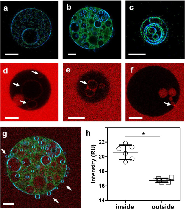
The activity-regulated cytoskeletal-associated protein (Arc, also known as Arg3.1) is an immediate early gene product induced by activity/experience and required for multiple modes of synaptic plasticity. Both long-term potentiation (LTP) and long-term depression (LTD) are impaired upon Arc deletion, as well as the ability to form long-term spatial, taste and fear memories. The best-characterized cellular function of Arc is enhancement of the endocytic internalization of AMPA receptors (AMPARs) in dendritic spines. Solution of the crystal structure of a C-terminal segment of Arc revealed a striking similarity to the capsid domain of HIV Gag. It was subsequently shown that Arc assembles into viral capsid-like structures that enclose Arc mRNA, are released into the extracellular space, and are internalized by neighboring cells. Thus, Arc is unique in participating in plasma membrane budding both into and out of the cell. In this report we study the interaction of Arc with membranes using giant unilamellar vesicles (GUVs). Using the fluorescent lipid probe LAURDAN, we find that Arc promotes the formation of smaller vesicles that penetrate into the GUV interior. Our results suggest that Arc induces negative membrane curvature and may therefore facilitate the formation of mRNA-containing extracellular vesicles from the plasma membrane.

Keywords: Arc; GUV; fluorescence; giant unilamellar vesicle; membrane budding; membrane remodeling.


Conflict of interest statement

The authors declare that the research was conducted in the absence of any commercial or financial relationships that could be construed as a potential conflict of interest.

Figures

FIGURE 1
FIGURE 1
Removal of nucleic acids from Arc preparations by ammonium sulfate precipitation. (A) Absorption spectra of Arc preparations obtained without AS treatment (A260/280: 0.96), and of supernatant (diluted 20-fold; A260/280: 2.08) and pellet (A260/280: 0.57) obtained after AS precipitation (see Materials and Methods). Arc concentrations were 17.6 µM (no AS) and 16 µM (after AS). (B) Coomassie blue-stained gel of Arc prepared without and with AS treatment. (C) Superdex 200 elution profiles of Arc prepared with or without AS treatment. One ml of 60 µM (without AS) or 80 µM (with AS) Arc was loaded. Arrows designate standards: thyroglobulin (RS: 19 nm), catalase (RS: 5.2 nm), and BSA (RS: 3.67 nm). Chromatography was performed at 4°C.
FIGURE 2
FIGURE 2
Exemplary fluorescence images of GUVs with and without protein addition (A–C) GUVs formed from DOPC and labeled with LAURDAN without protein addition (D–F) LAURDAN-labeled vesicles 2–60 min after addition of Arc (G–I) LAURDAN-labeled vesicles 2–60 min after addition of Arc-Alexa 594 (J) LAURDAN and (K) Arc-Alexa 594 fluorescence image of the same field of view (L) Fluorescence intensity of image pixels located on the membrane and outside the GUV shown in panel (K); bars with whiskers indicate mean ± standard error (SE). Groups were compared with the Mann-Whitney U test (*) p < 0.05. Scale bars, 20 µm.
FIGURE 3
FIGURE 3
Percentages of GUVs showing internal structures with and without addition of Arc, horizontal lines represent the averages, boxes represent standard errors (± SE), and whiskers indicate standard deviations (± SD). No treatment controls were carried out at 0–20 min time (initial control) and at 60 + min. GUVs treated with Arc or Arc-Alexa594 (2–9 µM) as well as buffer and EGFP (5 µM) controls were examined from 2–60 min after treatment. Groups were compared with the Mann-Whitney U test, p values were < 0.01 (**) for Arc vs control +60 min, Arc vs buffer, and Arc vs EGFP, and < 0.001 (***) for Arc vs control, Arc-Alexa594 vs control, Arc-Alexa594 vs control + 60 min, Arc-Alexa594 vs buffer, and Arc-Alexa594 vs EGFP. Each data point represents the fraction of GUVs with internal structures averaged over 2–4 field of views (each 354 µm across). The total number of GUVs imaged was N = 217, comprised of N = 91 for the control, N = 22 for Arc, N = 36 for Arc-Alexa594, N = 32 for control + 60 min, N = 21 for buffer, and N = 15 for EGFP. For each group, at least three independent experiments were carried out.
FIGURE 4
FIGURE 4
(A–C) Fluorescence intensity images of LAURDAN-labeled GUV composed of a ternary lipid mixture. Blue channel: 449/10 nm, green channel: 527/10 nm (D–F) Arc-Alexa647 fluorescence in the presence of GUVs. Arrows indicate Alexa 647-labeled Arc bound to the membranes of internal vesicles (D,E) and Arc protein populating the GUV interior (F). (G) Red-green-blue overlay of LAURDAN and Alexa 647 fluorescence (red channel: 683/10 nm) of the GUV shown in panel (B). (H) The Alexa 647 fluorescence was significantly higher in pixels inside compared to pixels outside the vesicle. Groups were compared with the Mann-Whitney U test (*) p < 0.05. Bars with whiskers indicate mean ± standard error (SE). Scale bars, 10 µm.

Similar articles

Cited by

References

    1. Angelova M. I., Dimitrov D. S. (1986). Liposome electroformation. Faraday Discuss. Chem. Soc. 81, 303–311. 10.1039/DC9868100303 - DOI
    1. Ashley J., Cordy B., Lucia D., Fradkin L. G., Budnik V., Thomson T. (2018). Retrovirus-like gag protein Arc1 binds RNA and traffics across synaptic boutons. Cell 172, 262–274. 10.1016/j.cell.2017.12.022 - DOI - PMC - PubMed
    1. Bacia K., Schuette C. G., Kahya N., Jahn R., Schwille P. (2004). SNAREs prefer liquid-disordered over “raft” (liquid-ordered) domains when reconstituted into giant unilamellar vesicles. J. Biol. Chem. 279, 37951–37955. 10.1074/jbc.M407020200 - DOI - PubMed
    1. Bagatolli L. A., Sanchez S. A., Hazlett T., Gratton E. (2003). Giant vesicles, laurdan, and two-photon fluorescence microscopy: evidence of lipid lateral separation in bilayers. Meth Enzymol. 360, 481–500. 10.1016/S0076-6879(03)60124-2 - DOI - PubMed
    1. Barylko B., Wilkerson J. R., Cavalier S. H., Binns D. D., James N. G., Jameson D. M., et al. (2018). Palmitoylation and membrane binding of Arc/Arg3.1: a potential role in synaptic depression. Biochemistry 57, 520–524. 10.1021/acs.biochem.7b00959 - DOI - PMC - PubMed

LinkOut - more resources