

Differential Signals From TNFα-Treated and Untreated Embryos in Uterine Tissues and Splenic CD4+ T Lymphocytes During Preimplantation Pregnancy in Mice
- PMID: 33763465
- PMCID: PMC7982469
- DOI: 10.3389/fvets.2021.641553
Differential Signals From TNFα-Treated and Untreated Embryos in Uterine Tissues and Splenic CD4+ T Lymphocytes During Preimplantation Pregnancy in Mice
Abstract
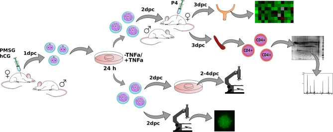
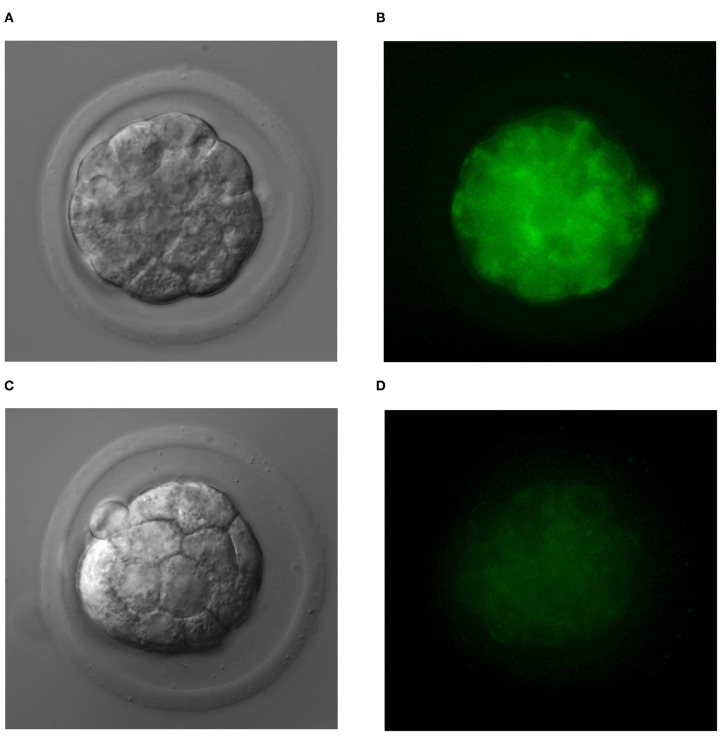
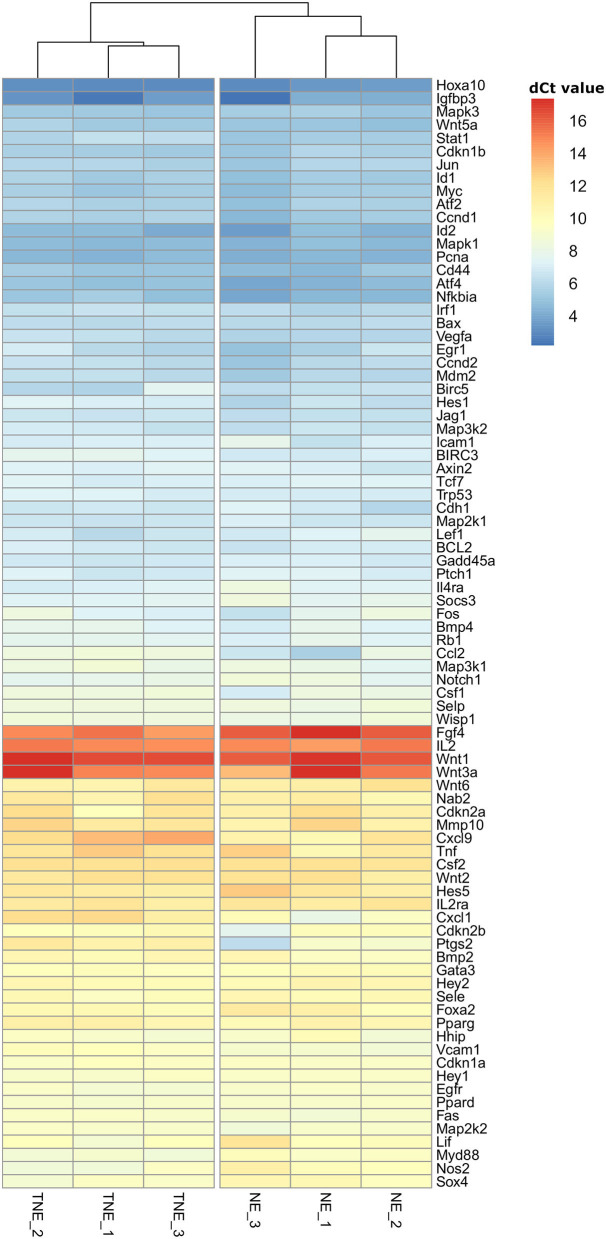
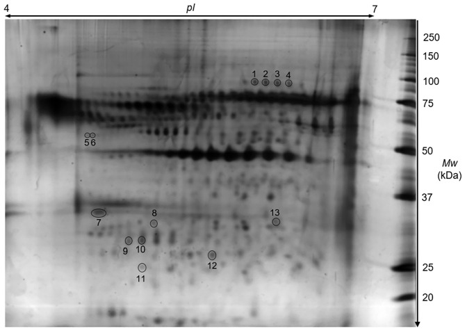
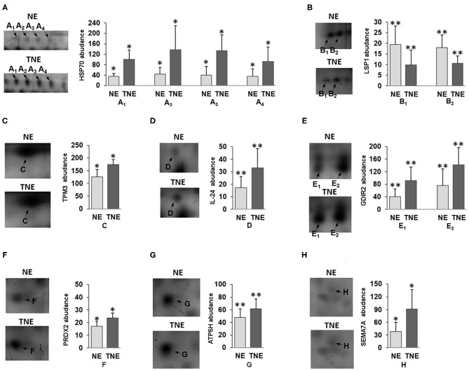
The main aim of this study was to examine if a female mouse body in preimplantation pregnancy can distinguish between embryos of normal and impaired biological quality in the local and peripheral compartments. Normal (control group) and TNFα (tumor necrosis factor-α)-treated embryos (experimental group) at the morula stage were non-surgically transferred into the uteri of CD-1 strain [Crl:CD1(Icr)] female murine recipients. Twenty-four hours after the embryo transfer, females were euthanised, and uteri and spleens were dissected. In uterine tissues (local compartment), we assessed the expression of 84 genes comprising nine signal transduction pathways, using a modified RT2 Profiler PCR Array. In the spleen (peripheral compartment), we determined the proteome of splenic CD4+ lymphocytes using 2D protein electrophoresis with subsequent protein identification by mass spectrometry. Sample clustering and differential gene expression analyses within individual signal transduction pathways revealed differential expression of genes in the uteri of females after transplantation of normal vs. TNFα-treated embryos. The most affected signal transduction cascade was the NFKB (Nuclear factor NF-kappa-B) pathway, where 87.5% of the examined genes were significantly differentially expressed. Proteomic analysis of splenic CD4+ T lymphocytes revealed significant differential expression of 8 out of 132 protein spots. Identified proteins were classified as proteins influenced by cell stress, proteins engaged in the regulation of cytoskeleton stabilization and cell motility, and proteins having immunomodulatory function. These results support the hypothesis that even before embryo implantation, the body of pregnant female mice can sense the biological quality of an embryo both at the local and peripheral level.
Keywords: CD4 lymphocytes; T cells; TNF; embryo; pregnancy; preimplantation; proteome; uterus.
Copyright © 2021 Buska-Mach, Kedzierska, Lepczynski, Herosimczyk, Ozgo, Karpinski, Gomulkiewicz, Lorek, Slawek, Dziegiel and Chelmonska-Soyta.
Conflict of interest statement
The authors declare that the research was conducted in the absence of any commercial or financial relationships that could be construed as a potential conflict of interest.
Figures
Similar articles
-
Proteome of spleen CD4 lymphocytes in mouse preimplantation pregnancy.J Physiol Pharmacol. 2014 Oct;65(5):719-31. J Physiol Pharmacol. 2014. PMID: 25371532
-
Global decrease in the expression of signalling pathways' genes in murine uterus during preimplantation pregnancy.Reprod Biol. 2017 Mar;17(1):89-96. doi: 10.1016/j.repbio.2017.01.003. Epub 2017 Feb 15. Reprod Biol. 2017. PMID: 28215431
-
Proteomic analysis of proteins differentially expressed in uterine lymphocytes obtained from wild-type and NOD mice.J Cell Biochem. 2009 Oct 1;108(2):447-57. doi: 10.1002/jcb.22271. J Cell Biochem. 2009. PMID: 19623579
-
Effect of tumour necrosis factor-alpha (TNF-alpha) on protein synthesis by mouse preimplantation stage embryos in vitro.Indian J Physiol Pharmacol. 2005 Apr;49(2):139-47. Indian J Physiol Pharmacol. 2005. PMID: 16170981
-
Proteome of the early embryo-maternal dialogue in the cattle uterus.J Proteome Res. 2012 Feb 3;11(2):751-66. doi: 10.1021/pr200969a. Epub 2011 Dec 23. J Proteome Res. 2012. PMID: 22148898
References
LinkOut - more resources
Full Text Sources
Other Literature Sources
Research Materials