Case Reports
. 2021 Mar 19;8(3):e00546.
doi: 10.14309/crj.0000000000000546. eCollection 2021 Mar.

Pancreatic Ganglioneuroma Presenting in an Octogenarian

Affiliations
Case Reports

Pancreatic Ganglioneuroma Presenting in an Octogenarian

Aciel Ahmed Shaheen et al. ACG Case Rep J. .

Abstract

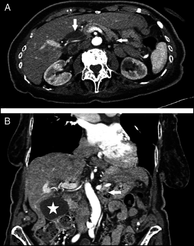
Pancreatic ganglioneuromas occur mostly in children and rarely in young adults, with no cases reported in adults older than 60 years. An 86-year-old-woman, with active advanced multiple myeloma, presented with epigastric pain for 2 days. Abdominal and pelvic computed tomography demonstrated a distended gallbladder, mildly dilated biliary tree, and a 13 × 8-mm hypodense mass in pancreatic body, without extrapancreatic invasion at endoscopic ultrasound. Fine-needle endoscopic ultrasound-guided core biopsy revealed characteristic histopathology of ganglioneuroma, as confirmed by immunohistochemical positivity for S100, SOX-10, and synaptophysin. This demonstrates novel finding of pancreatic ganglioneuroma occurring in the elderly. Lesion inclusion in the differential diagnosis may mandate tissue for pathologic diagnosis and complete lesion resection.


Figures

Figure 1.
Figure 1.
Abdominal computed tomography (A) axial section showing 13 × 8-mm hypodense pancreatic mass (horizontal arrow), and dilated CBD at 13 mm wide (vertical arrow) and (B) coronal section showing hypodense pancreatic mass (arrow), and dilate gallbladder (star).
Figure 2.
Figure 2.
Endoscopic ultrasound showing irregular, hypoechoic mass in the pancreatic body with well-defined borders (arrow). The mass measures 14 mm × 9 mm as indicated by measuring dots.
Figure 3.
Figure 3.
Histopathology of ganglioneuroma tumor (A) medium power photomicrograph of hematoxylin and eosin–stained section of tumor shows an admixture of small-sized, prominent, spindle-shaped Schwann cells (*), mostly present on the left, and clusters of large ganglion cells (arrows), mostly present on the right. (B) S-100 immunohistochemical positivity (brown color) present in nuclei and cytoplasm of Schwann cell component (*), as well as in nuclei of the larger ganglion cells (arrows). (C) SOX-10 immunohistochemical positivity (brown color) present in nuclei and cytoplasm of Schwann cell component (*). The ganglion cell component of the tumor lacks immunohistochemical positivity for SOX-10.

References

    1. Decarolis B, Thorsten S, Krug B, et al. . Treatment and outcome of ganglioneuroma and ganglioneuroblastoma intermixed. BMC Cancer. 2016;16:542. - PMC - PubMed
    1. Ikoma N, Santamaria-Barria JA, Wray C, Tsao K. Ganglioneuroma of the pancreas in a 4-year-old girl. BMJ Case Rep. 2016;2016:bcr2016217425. - PMC - PubMed
    1. Mazzola M, Bertoglio C, Achilli P, et al. . Pancreatic ganglioneuroma: A rare entity with a difficult approach: A case report and systematic review. Dig Med Res. 2019;2 (doi: 10.21037/dmr.2019.11.02).
    1. Fang CW, Wang JS, Wu TT, Lin JT. Occurrence of paratesticular ganglioneuroma 18 years after concurrent adrenal ganglioneuroma and papillary thyroid carcinoma – A case report. BMC Cancer. 2019;19:1265. - PMC - PubMed
    1. Adas M, Koc B, Adas G, Ozulker F, Aydin T. Ganglioneuroma presenting as an adrenal incidentaloma: A case report. J Med Case Rep. 2014;8:131. - PMC - PubMed

Publication types